MRI t1 contrasting agent comprising manganese oxide nanoparticle

a manganese oxide nanoparticle and contrasting agent technology, applied in the field of mno nanoparticles as mri t1 contrasting agent, can solve the problems of insufficient amount of contrasting brain or other, insufficient amount of mri, and toxicity of gadolinium ion, so as to remove the toxicity of mn2+, and improve the effect of contrasting brain and other aspects

Inactive Publication Date: 2012-05-10
SEOUL NAT UNIV R&DB FOUND
View PDF0 Cites 15 Cited by
  • Summary
  • Abstract
  • Description
  • Claims
  • Application Information

AI Technical Summary

Benefits of technology

[0062]Firstly, the MnO nanoparticles according to the present invention make it possible to produce bright T1 weighted imaging of various organs such as brain, liver, kidney, spinal cord, etc.; to visualize anatomic structures of brain due to high intracellular uptake, particularly due to the passage through blood brain barrier (BBB); and to image human cells and blood vessels by removing the toxicity of Mn2+.
[0063]Secondly, the conjugation of the MnO nanoparticle with targeting agents allows the target imaging of cells such as cancer, tumors, etc.; monitoring of expression and migration of cells such as stem cells, in cytotherapy since it is easy to modify the surface of the MnO nanoparticles of the present invention.

Problems solved by technology

However, Gadolinium ion is very toxic, and thus in order to prevent this, Gadolinium ion is used in the form of a chelate or a polymer-bound compound.
Consequently, MEMRI has intrinsic limitations to be further developed for human brain application.
Ordinarily, approximately 5 μmol / kg (0.5 ml / kg) can be administered to humans, however this amount is completely insufficient for contrasting the brain or other organs (ref.
However, the conventional positive contrasting agents have limitations in human application since the conventional positive contrasting agents composed of paramagnetic metal ions for derivatives thereof are toxic.
Also, the conventional positive contrasting agents have a short residence time in blood.
Furthermore, it is difficult to conjugate targeting agents with he conventional positive contrasting agents due to steric hindrance of the ligand of the complex.
However, the inherent magnetism of the SPIO nanoparticle shortens its T2 relaxation time, and thus produces the magnetic field which distorts MRI image.
Moreover, the inherent magnetism of the SPIO nanoparticle causes a blooming effect on the magnetic field near the SPIO nanoparticle and thus produces signal loss or distortions in the background image, which makes it impossible to obtain the proximate anatomical images.

Method used


Image

  • MRI t1 contrasting agent comprising manganese oxide nanoparticle
  • MRI t1 contrasting agent comprising manganese oxide nanoparticle
  • MRI t1 contrasting agent comprising manganese oxide nanoparticle

Examples


example 2

Biocompatibility and Contrast Ability of MnO Nanoparticles Coated with PEG

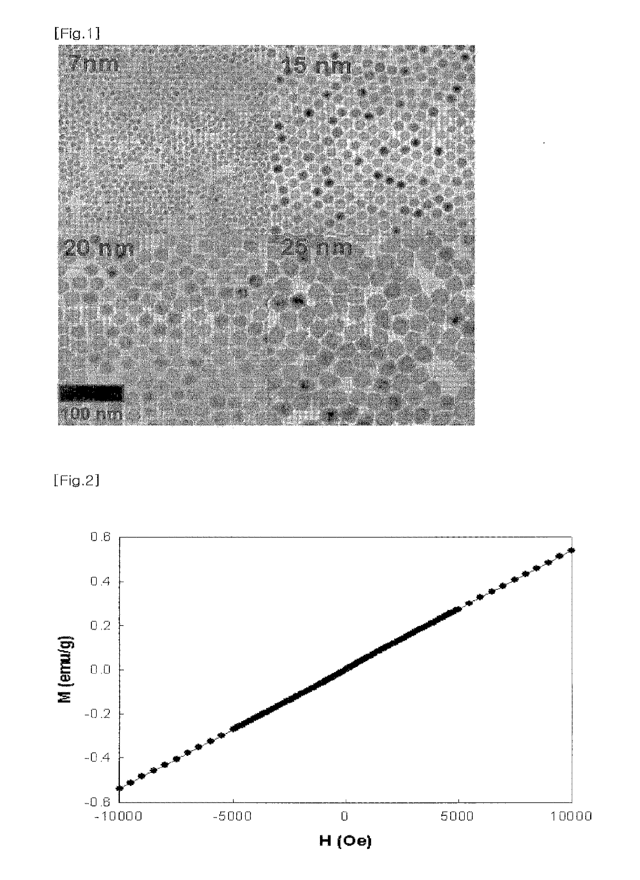
[0082]The sizes of nanoparticles prepared in Example 1 were very uniform and could be controllable. Also, the nanoparticles were biocompatible due to the coating with PEG, and stable over several months.

[0083]When the size of the MnO nanoparticle including a biocompatible material layer was more than 500 nm, the MnO nanoparticle coated with a biocompatible material was degraded by the immune system or in the liver, and thus residence time of the MnO nanoparticle in a living organism was decreased, resulting in decrease in MRI scanning time. Therefore, the size of the MnO nanoparticle including a biocompatible material layer should be preferably no more than 500 nm and more preferably no more than 100 nm.

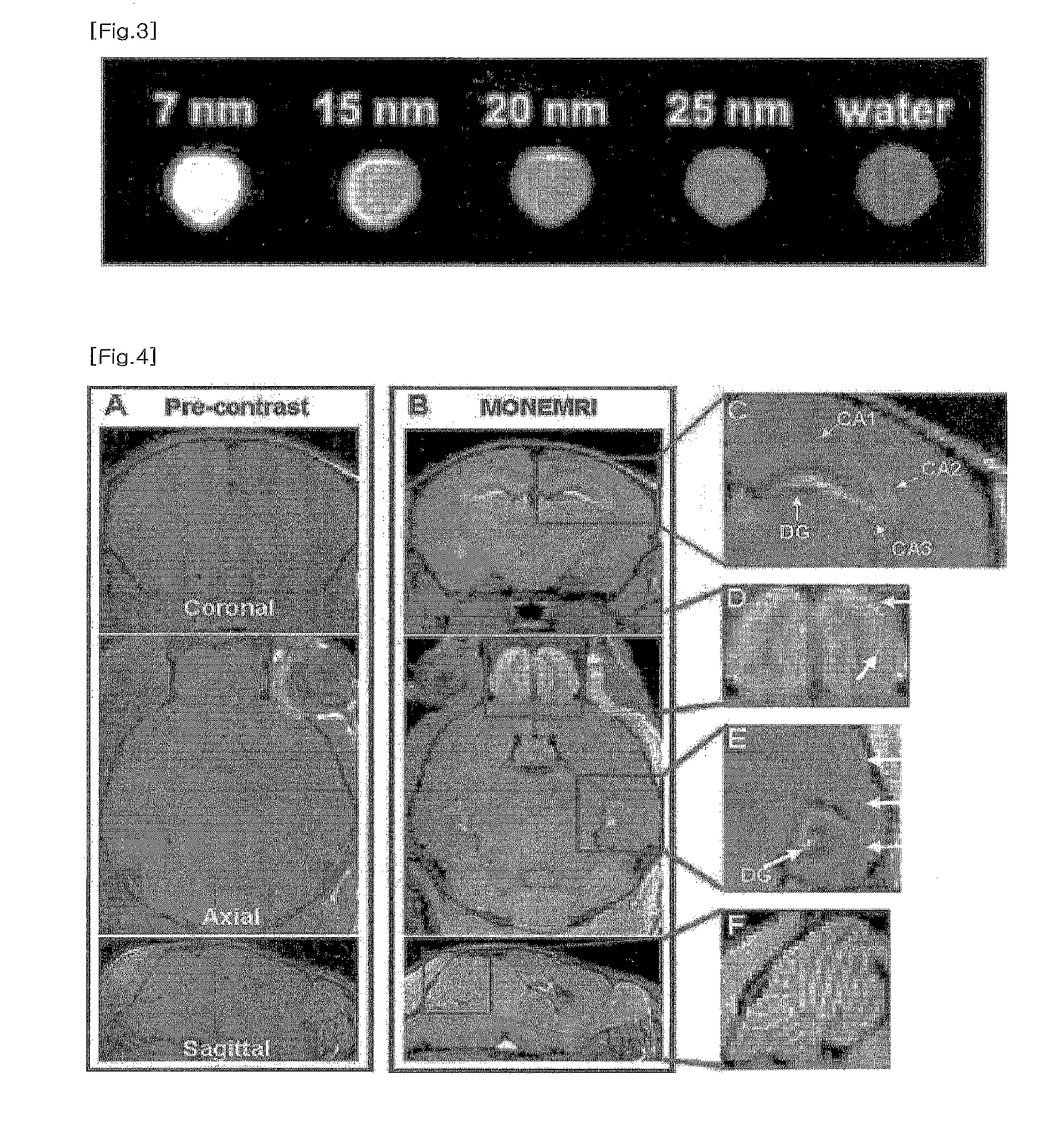
[0084]The contrast ability of MnO nanoparticles for MRI were tested with 3.0 T clinical MRI system. As shown in FIG. 2, the MnO nanoparticles at the concentration of 5 mM clearly showed bright signal enhancemen...

example 3

Manganese Oxide Nanoparticles Enhanced MR Imaging (MONEMRI)

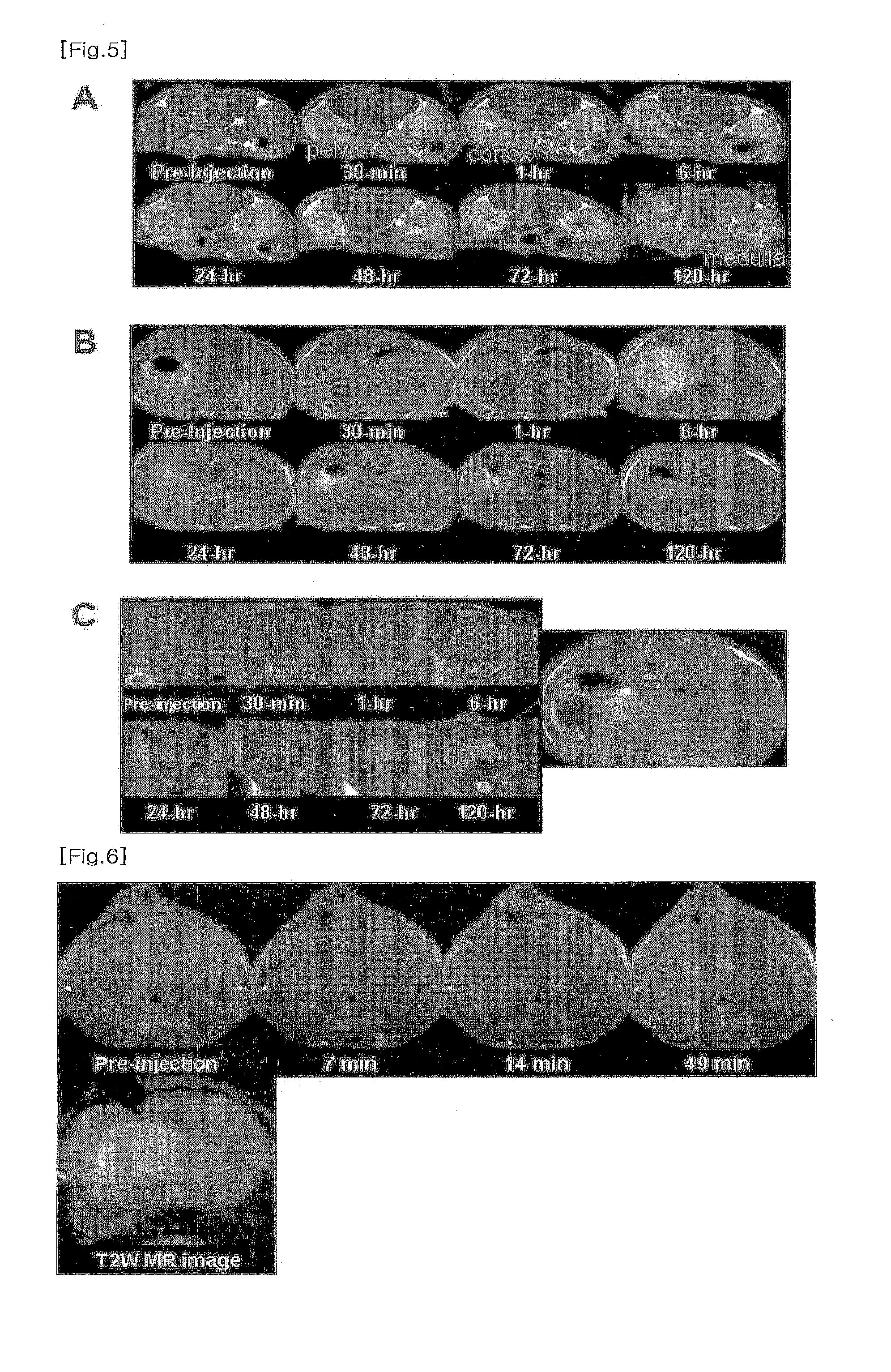
[0085]MONEMRI of a mouse was observed by using the MnO nanoparticles of the present invention. The MRI experiment was carried on a 4.7T / 30 MRI system (Brucker-Biospin, Fallanden, Switzerland). The 25 nm sized MnO nanoparticles were bolus injected to a mouse through a tail vein, for the in vivo MRI imaging. The experimental conditions were as follows:

[0086]3-1. MRI Imaging Conditions of Brain

[0087]fast spin-echo T1-weighted MRI sequence

[0088]TR / TE=300 / 12.3 ms

[0089]echo train length=2

[0090]140 m 3D isotropic resolution

[0091]FOV=2.56×1.28×1.28 cm3

[0092]matrix size=256×128×128

[0093]3-2. MRI Imaging Conditions of Abdomen

[0094]fast spin-echo T1-weighted MRI sequence

[0095]TR / TE=400 / 12 ms

[0096]NEX=16

[0097]slice thickness=1.5 mm

[0098]FOV=2.78×168 cm2

[0099]matrix size=192×192

[0100]The resulting excellent MRI images of the mouse brain (FIG. 4) depicting fine anatomic structure were obtained, comparing with the MRI images without the ...

example 4

Preparation of Targeting Probe Conjugated MnO Nanoparticles

[0102]Target specific probe conjugated MnO nanoparticles were prepared by the following two steps.

[0103]4.1 Synthesis of MnO Nanoparticles Having Reactive Functional Groups

[0104]At the step of coating the MnO nanoparticles dispersed in an organic solvent with biocompatible poly(ethylene glycol) in Example 1, the MnO nanoparticles were coated with phospholipids including PEG of which end was functionalized by reactive groups such as amine (—NH2), thiol (—SH), carboxylate (—CO2—), etc. For example, the MnO nanoparticles were coated with a mixture of 1,2-distearoyl-sn-glycero-3-phosphoethanolamine-N-[methoxy(polyethylene glycol)-2000] (mPEG-2000 PE, Avanti Polar Lipids, Inc.) and 1,2-distearoyl-sn-glycero-3-phosphoethanolamine-N-[maleimide(polyethylene glycol)-2000] (DSPE-PEG(2000) Maleimide, Avanti Polar Lipids, Inc.) in order to endow the MnO nanoparticles with maleimide. The method was similar to that of Example 1.

[0105]4.2 ...


PUM

PropertyMeasurementUnit
diameteraaaaaaaaaa
diameteraaaaaaaaaa
diameteraaaaaaaaaa

Abstract

The present invention relates to the use of and method for using MnO nanoparticles as MRI T1 contrasting agents which reduces T1 of tissue. More specifically, the present invention is directed to MRI T1 contrasting agent comprising MnO nanoparticle coated with a biocompatible material bound to a biologically active material such as a targeting agent, for example tumor marker etc., and methods for diagnosis and treatment of tumor etc. using said MRI T1 contrasting agent, thereby obtaining more detailed images than the conventional MRI T1-weighted images. The MRI T1 contrasting agent of the present invention allows a high resolution anatomic imaging by emphasizing T1 contrast images between tissues based on the difference of accumulation of the contrasting agent in tissues. Also, the MRI T1 contrasting agent of the present invention enables to visualize cellular distribution due to its high intracellular uptake. The MRI T1 contrasting agent of the present invention can be used for target-specific diagnosis and treatment of various diseases such as tumor etc. when targeting agents binding to disease-specific biomarkers are conjugated to the surface of nanoparticles.

Description

TECHNICAL FIELD[0001]The present invention relates to the use of and method for using MnO nanoparticles as MRI T1 contrasting agents which reduces T1 of tissue. More specifically, the present invention is directed to MRI T1 contrasting agent comprising MnO nanoparticle coated with a biocompatible material bound to a biologically active material such as a targeting agent, for example tumor marker etc., and methods for diagnosis and treatment of tumor etc. using said MRI T1 contrasting agent, thereby obtaining more detailed images than the conventional MRI T1-weighted images.[0002]The MRI T1 contrasting agent of the present invention allows a high resolution anatomic imaging by emphasizing T1 contrast images between tissues based on the difference of accumulation of the contrasting agent in tissues. Also, the MRI T1 contrasting agent of the present invention enables to visualize cellular distribution due to its high intracellular uptake. The MRI T1 contrasting agent of the present inv...

Claims


Application Information

Patent Timeline
Patent Type & Authority Applications(United States)
IPC IPC(8): A61K49/12A61K49/16C01G45/02A61K49/10A61K49/14B82Y5/00B82Y40/00
CPCA61K49/08A61K49/126A61K49/1854Y10T428/2982A61K49/186A61K49/1863B82Y5/00A61K49/1857A61K49/06A61K49/10
Inventor HYEON, TAEGHWANAN, KWANGJINNA, HYON BINLEE, JUNGHEE
Owner SEOUL NAT UNIV R&DB FOUND
Why Eureka
Try Eureka