
Magnetic resonance compatible and susceptibility-matched apparatus and method for mr imaging & spectroscopy

a technology of susceptibility matching and magnetic resonance, applied in the field of magnetic resonance compatible and susceptibility matching apparatus and method for mr imaging & spectroscopy, can solve the problems of image distortion, poor conductivity of fused-silica, and fluctuation in the local magnetic field at the tissue-electrode interface, so as to minimize the introduction of mr image and spectral distortion, and reduce the perturbation of the local magnetic field

Inactive Publication Date: 2012-05-31
UNIV OF FLORIDA RES FOUNDATION INC
View PDF14 Cites 3 Cited by
  • Summary
  • Abstract
  • Description
  • Claims
  • Application Information

AI Technical Summary

Benefits of technology

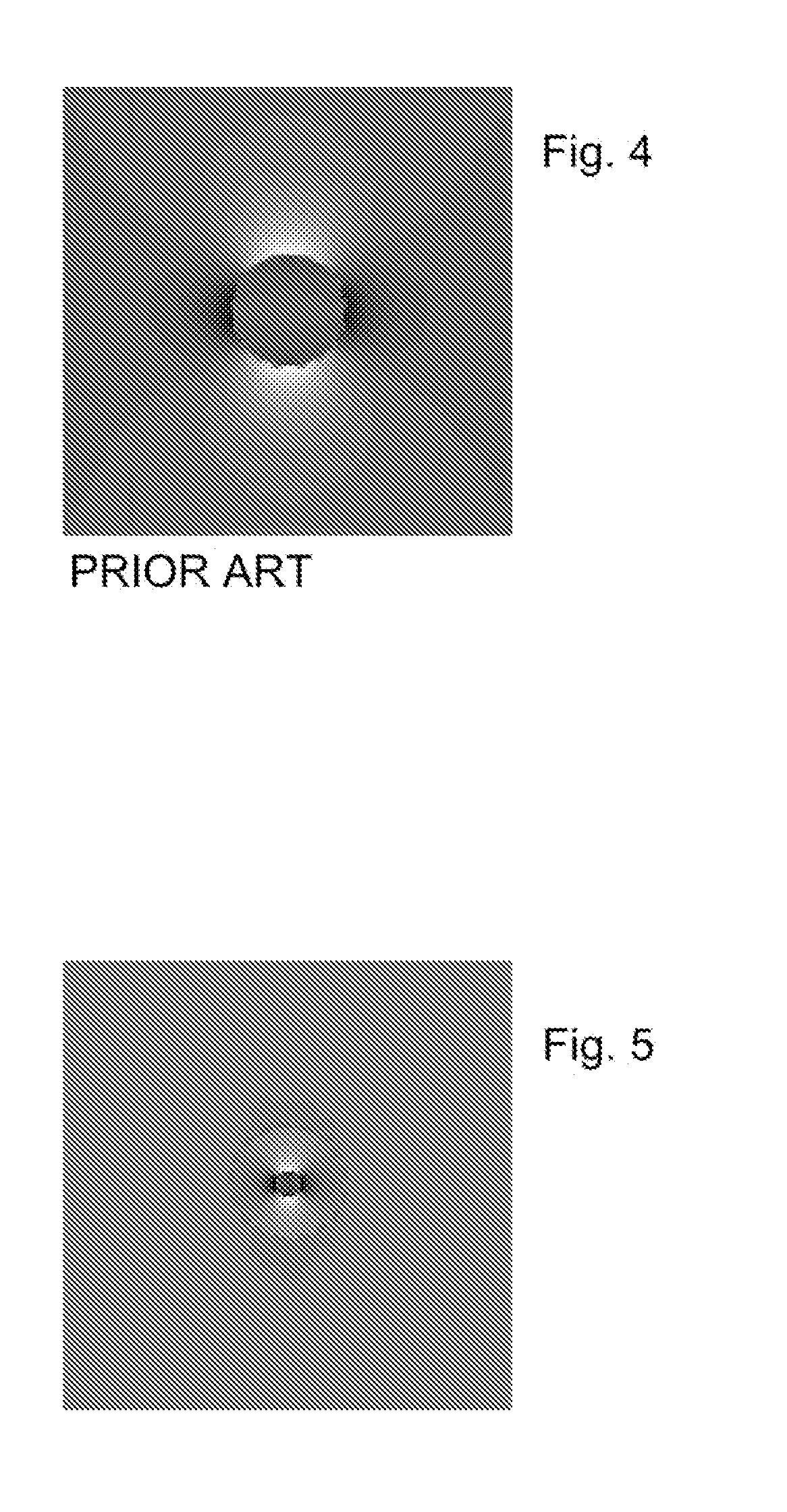
[0005]The invention provides non-magnetic, susceptibility-matched metal devices for magnetic resonance imaging (MRI) and MR spectroscopy, that have magnetic susceptibility properties matched to materials in which the device is used (e.g. biological tissues). Susceptibility-matching the metal devices to the surrounding material reduces perturbations in the local magnetic field, thus minimizing MR image and spectral distortions introduced near the interface between device and the surrounding material.
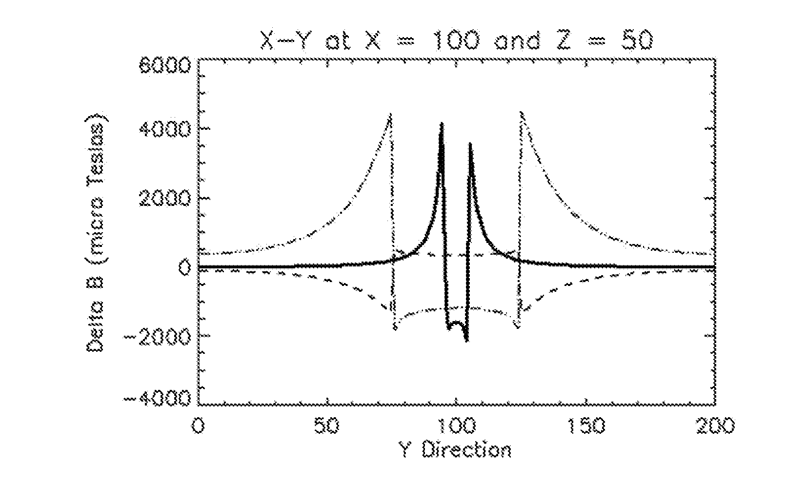
[0015]In accordance with the invention, experimental measurements, as well as modeling using Maxwell's equations, as further explained in the incorporated references, are carried out to obtain the optimal choice of geometry and metals, such that fluctuations in the local magnetic field near the interface between the device and surrounding material are minimized, thus reducing image and spectral distortions. In part, the invention achieves this result by providing devices whose magnetic susceptibility matches or nearly matches that of the material in which the device is inserted, wherein the devices may therefore be considered to be susceptibility-matched.
[0017]Moreover, a susceptibility-matched metal cannula, in accordance with the invention, may be left in the target tissue before, during, and after MR imaging and spectroscopy, without creating significant distortion. More particularly, cannulas in accordance with the invention can be used to significantly reduce distortion of resulting images and spectra. In accordance with the invention, these cannulas may be used in vivo for infusions of therapeutic agents into tissue during MR sessions. Alternatively, the cannulas may be used for selective illumination of optically responsive cells in light activated cell channel control applications. The lumen of the cannula can be left hollow for non-optical uses. Alternatively, optical fibers may be inserted within the lumen, to thereby expose selected tissue regions to light. Because the cannula is constructed with metal, it may also function as a stimulating / recording electrode. Electrophysiology information, gathered from the recording function of the cannula of the invention, could be used to monitor physiology or target regions of tissue based on differences in electrophysiology.
[0018]The reduction in distortion provided by the invention enables electrodes to be positioned with greater precision, and for tissue near the electrodes to be properly characterized using MR imaging.

Problems solved by technology

These electrodes have magnetic susceptibility properties that are significantly different than biological tissue, and this leads to fluctuations in the local magnetic field at the tissue-electrode interface.
Local magnetic field fluctuations can result in signal loss and affect the spatial encoding of the MR image, resulting in image distortions.
Unfortunately, fused-silica exhibits poor conductivity, which limits its use in a recording electrode.
Moreover, fused-silica is very flexible, and can therefore be difficult to control during insertion.

Method used


Image

  • Magnetic resonance compatible and susceptibility-matched apparatus and method for mr imaging & spectroscopy
  • Magnetic resonance compatible and susceptibility-matched apparatus and method for mr imaging & spectroscopy
  • Magnetic resonance compatible and susceptibility-matched apparatus and method for mr imaging & spectroscopy

Examples


Embodiment Construction

[0058]In the description which follows, the particular embodiments described herein are not to be considered as limiting of the present invention.

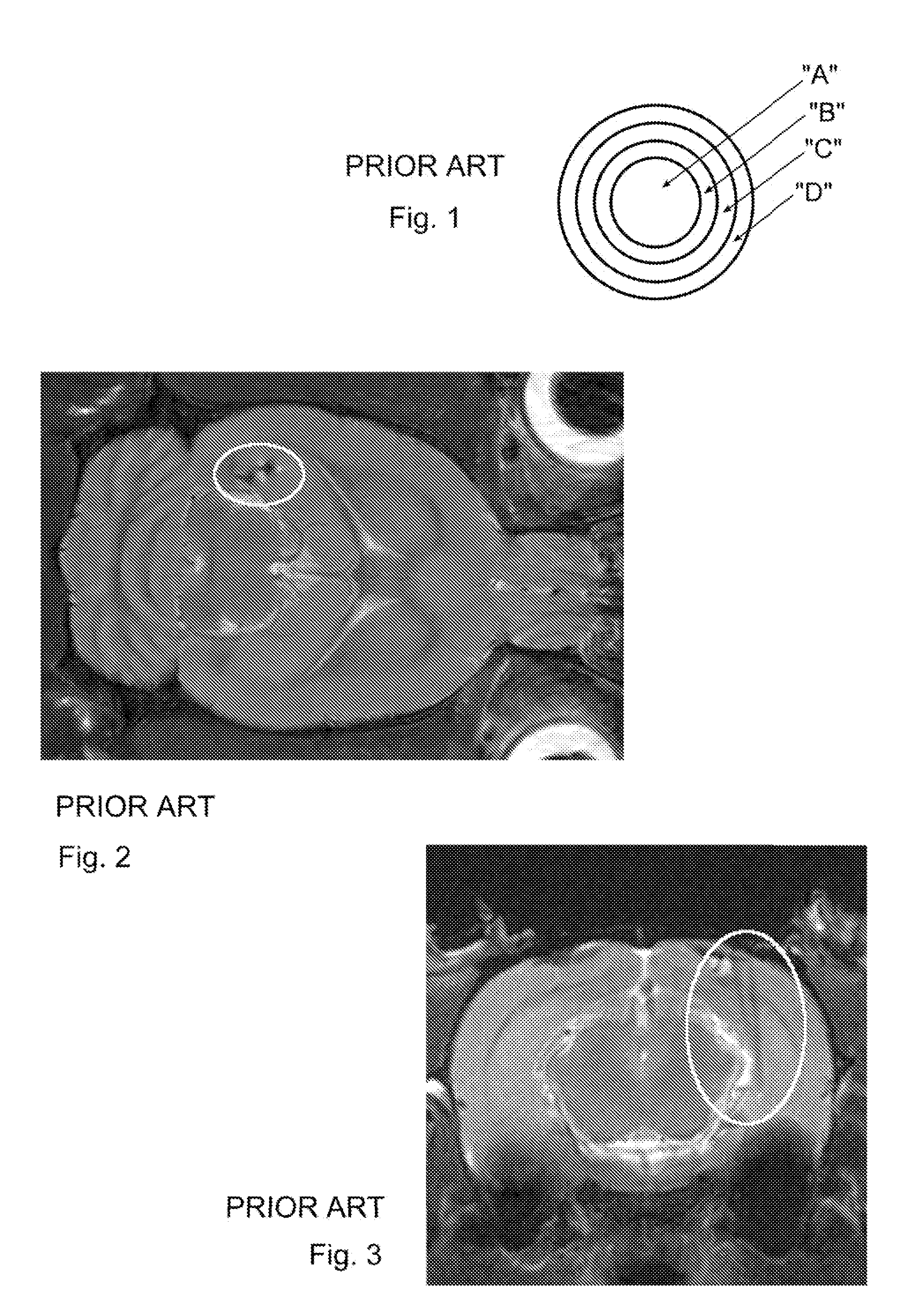
[0059]Prior art devices used during Magnetic Resonance Imaging (MRI) and MR spectroscopy (hereafter MR), that have magnetic susceptibility that is poorly matched to surrounding tissue, can produce perturbations in the local magnetic field, creating MR image and spectral distortions introduced near the interface between the device and the surrounding material. A schematic illustration of a typical prior art electrode is provided in FIG. 1, in which “A” represents a 50 μm diameter tungsten core; “B”, a 0.13-0.38 μm thick nickel flash; “C”, a 1.3 μm gold layer; and “D”, a 58-78 μm polyimide coating.

[0060]With reference to FIGS. 2-3, the chronic limbic epilepsy rat model requires the placement of chronic electrodes in a specific brain region. To minimize the impact, extremely small electrodes (e.g. 50 μm diameter wire) are used. However, elect...


PUM


Abstract

Electrodes, infusion cannula, and interventional MRI instrumentation, are constructed with multiple layers or mixtures of metals or alloys that allow the diamagnetic behavior of some metals to combine with the paramagnetic behavior of others, wherein the devices have a magnetic susceptibility which is close to that of the material, for example body tissue, being im The material may thereby be imaged using MRI with resultant images having greatly reduced distortion. Optimal metal composites are determined through mathematical modeling and measurements. In particular, MR compatible susceptibility-matched electrodes may be used for stimulation and for acquiring electroencephalography (EEG) data before, during and after MR image and spectroscopy measurements, with a significant reduction in distortion of the resulting images and spectra. In accordance with the invention, these electrodes may further be incorporated into micro-electrode arrays. In addition, MR compatible susceptibility-matched cannula can implanted before, during and after MR image and spectroscopy measurements, with a significant reduction in distortion of the resulting images and spectra.

Description

CROSS REFERENCE TO RELATED APPLICATIONS[0001]This application claims the benefit of priority to U.S. Provisional Application Ser. No. 61 / 232,288, filed Aug. 7, 2009, the contents of which is hereby incorporated by reference in its entirety.FIELD OF THE INVENTION[0002]The invention relates to minimizing magnetic resonance image and spectral distortions introduced near the interface between a device and the surrounding material.BACKGROUND OF THE INVENTION[0003]Known stimulating / recording electrodes typically consist of stainless steel or tungsten wires, with an insulating layer of polyimide or Teflon (a registered trademark of I.E. Dupont De Nemours and Co., Wilmington, Del.). These electrodes have magnetic susceptibility properties that are significantly different than biological tissue, and this leads to fluctuations in the local magnetic field at the tissue-electrode interface. Local magnetic field fluctuations can result in signal loss and affect the spatial encoding of the MR ima...

Claims


Application Information

Patent Timeline
IPC IPC(8): G01R33/48A61M25/00A61B5/04
CPCA61B5/055A61B5/0478A61B5/291
Inventor MARECI, THOMAS HAROLDCARNEY, PAUL RICHARDASTARY, GARRETT WILLIAMSEPULVEDA, HECTORMANUEL, MICHELE VIOLAFISHER, CHARLES ROBERT
Owner UNIV OF FLORIDA RES FOUNDATION INC
Learn More