
Aptamer-loaded, biocompatible nanoconstructs for nuclear-targeted cancer therapy

Active Publication Date: 2013-05-09
NORTHWESTERN UNIV
View PDF5 Cites 3 Cited by
  • Summary
  • Abstract
  • Description
  • Claims
  • Application Information

AI Technical Summary

Benefits of technology

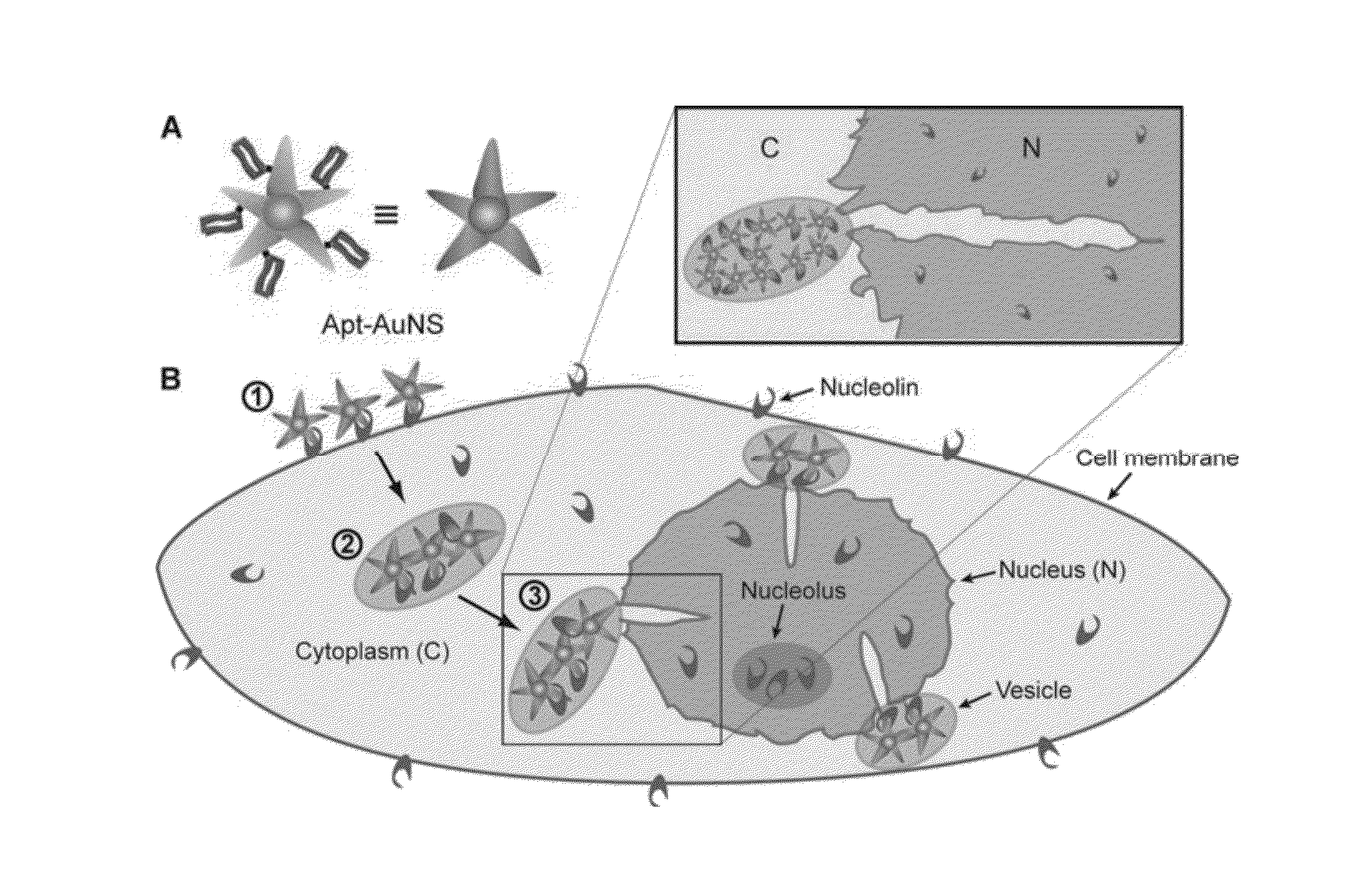
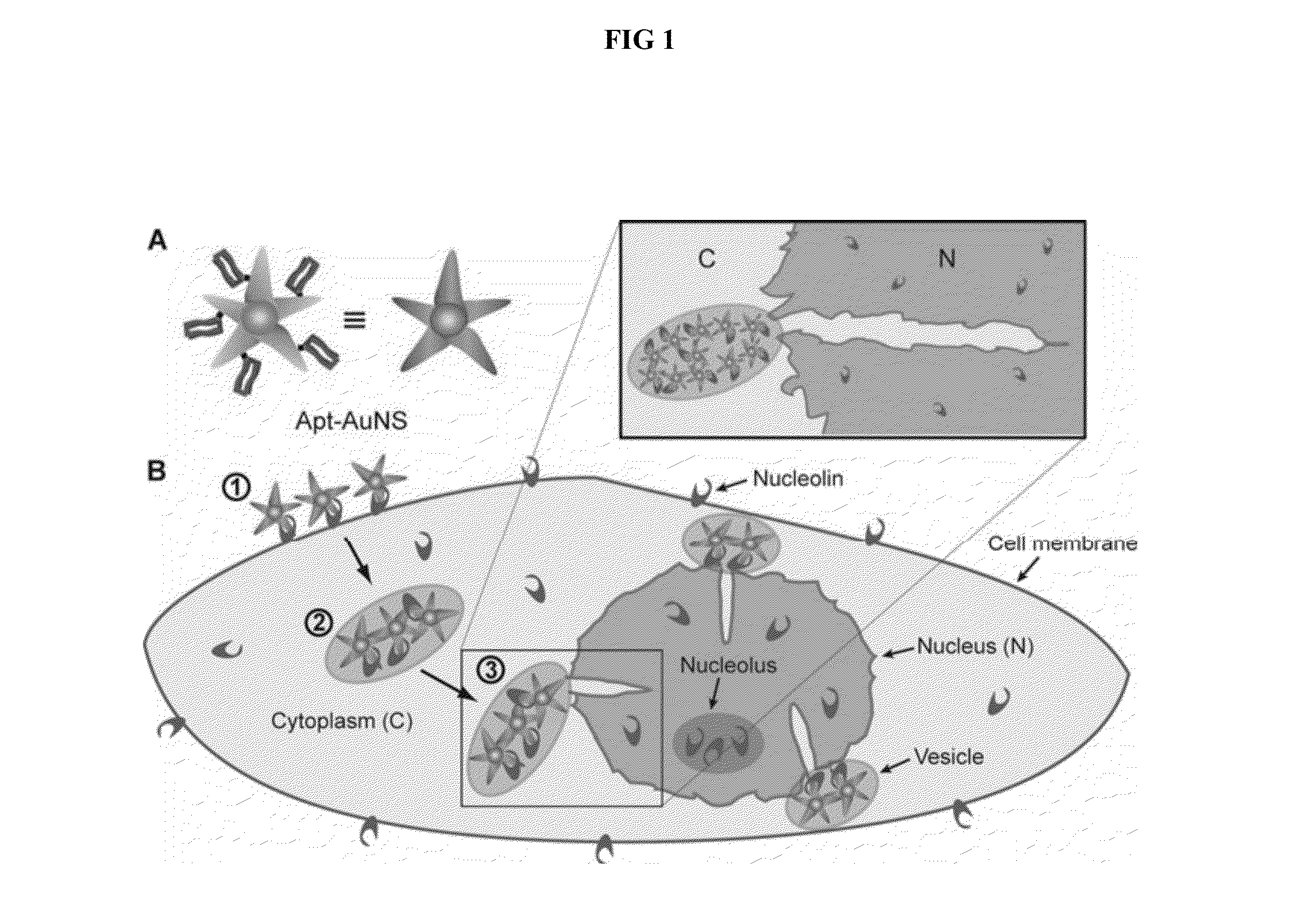
The invention describes a two-component nanoconstruct consisting of an aptamer and a gold nanostar. This construct has been shown to increase the therapeutic and delivery efficiency of the aptamer in treating conditions like cancer. The nanoconstruct can reduce cell proliferation by inducing apoptosis and can also be targeted to the nucleus of cancer cells to provide deformations in the nuclear envelope and alter the nuclear phenotype. The method is selective towards cancer cells and can be applied to a wide range of metastic and primary tumor cells.

Problems solved by technology

One major issue of these treatments is that not all cancer cells in the same tumor express the target receptor.
Hence, these treatments become cell-type dependent and frequently cannot eradicate the entire population of tumor cells (Brown, K. C., Current opinion in chemical biology 2000, 4, 16-21).

Method used


Image

  • Aptamer-loaded, biocompatible nanoconstructs for nuclear-targeted cancer therapy
  • Aptamer-loaded, biocompatible nanoconstructs for nuclear-targeted cancer therapy
  • Aptamer-loaded, biocompatible nanoconstructs for nuclear-targeted cancer therapy

Examples


example 1

Cell Culturing

[0051]The human cervical carcinoma HeLa cell line (ATCC) is maintained in Dulbecco's Modified Eagle Medium (DMEM) (Gibco) supplement with 10% fetal bovine serum (FBS)(Invitrogen). The human epithelial cell line MCF-10A (TBC) is maintained in DMEM / F12 medium (Gibco) supplement with 10% horse serum (Invitrogen), 20 ng / m Lepidermal growth factor (EGF) (Sigma Aldrich), 0.5 mg / mL hydrocortisone (Sigma Aldrich), 100 μg / mL cholera toxins (Sigma Aldrich), and 10 μg / mL insulin (Sigma Aldrich). The cells are cultured at 37° C. with 5% CO2 and plated in T25 flasks (VWR) with aforementioned media.

example 2

Synthesis of Biocompatible Gold Nanoparticles with Optical Properties in the NIR

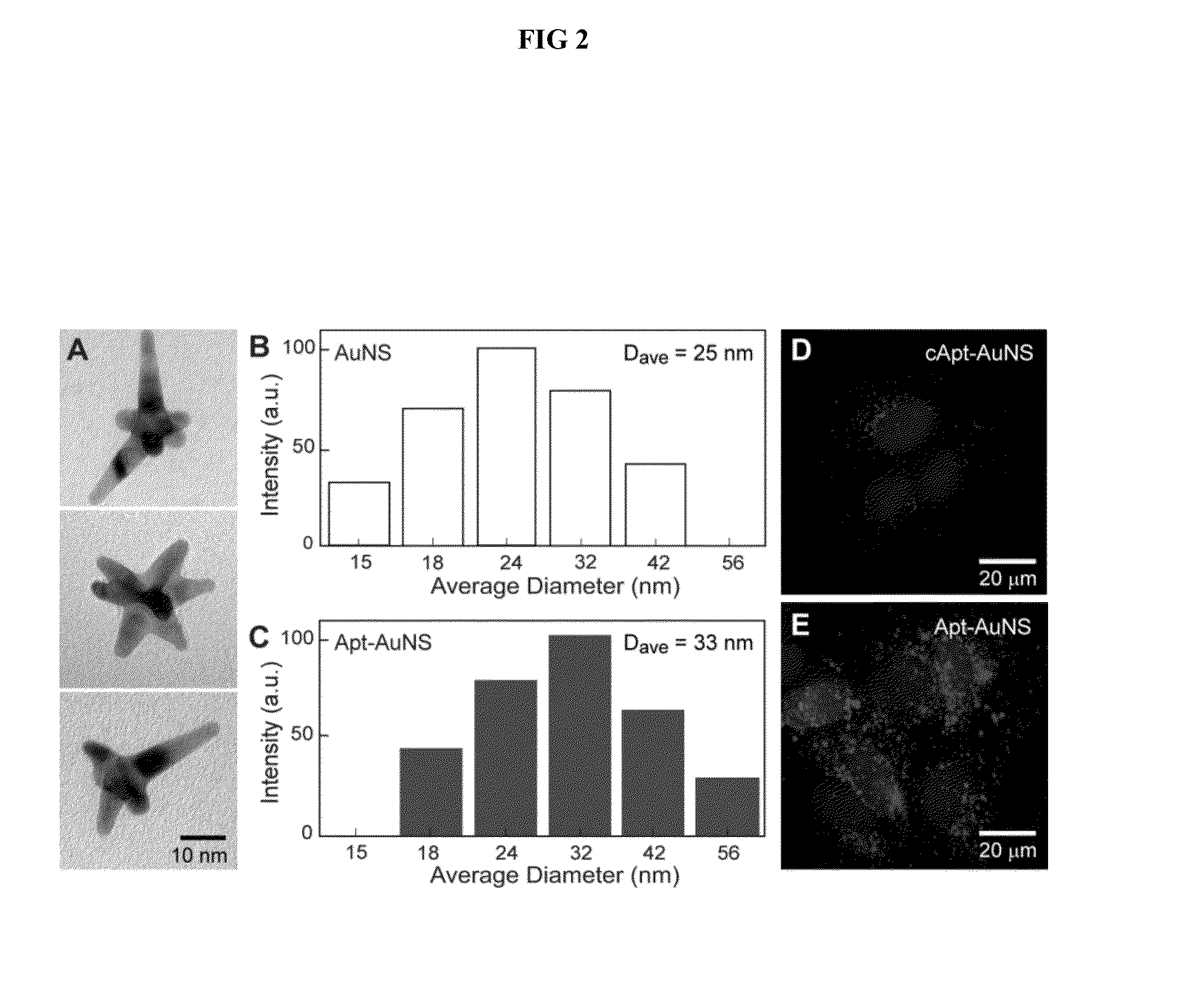
[0052]By adapting a protocol from Xie et at (Xie, J. P. et al., 2007 Chem Mater 19, 2823, incorporated herein by reference), gold nanostars (AuNSs) are synthesized by reducing Au(III) chlorate in HEPES buffer to create biocompatible, surfactant-free gold nanoparticles (AuNPs) for in vitro studies. The AuNSs are prepared by mixing 5 μL of 40 mM HAuCl4 (Sigma Aldrich) with 1 mL of 140 mM HEPES buffer. The resonance wavelength of the AuNS is measured using UV-vis spectroscopy. The size of the particles is determined using both high resolution transmission electron microscopy (HR-TEM) and dynamic light scattering (DLS).

[0053]To quantify the AuNS concentration, the Au content is measured in a specified volume of AuNS with inductively coupled plasma-mass spectrometry (ICP-MS). Next, by assuming similar numbers of Au atoms in a AuNS and a spherical 30-nm Au particle, the number of AuNS in a specific solution ba...

example 3

Loading Aptamer Drugs on the Gold Nanostars

[0054]Formation of Apt-AuNS Nanoconstructs

[0055]AS-1411 aptamer with a disulfide modification at the 5′-end and a control aptamer (cApt), where all the guanines are replaced with cytosines, are purchased from TriLink Biotechnologies, Inc. HPLC purified aptamers are dissolved in Millipore water (18.2 MΩ-cm) to make 1 mM solutions. The disulfide bond is cleaved by adding 2.5 μL of 25 mM tris(2-carboxyethyl)phosphine (TCEP) (Sigma Aldrich) to 10 μL of the 1 mM aptamer solution. After 30 minutes, the thiolated aptamer solution is added to 10 mL of 0.2 nM solution of AuNS and left overnight to form the nanoconstruct (Apt-AuNS). To increase the surface concentration of aptamers on AuNS (Hill, H. D. et al., 2009 Acs Nano 3, 418, incorporated herein by reference), the mixture solution is salted with 2.5 mL of a 500-mM solution of NaCl twice, separated by 4 hours. The size of the nanoconstructs is characterized using DLS.

[0056]Calculation of the Num...


PUM

PropertyMeasurementUnit
Cell proliferation rateaaaaaaaaaa
Cell viabilityaaaaaaaaaa

Abstract

Disclosed herein is a nanoconstruct comprising an aptamer and a gold nanostar. The nanoconstruct can be used in a method of inducing changes to a nuclear phenotype of a cell comprising transporting the nanoconstruct to a nucleus of a cell, and releasing the aptamer from a surface of the gold nanostar into the nucleus of the cell to afford deformations or invaginations in the nuclear membrane, thereby inducing changes to the nuclear phenotype. The method can be used to treat certain hyperproliferative disorders such as cancer.

Description

[0001]This invention was made with government support under grant numbers DP1OD003899, U54 CA119341, CA060553 awarded by the National Institutes of Health; grant numbers DMR1121262, EEC0647560, CHE9810378 awarded by the National Science Foundation; and grant number NNA06CB93G awarded by the National Aeronautics Space Administration. The government has certain rights in the invention.[0002]The present invention relates generally to interactions between drug-loaded nanoparticles and a cell nucleus. Specifically, the invention relates to a method of inducing changes in nuclear phenotype of a cell by shuttling drug-loaded nanoparticles directly to a cell nucleus, the changes in nuclear phenotype correlated with cell activity.BACKGROUND OF THE INVENTION[0003]The nucleus is the most important organelle in the proliferation, angiogenesis, and apoptosis of a cell (Zink, D. et al., 2004 Nat Rev Cancer 4, 677; Torchilin, V. P., 2006 Annu Rev Biomed Eng 8, 343). Controlling the functions of th...

Claims


Application Information

Patent Timeline
IPC IPC(8): A61K9/14A61K31/711
CPCA61K9/14A61K31/711Y10S977/773Y10S977/906B82Y5/00A61K41/0042A61K47/6923
Inventor ODOM, TERI W.DAM, DUNCAN-HIEU M.LEE, JUNG HEON
Owner NORTHWESTERN UNIV
Learn More