Functionalization of and use of functionalized second harmonic generating nanoprobes

a technology of functionalized nanoprobes and nano-probes, which is applied in the field of functionalized shg nanoprobes, can solve the problems of high cost of techniques, impede widespread use, and the ultimate need of characterizing biological targets is largely unm

Active Publication Date: 2013-05-23
CALIFORNIA INST OF TECH
View PDF1 Cites 17 Cited by
  • Summary
  • Abstract
  • Description
  • Claims
  • Application Information

AI Technical Summary

Benefits of technology

[0018]In still yet another embodiment, the functionalization molecule renders the probe nanostructure acceptable for use in conjunction with a technique selected from SHG imaging, direct nucleic acid sequencing in a Multi-SHG Detection Imaging modality, FRESH, optical monitoring of electric fields, imaging / detecting medical conditions or neoplasm, detection or tracking of therapeutic agents, and rapid detection systems.

Problems solved by technology

One of the grand open challenges in modern science is to identify cells or probe molecules and understand the mechanism and dynamics of biological processes at the molecular level with high spatiotemporal resolution, and particularly inside living cells and tissue.
However, these techniques require high costs and some fundamental technological barriers hinder their widespread use.
However, the ultimate need of characterizing biological targets is largely unmet due to fundamental deficiencies associated with the use of fluorescent agents.
For example, fluorescent probes face two major limitations that have a significant impact on the signal strength: 1) dye saturation, because the number of photons emitted by the fluorophore in a given time is restricted by the excited state lifetime, and 2) dye bleaching, which limits the total number of photons produced per dye.
In addition, autofluorescence from tissue organic components after illumination absorption can severely limit the signal-to-noise ratio of fluorescence imaging experiments.
Finally, fluorescence is fundamentally an optically incoherent process, and as a result extracting 3D information from the source is inherently difficult.
However, because inherent material properties targeting mechanism do not provide the targeting or delivery characteristics desired, methods to modify SHG nanoprobes with relevant chemical and biological agents are needed.

Method used


Image

  • Functionalization of and use of functionalized second harmonic generating nanoprobes
  • Functionalization of and use of functionalized second harmonic generating nanoprobes
  • Functionalization of and use of functionalized second harmonic generating nanoprobes

Examples


example 1

Functionalization of BaTiO3

[0111]In one exemplary embodiment, BaTiO3 was functionalized in accordance with the functionalization method described above. The specific chemical schematic used in this example is shown in FIG. 6, and described in detail below.

Materials and Methods

[0112]Chemicals were obtained from the following sources and used without further purification: barium titanate (BaTiO3, Nanostructured & Amorphous Materials, Inc., 99.9%, 200 nm), N-aminoethyl-2,2,4-trimethyl-1-aza-2-silacyclopentane (Gelest), hydrogen peroxide solution (Alrich, 30%, aq), anhydrous toluene (Aldrich), 6-azidohexanoic acid, biotin-cyclooctyne, Alexa Fluor 488 conjugated streptavidin (Invitrogen, 2 mg / mL), anhydrous dichloromethane (DCM, Aldrich, 99.8%), 1-ethyl-3-(3-dimethylaminopropyl)carbodiimide (EDC, Thermo Scientific), Nhydroxysuccinimide (NHS, Thermo Scientific), methoxy-Poly(Ethylene Glycol)-succinimidyl carboxymethyl (mPEG2k-SCM, Laysan Bio, MW 2,000), acetone 5-(succinimidyloxycarbonyl...

example 2

Zebrafish Imaging

[0126]In this exemplary embodiment, a protocol for the preparation and use of a particular SHG nanoprobe label, barium titanate (BaTiO3 or BT), for in vivo imaging in living zebrafish embryos was performed. Chemical treatment of the BaTiO3 nanoparticles results in surface coating with amine-terminal groups, which act as a platform for a variety of chemical modifications for biological applications. Here cross-linking of BaTiO3 to a biotin-linked moiety is described using click chemistry methods and coating of BaTiO3 with nonreactive poly(ethylene glycol) (PEG). Details for injecting PEG-coated SHG nanoprobes into zygote-stage zebrafish embryos, and in vivo imaging of SHG nanoprobes during gastrulation and segmentation are also provided.

Methods and Materials

[0127]To prepare a 300× Danieau stock solution (which should be diluted to 30× in water before using with embryos), add 34.8 ml of 5 M NaCl, 2.1 ml of 1 M KCl, 1.2 ml of 1 M MgSO4, 1.8 ml of 1 M Ca(NO3)2 and 15 ml...

example 3

BaTiO3-Biotin Functionalization

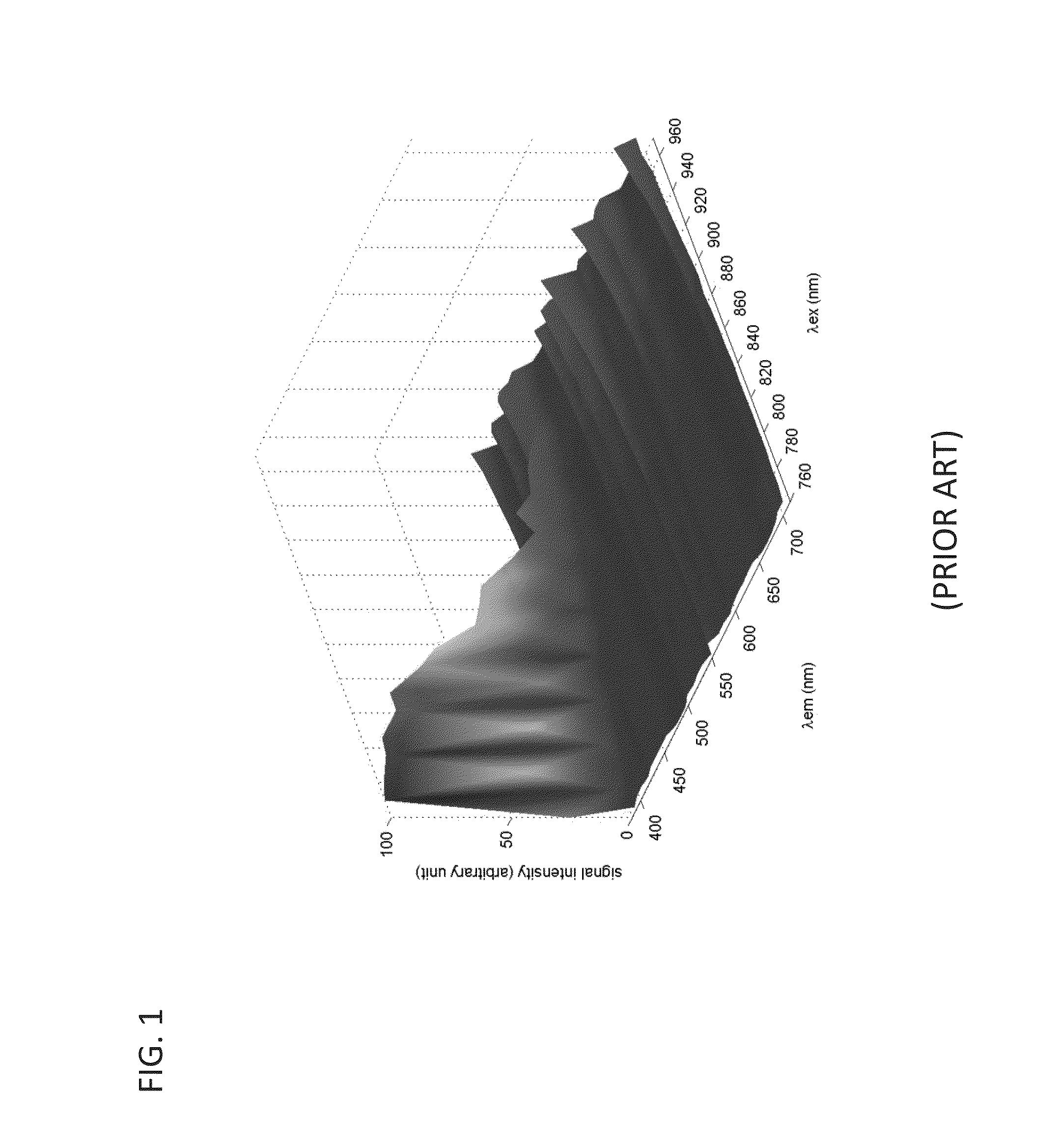
[0141]To test for proper functionalization of the barium titanate conjugated to biotin using click chemistry, the BaTiO3-biotin was first bound to Alexa Fluor (AF) 488 dye-linked Streptavidin as mentioned above. Then, using centrifugation, the BaTiO3-biotin and BaTiO3—OH control solutions were washed 5 times with DI H2O to remove any excess AF-488-linked Streptavidin that was not tightly bound (i.e. that was not bound to biotin, which is a high-affinity binding partner of streptavidin). A drop (˜200 μL) of the washed BaTiO3-Biotin conjugated to AF-488-linked Streptavidin was placed in one well of an 8-well coverslip chamber and a drop (˜200 μL) of the washed control BaTiO3—OH solution was placed into a second adjacent well. Crystals that sedimented near the coverslip were imaged at an illumination wavelength of 965 nm. By taking spectral data over a wide wavelength range (421 nm-587 nm), co-localization of Alexa-488 signal could be seen with properly f...


PUM

PropertyMeasurementUnit
thicknessaaaaaaaaaa
depthaaaaaaaaaa
thicknessaaaaaaaaaa

Abstract

Functionalized second harmonic nanoprobes for imaging samples and a method of using such probes to monitor the dynamics different processeses using a variety of imaging techniques are provided. The functionalized second harmonic generating (SHG) nanoprobes are comprised of various kinds of nanocrystalline materials that do not possess an inversion symmetry and therefore are capable of generating second harmonic signals that can then be detected by conventional two-photon microscopy, and are provided with functional surface modifications that allow for targeted imaging of a variety of biological and non-biological processes and structures such as cell signaling, neuroimaging, protein conformation probing, DNA conformation probing, gene transcription, virus infection and replication in cells, protein dynamics, tumor imaging and cancer therapy evaluation and diagnosis as well as quantification in optical imaging.

Description

CROSS-REFERENCE TO RELATED APPLICATIONS[0001]The current application claims priority to U.S. Provisional Application No. 61 / 562,289, filed Nov. 21, 2011, the disclosure of which is incorporated herein by reference.STATEMENT OF FEDERAL RIGHTS[0002]This invention was made with government support under RO1 HD043897 and P50HGD04071 awarded by the National Institutes of Health.FIELD OF THE INVENTION[0003]The current invention is directed to functionalized SHG nanoprobes, and methods for functionalizing and targeting SHG nanoprobes.BACKGROUND OF THE INVENTION[0004]One of the grand open challenges in modern science is to identify cells or probe molecules and understand the mechanism and dynamics of biological processes at the molecular level with high spatiotemporal resolution, and particularly inside living cells and tissue. As a result of the wealth of information potentially accessible from such biological targets, there has been a growing demand for imaging tools for biomedical researc...

Claims


Application Information

Patent Timeline
Patent Type & Authority Applications(United States)
IPC IPC(8): C07K17/14C07F7/28C08G65/48
CPCC07F7/10C07F7/1836C07F7/28C08G65/33303C08G65/48Y10S977/773Y10S977/902C07K17/14C07F7/1804A61K49/0063A61K49/0093G01N33/588
Inventor PANTAZIS, PERIKLISCULIC-VISKOTA, JELENADEMPSEY, WILLIAM P.FRASER, SCOTT E.
Owner CALIFORNIA INST OF TECH
Why Eureka
Try Eureka