

Device and method for removal of blood-borne pathogens, toxins and inflammatory cytokines
- Summary
- Abstract
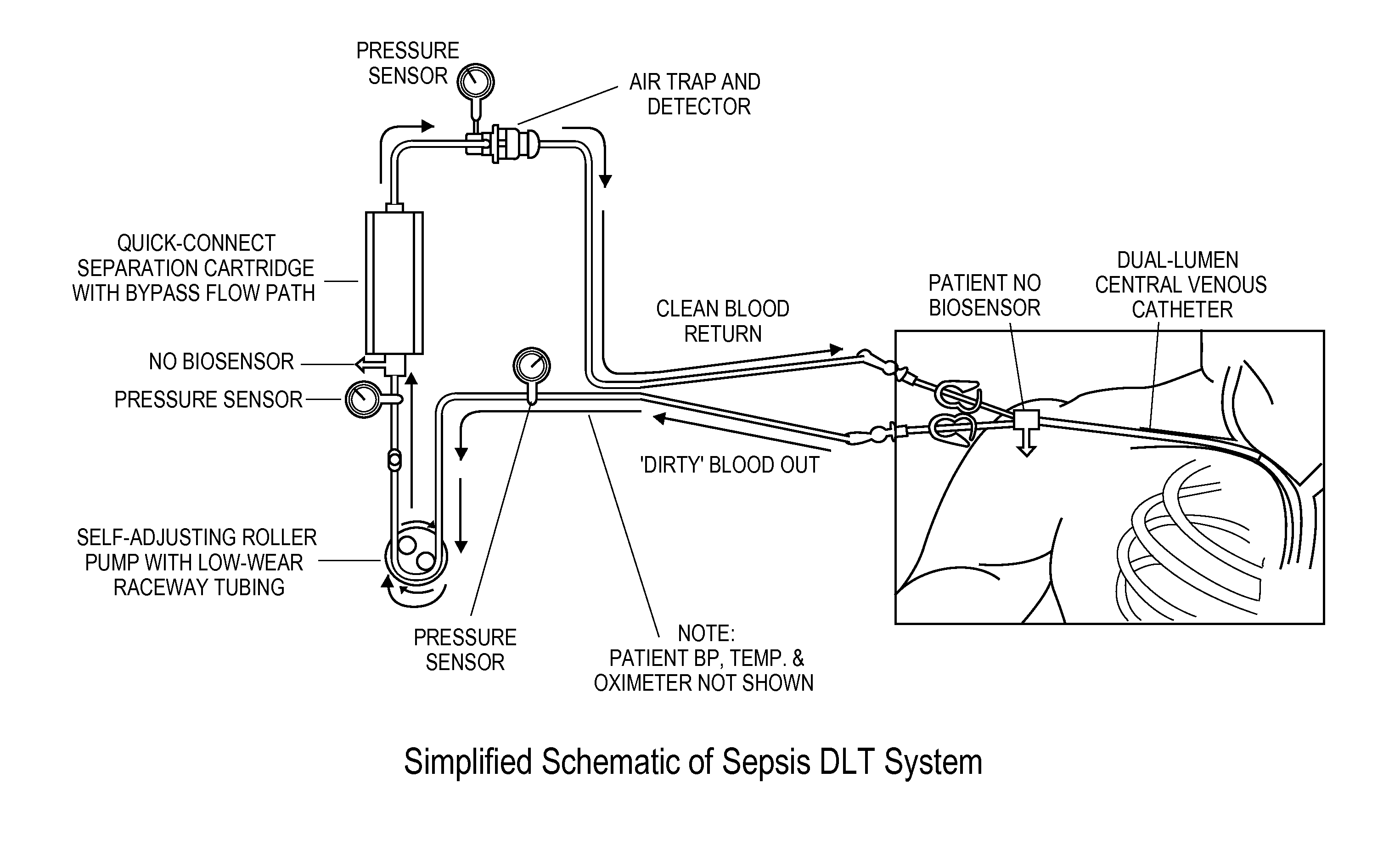
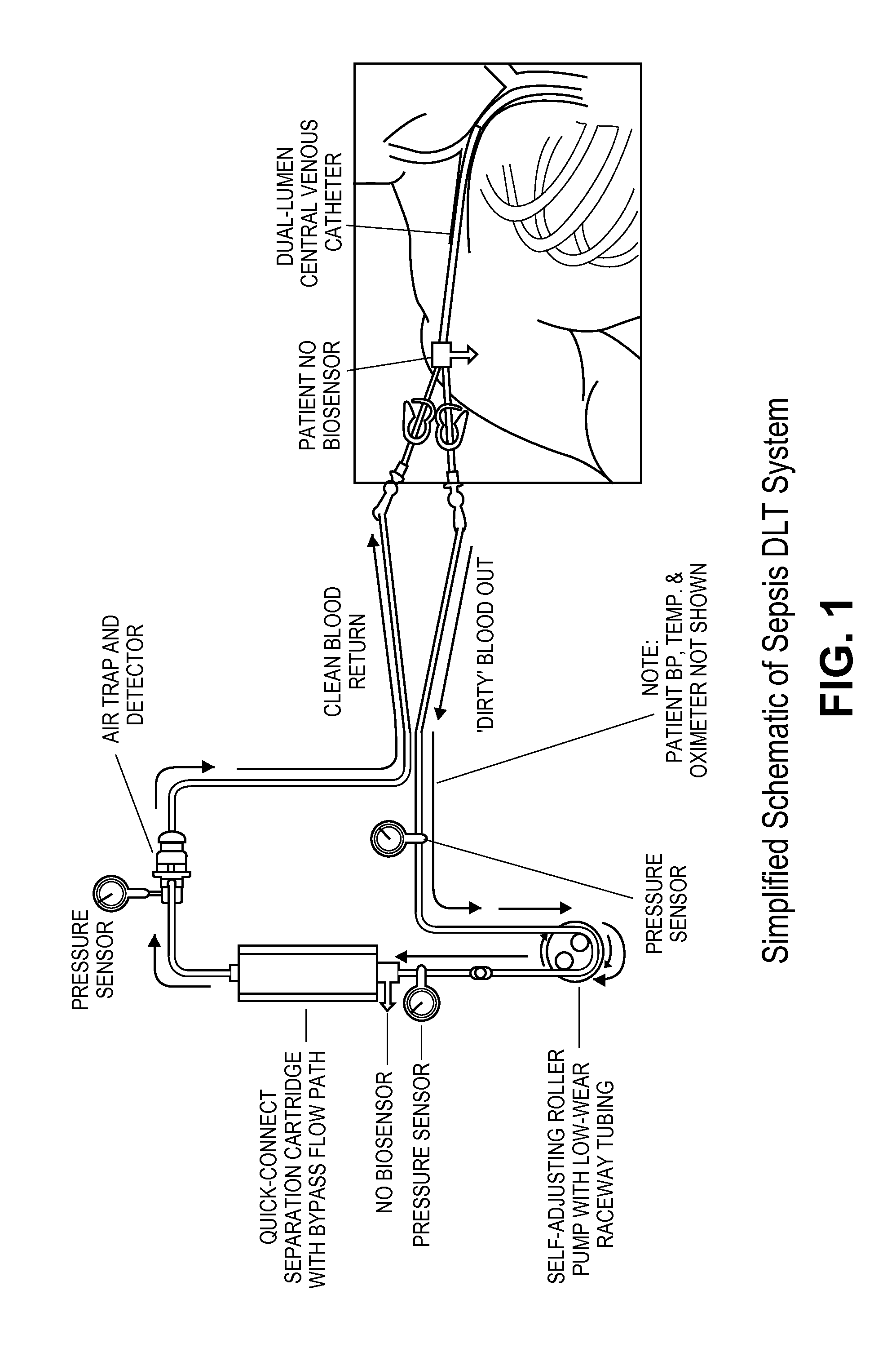
- Description
- Claims
- Application Information
AI Technical Summary
Benefits of technology
Problems solved by technology
Method used
Image

Examples
example 1
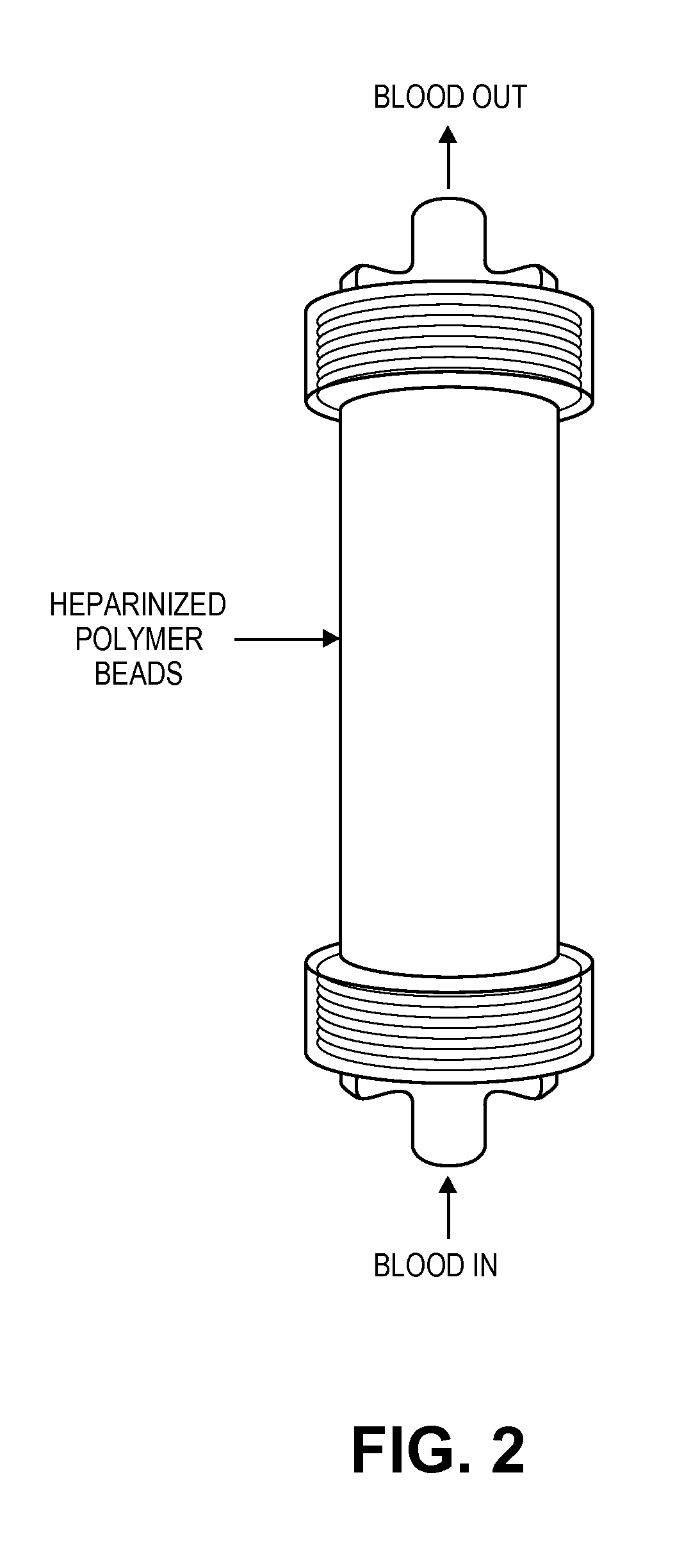
Preparation of Heparin Column
[0195]Polyethylene (PE) beads, with an average diameter of 0.3 mm (lot no. 180153), are supplied by the Polymer Technology Group (Berkeley, USA) and the columns (Mobicol, 1 mL) are obtained from MoBiTec (Germany). Heparin and polyethyleneimine (PEI) are purchased from Scientific Protein Laboratories (Waunakee, Wis., USA) and BASF (Ludwigshafen, Germany) respectively. All chemicals used are of analytical grade or better.
[0196]Immobilization of heparin onto the beads was performed as described by Larm et al. (Larm O, Larsson R, Olsson P. A new non-thrombogenic surface prepared by selective covalent binding of heparin via a modified reducing terminal residue. Biomater Med Devices Artif Organs 1983; 11: 161-173).
[0197]The polymeric surface was heparinized using the general procedure described below.
[0198]The polymeric surface is etched with a oxidizing agent (potassium permanganate, ammoniumperoxidisulfate) in order to introduce hydrophilic characteristics t...
example 2
Patients
[0207]The study protocol was approved by the local ethics committee at the Karolinska University Hospital and signed informed consent was obtained from each patient. Arterial blood was drawn from the hemodialyzers of three septic (fever>38° C., chills, leukocytes>12×109 cells / L) patients (2M / 1F, aged 43, 56 and 68 years; Table 1).
TABLE 1Clinical characteristics of patients donating blood.WhiteMeanbloodBodyarterialRespiratorycellAgetemperatureHeart ratepressureratecountPatientSex(years)(° C.)(beats / min)(mmHg)(breaths / min)(109 / L)1M4339.2°110762019.02M5638.6°90951817.53F6838.5°100892119.5
[0208]The patients are previously administered with broad-spectrum antibiotics (ceftazidime or cefuroxime together with an aminoglycoside; one dose of each) and heparin (200 IU / kg body weight at the start of the dialysis). The blood was collected in EDTA vacuum tubes and immediately transferred to an adjacent room where 1 mL was applied to the previously prepared columns and passed through usin...
example 3
Quantitative Analysis of Cytokines
[0209]The amounts of cytokines are determined using photoluminescence with a plate reader (Multiskan Ascent). Each sample was measured in three wells, and the geometric mean used for analysis. The intraassay coefficient of variation was below 8% for all kits. We used Coamatic antithrombin kit (Haemochrom, cat #8211991), Quantikine Human IL-6 (R&D Systems, cat #D6050), Quantikine Human IL-10 (R&D Systems, cat #D1000B), Protein C Antigen Test 96 (HL Scandinavia AB, product #H5285), Human CCL5 / RANTES Quantikine (R&D Systems, cat #DRN00B), Quantikine Human sVCAM-1 (CD106) (R&D Systems, cat #DVC00), Quantikine Human IFN-gamma (R&D Systems, cat #DIF50), Quantikine HS TNF-alpha / TNFSF1A (R&D Systems, cat #HSTA00D) and BD OptEIA Human C5a ELISA Kit II (BD Biosciences, cat #557965).
Statistical Evaluation
[0210]Paired Kruskal-Wallis test was used to compare the before and after column blood concentrations of each cytokine, with a two-tailed p-value below 0.05 i...
PUM
| Property | Measurement | Unit |
|---|---|---|
| Time | aaaaa | aaaaa |
| Pressure | aaaaa | aaaaa |
| Power | aaaaa | aaaaa |
Abstract
Description
Claims
Application Information