Extended depth of field three-dimensional nano-resolution imaging method, optical component, and imaging system

a three-dimensional nano-resolution, nano-resolution technology, applied in the field of micro-image processing, can solve the problems of small imaging extend of these methods, inability to improve axial resolution, and inability to and achieve high resolution of double helix imaging. , the effect of extending the depth of field

Inactive Publication Date: 2014-11-27
SHENZHEN UNIV
View PDF2 Cites 21 Cited by
  • Summary
  • Abstract
  • Description
  • Claims
  • Application Information

AI Technical Summary

Benefits of technology

[0010]The other purpose of the present invention is to provide an extended depth of field super-resolution fluorescence microscopic imaging and detecting system, the system comprises elements arranged in order along the transmission direction of the optical path: a probing objective lens used for receiving fluorescence beams emitting from the molecule to be measured; a light filter, used for filtering the beams and then output the fluorescence; a dichroic mirror, used for reflecting the fluorescence; an imaging component, adopting the above optical component, used for converting the fluorescence beams to imaging beams with double helix and multi-stage imaging properties; a tube lens used for focusing the reflected fluorescence and transferred to the imaging component; a detector used for receiving the imaging beams and then making double helix and multi-stage imaging.
[0011]In the present invention, the optical module of the present embodiment combines the double helix imaging and dual effect of the multi-stage image of the defocus optical grating, the depth of field of the multi-stage imaging is large, the resolution of the double helix imaging is high and has a certain depth of field, during the optical module imaging, on one hand, multi-stage imaging can greatly extend the depth of field, and can be clearly imaged both sides of the object surface, but also the scope of the axial position is further expanded due to double helix effect and further expand the depth of field; on the other hand, the resolution of the double helix imaging is high. In this invention, the image depth of field is up to ten microns or more so as to use for dynamic range imaging of any depth subcellular in the intact cells and obtaining dynamic function images of multiple movement molecules, it has significance meaning for understanding the change relationships and laws of subcellular structure and cell function at a higher level.

Problems solved by technology

Meanwhile, making nano-resolution three-dimensional structure and function imaging with the intact cell so as to understand the change relationships and laws of subcellular structure and cell function at a higher level is the urgent needs of the life sciences and also a major challenge for imaging science.
However, making nano-resolution three-dimensional imaging to a cell of diameter of 10 μm or more by single molecule localization technique still has many problems.
Firstly, the single molecule localization does not improve the axial resolution and needs to combine certain methods of improve the axial resolution, such as cylindrical mirror astigmatism method, double helix point spread function method (DH-PSF), double plane detection method, the virtual space super resolution microscopic (VVSRM), which can achieve a three-dimensional imaging of horizontal spatial resolution of about 20-30 nm, and a axial resolution of 40-70 nm, at present, the extend of imaging of these methods is only 2 μm.
In addition, the interference photosensitive interferometric photoactivated localization microscopy (iPALM) may improve the three-dimensional resolution to 20 nm or less, but the imaging range is only limited under the 500 nm of the cover glass, therefore, the imaging extend of these methods are small.
Although the SPT method of wide field detection has been developed a variety of axial resolution method such as image stack, defocused imaging, surround the particle movement with a focused laser beam, Fresnel particle tracking (FPT), as well as cylindrical mirror astigmatism method and so on, these methods already achieve three-dimensional nano positioning, but only achieve 3 μm imaging depth, while the thickness of intact cells generally are ten microns, therefore, current methods can not meet the demand of extended depth of field of the tracking of a plurality of molecules within the cell.

Method used


Image

  • Extended depth of field three-dimensional nano-resolution imaging method, optical component, and imaging system
  • Extended depth of field three-dimensional nano-resolution imaging method, optical component, and imaging system
  • Extended depth of field three-dimensional nano-resolution imaging method, optical component, and imaging system

Examples


embodiment one

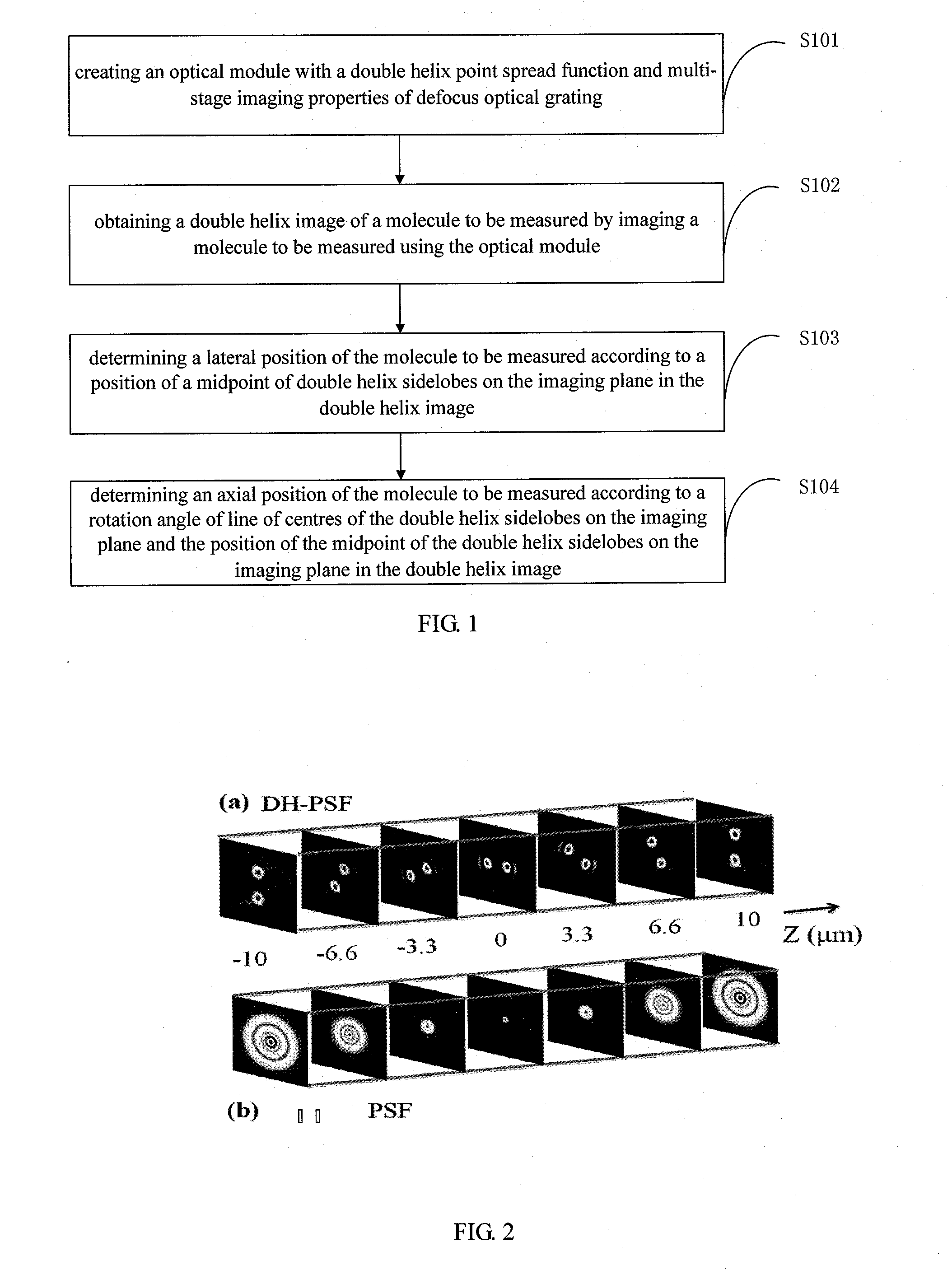
[0025]FIG. 1 is a flow chart of the extended depth of field three-dimensional nano-resolution imaging method in accordance with the first embodiment of the present invention, for convenience of description, only relevant parts of the embodiment are shows.

[0026]Referring to FIG. 1, the method includes the following steps:

[0027]Step S101: creating an optical module with a double helix point spread function and multi-stage imaging properties of defocus optical grating;

[0028]Step S102: obtaining a double helix image of a molecule to be measured by imaging a molecule to be measured using the optical module;

[0029]Step S103: determining a lateral position of the molecule to be measured according to a position of a midpoint of double helix sidelobes on the imaging plane in the double helix image;

[0030]Step S104: determining an axial position of the molecule to be measured according to a rotation angle of line of centers of the double helix sidelobes on the imaging plane and the position of ...

embodiment two

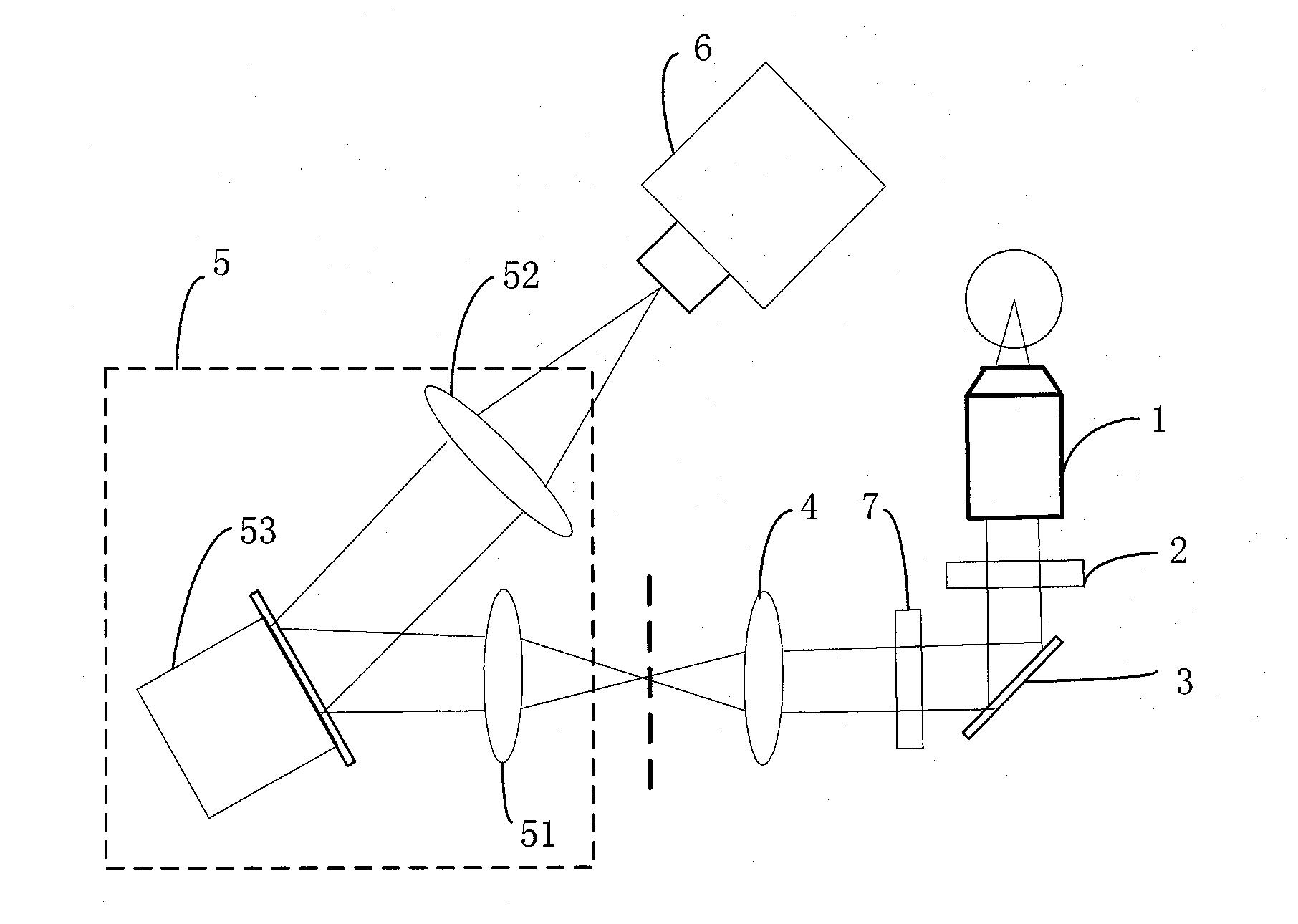
[0049]FIG. 9 is a schematic view of an optical component used for extended depth of field three-dimensional nano-resolution imaging in accordance with the second embodiment of the present invention, for convenience of description, only relevant parts of the embodiment are shows.

[0050]Based on the extended depth of field three-dimensional nano-resolution imaging method, in the present embodiment, further an optical component is provided for extended depth of field three-dimensional nano-resolution imaging. This component is mainly used for three-dimensional imaging system in order to achieve an extended depth of field and high-resolution of three-dimensional imaging of the cell.

[0051]The optical component includes a first lens 901, an optical module 902 and a second lens 903 setting in order along the transmission direction of the optical path. Wherein the optical module 902 has a double helix point spread function and multi-stage imaging properties of defocus optical grating, and is...

embodiment three

[0053]FIG. 10 is a schematic view of an extended depth of field super-resolution fluorescence microscopic imaging and detecting system in accordance with the third embodiment of the present invention, FIG. 11 is a schematic view of another extended depth of field super-resolution fluorescence microscopic imaging and detecting system in accordance with the third embodiment of the present invention, for convenience of description, only a relevant part of the embodiment are shows.

[0054]The embodiment of the invention provides an extended depth of field super-resolution fluorescence microscopic imaging and detecting system based on the above imaging method and optical components, the imaging method of the present invention is combined with the super-resolution fluorescence microscopic imaging methods (such as PALM, STORM) to achieve extended depth of field three-dimensional nano-resolution fluorescence microscopic imaging and detecting.

[0055]Referring to FIG. 10, the extended depth of f...


PUM

PropertyMeasurementUnit
diameteraaaaaaaaaa
thicknessaaaaaaaaaa
thicknessaaaaaaaaaa

Abstract

An extended depth of field three-dimensional nano-resolution imaging method includes: creating an optical module with a double helix point spread function and multi-stage imaging properties of a defocus optical grating; obtaining double helix image of a molecule by imaging a molecule using the optical module; determining a lateral position of the molecule according to a position of a midpoint of double helix sidelobes on the imaging plane in the double helix image; determining an axial position of the molecule according to a rotation angle of a line of centers of the double helix sidelobes on the imaging plane and the position of the midpoint of the double helix sidelobes on the imaging plane in the double helix image. The double helix point spread function and the defocus optical grating multi-stage imaging are combined to implement three-dimensional imaging to extended the depth of field and to improve the resolution.

Description

CROSS-REFERENCE TO RELATED PATENT APPLICATIONS[0001]This application is a U.S. National Phase Application under 35 U.S.C. §371 of International Patent Application No. PCT / CN2013 / 078029, filed Jun. 26, 2013, and claims the benefit of Chinese Patent Application No. 201210467807.7, filed Nov. 19, 2012, all of which are incorporated by reference herein.FIELD OF THE INVENTION[0002]The present invention relates to microscopic imaging technology, and more particularly, to an extended depth of field three-dimensional nano-resolution imaging method, optical component, and imaging system.BACKGROUND OF THE INVENTION[0003]A cell is the basic unit of organisms and life activities, in-depth study on cell is the key to uncover the mysteries of life, improve life and conquer disease. Molecule imaging under intact cell to obtain the subcellular fine structure and even the molecule profiling and to obtain information of the structural changes and molecular dynamic process under the living cell is alw...

Claims


Application Information

Patent Timeline
Patent Type & Authority Applications(United States)
IPC IPC(8): G02B5/18G01N21/64G01N21/84
CPCG02B5/1842G01N21/84G02B5/189G01N21/6486G01N21/6447G01N21/47G01N21/6458
Inventor NIU, HANBENYU, BINCHEN, DANNILI, HENG
Owner SHENZHEN UNIV
Why Eureka
Try Eureka