Model and method for a transgenic bovidae expressing cardiac fibrosis and associated pathology

Active Publication Date: 2015-01-29
THE UNIV OF UTAH +1
View PDF2 Cites 4 Cited by
  • Summary
  • Abstract
  • Description
  • Claims
  • Application Information

AI Technical Summary

Benefits of technology

[0008]The Applicants herein have determined that a need exists for a model and method for expressing cardiac fibrosis in an animal heart exhibiting sufficient similarity

Problems solved by technology

However, due to the small heart size, functional electrophysiology of transgenic mice is problematic.
Mouse models are also not suitable for advancing the development of implantable devices, ablation therapies, or improved imaging techniques.
Additionally,

Method used


Image

  • Model and method for a transgenic bovidae expressing cardiac fibrosis and associated pathology
  • Model and method for a transgenic bovidae expressing cardiac fibrosis and associated pathology
  • Model and method for a transgenic bovidae expressing cardiac fibrosis and associated pathology

Examples


example 1

Vector Construction

[0042]Mouse α MHC Promoter

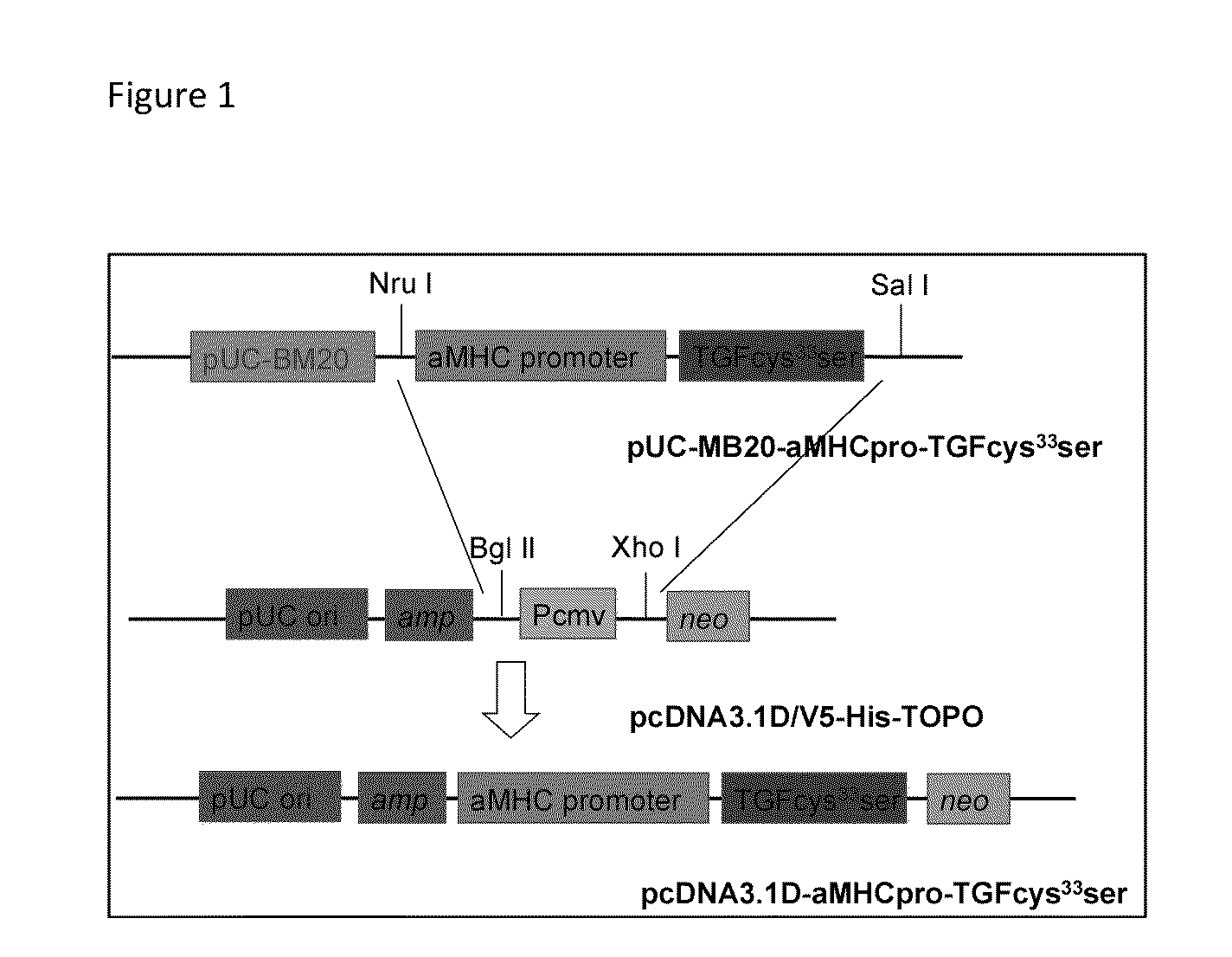
[0043]A pcDNA3.1DV5-MHC-TGF-β1cys33ser vector was constructed by subcloning the MHC-TGF-β1 fragment from the plasmid pUC-BM20-MHC-TGF-β1 (Nakajima et al., 2000[1]) into the pcDNA3.1D V5 vector (FIG. 1). A cysteine-to-serine substitution at amino acid residue 33 prevented covalent binding between the small latent complex and LTBP-1. This greatly increased the amount of active TGF-β1. The Neon™ transfection system was used to electroporate primary goat fetal fibroblasts. After two weeks of G418 selection, the resulting G418 resistant colonies were screened by PCR to confirm transgene integration into goat genomic DNA. PCR positive cells were used for SCNT.

The Goat α-MHC promoter sequence is as follows (SEQ ID NO. 1):

TTTGGAAGAAGCAGAATAAAGCAATTTTCCTTGAAGTGAGATCCTGCCTCTAGACTCTTCTTCACAGCCTGCCAGCCACAGGAACACAAGGACATGACCACGGGACGGGGAGGGGGGCTCCAGGGGAGGAGGCCAGACCCAGGAGGCCTCCCTGGGGAGCCTGGGAGGCTCCGAGCATCCTTGGTGCGGCACTGCCATGGTCTCCCGTCACCTCCTCAGCAGATGACA...

example 2

TALEN-Mediated Targeted Insertion of Transforming Growth Factor-131 (TGF-β1) Gene into the Goat AAVS1 Locus

[0044]AAVS1 locus, an integration site of adeno-associated virus 2, is used in the art as a nonpathogenic “safe harbor” for site-specific integration to achieve persistent and strong transgene expression [3]. Through TALEN-mediated gene targeting strategy, the hTGF-β1 gene was successfully integrated into the goat AAVS1 locus. As disclosed herein TALEN-AAVS1 can efficiently induce a double-strand break with cleavage efficiency of 2% in transfected goat fibroblast cells. The α-MHC-TGF-β1 expression cassette (8.5 kb) was knocked into the goat AAVS1 locus by co-transfecting goat fibroblast cells with TALEN-AAVS1 and α-MHC-TGF-β1 donor vector. Single cell fibroblast colonies were isolated from the transfected cells using puromycin-mediated positive selection. Among the 8 puromycin resistant clones, 6 (75%) were confirmed as correctly targeted by PCR and sequence analysis. These cel...

example 3

Production of Transgenic Cloned Goats

[0045]Somatic donor cells were adult, neonatal or fetal fibroblasts. Adult fibroblasts were obtained from a transgenic doe skin biopsy and neonatal fibroblasts from 1-5 day old transgenic kid skin biopsies. Fetal fibroblasts were isolated from 25-35 day-old fetus, then electroporated with a pcDNA3.1DV5-MHC-TGF-β1cys33ser vector, followed by G-418 selection, screening and subsequent use for SCNT. SCNT procedure is illustrated in FIG. 2. Oocytes with >4 layers of cumulus cells were collected by slicing abattoir ovaries and matured in vitro for 20-24 h. After being denuded, oocytes presenting a 1st polar body were enucleated and received a donor cell. Fused embryos were then activated for 5 min in 5 μM ionomycin followed by 4 h in 2 mM DMAP with 5 μg / ml cyclo-heximide. Activated embryos were cultured in G1.2 medium with 5 mg / ml BSA for either 12 or 60 hours post activation, followed by surgical transfer into the oviducts of recipients synchronized t...


PUM


Abstract

Herein provided are a model and method for a transgenic a bovidae having an TGF-β1 gene inserted into the bovidae genome and capable of expressing higher than normal levels of TGF-β1 in cardiac muscle.

Description

CROSS-REFERENCES TO RELATED APPLICATIONS[0001]This claims priority to U.S. Provisional Patent Application No. 61 / 858,430 entitled “MODEL AND METHOD FOR A TRANSGENIC GOAT EXPRESSING CARDIAC FIBROSIS AND ASSOCIATED PATHOLOGY” and filed on Jul. 25, 2013 for Irina Polejaeva which is incorporated herein by reference.BACKGROUND[0002]1. Field of the Invention[0003]The disclosure provided herein relates to a genetically modified bovidae, specifically a goat, as a model for studying cardiac fibrosis and fibrosis associated cardiac pathologies in human-sized hearts. The disclosure provided further relates to methods by which genetically modified goats and other bovidae may be produced.[0004]2. Description of the Related Art[0005]Human and animal studies indicate that fibrosis plays a central role in the cardiac remodeling observed in various cardiac diseases. In fibrosis, profibrotic factors act on cardiac cells to increase the deposition of extracellular matrix (ECM). These changes alter the...

Claims


Application Information

Patent Timeline
IPC IPC(8): A01K67/027A61D19/04
CPCA01K67/0278A01K2267/0375A01K2227/10A61D19/04C07K14/495A01K2217/072A01K2217/206A01K2227/102A01K67/0275
Inventor POLEJAEVA, IRINAWANG, ZHONGDEHU, SHENGWEIRANJAN, RAVITHOMAS, AARON JAMESDAVIES, CHRISTOPHER JOSEPHWHITE, KENNETH L.
Owner THE UNIV OF UTAH
Why Eureka
Try Eureka