
Eureka
For R&D, Eureka makes reading and utilizing patents & technical documents easy.
Eureka AIR
Designed for self-driven R&D workflows. Generate viable solutions, solve complex R&D challenges, empower your innovation with AI.

Eureka Materials
Designed for material experts only. Revolutionize your material R&D, from search, analyze, to developing new materials.

TechResearch
Generate reliable direction feasibility study reports for your R&D in just a few steps.

TechSeek
Discover and master advanced knowledge NOW. Basics, ideas, possibilities, all at once.

TechMind
As an expert in R&D Theories, TechMind can generates customized viable solutions instantly.

TechRisk
Analyze your overall solution with one click, know your potential R&D risks in advance.

TechMonitor
Get weekly tech updates, stay abreast of the latest tech innovations and key insights.
Magnetic Nanoparticles for Disease Diagnostics
- Summary
- Abstract
- Description
- Claims
- Application Information
AI Technical Summary
Benefits of technology
Problems solved by technology
Method used
Image

Examples
example 1
[0095]Synthesis and Characterization of Metal-curcumin Complexes
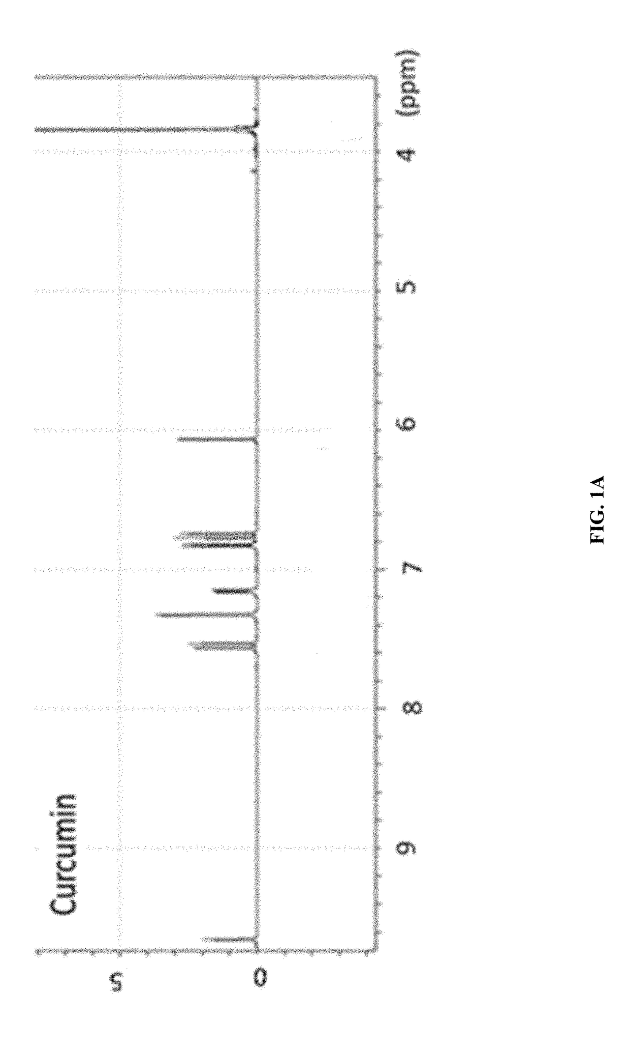
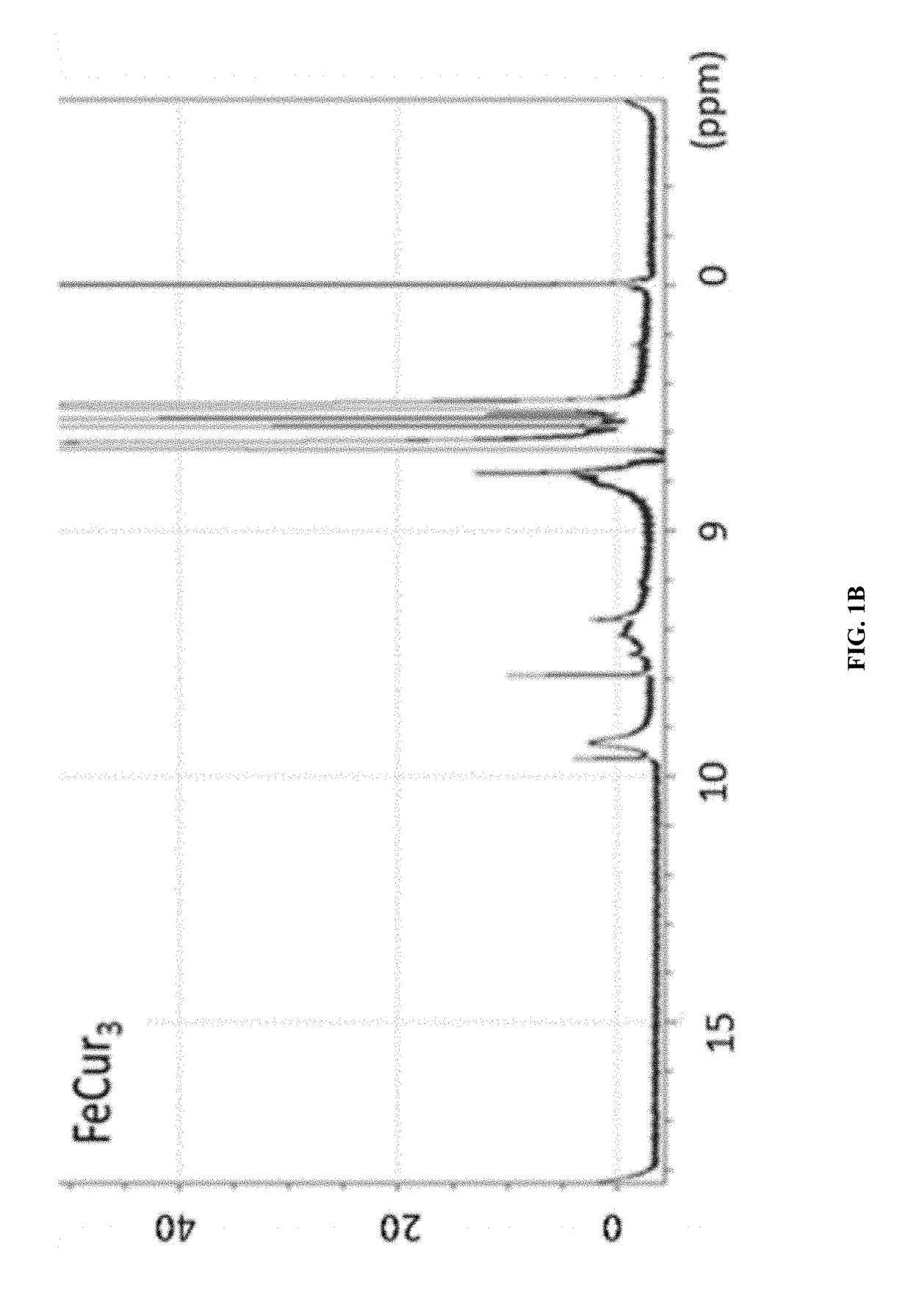
[0096]The synthetic strategy is similar to the reported literature (Sigurdsson et al., 2008; Wadghiri et al., 2003) with modification. Iron-curcumin complex will be described as an example for the synthetic procedure. Iron-curcumin complex was synthesized by dissolving curcumin with minimal amount of dimethylformamide. The iron solution (e.g., Fe(NO3)3 or FeCl3) with one-third mole ratio of curcumin was pre-dissolved in dimethylformamide and added dropwise to the curcumin solution with stirring. The solution was turned to deep red and stirred for overnight under darkness at room temperature. The solution was lyophilized and the powder was washed with Milli-Q water and minimal amount of dichloromethane. FIG. 1 shows the 1H-NMR spectrum of iron-curcumin complex showed broad peaks of curcumin demonstrated the conjugated curcumin to the paramagnetic iron. The lower frequency of the C═O and C═C bond observed in the metal com...
example 2
[0097]Encapsulation of Iron-curcumin Complex by Flash Nanoprecipitation (FNP) Method—MIVM
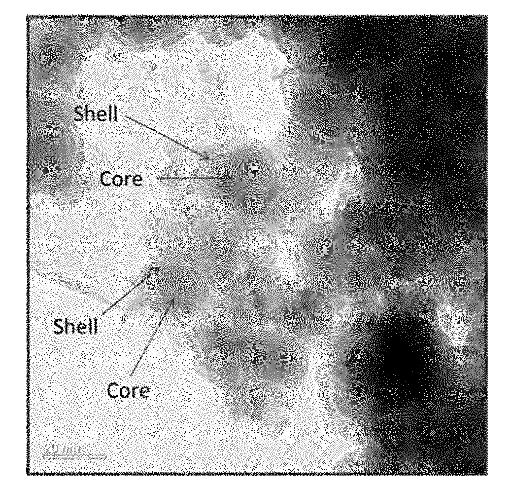
[0098]The synthesized iron-curcumin complex is barely soluble in water due the hydrophobic nature of curcumin. In order to enhance its aqueous solubility, blood circulation time and prevent non-specific uptake by immune system, the iron-curcumin complex was further encapsulated inside polymeric micelle. PEG(2 k amu)-PLA (10 k amu) co-block polymer and iron-curcumin complex were dissolved in organic phase (e.g., DMF or acetone) while co-stabilizer polyvinyl pyrrolidone (PVP) (30 k amu) was dispersed in aqueous phase. The injection ratio is iron-curcumin complex: coblock polymer: PVP=1:2:4. Both streams were co-injected into Multi-inlet vortex mixer (MIVM) with injection rate at 45 ml / min and 5 ml / min for aqueous phase and organic phase, respectively. The PLA tail of PEG-PLA co-block polymer was in favor to adhere on the iron-curcumin complex surface and formed micelle structure. Also, due to the ...
example 3
[0102]Potential MRI Agent of Iron-curcumin Nanoparticles
[0103]The potential of iron-curcumin nanoparticles to be a magnetic resonance imaging agent was investigated by scanning the MRI signal of iron-curcumin nanoparticles in agarose gel. Agarose gel with different concentration of iron-curcumin nanoparticles were prepared and stacked inside 50 mL centrifugal tube. It was then scanned by the Mill instrument to analyze the MRI signal (FIG. 9). The magnetic moment was also measured by vibrating sample magnetometer (FIG. 10A and FIG. 10B)
PUM
| Property | Measurement | Unit |
|---|---|---|
| Hydrodynamic diameter | aaaaa | aaaaa |
| Hydrodynamic diameter | aaaaa | aaaaa |
| Molecular weight | aaaaa | aaaaa |
Abstract
Description
Claims
Application Information
 - R&D Engineer
- R&D Manager
- IP Professional
- Industry Leading Data Capabilities
- Powerful AI technology
- Patent DNA Extraction
Browse by: Latest US Patents, China's latest patents, Technical Efficacy Thesaurus, Application Domain, Technology Topic, Popular Technical Reports.
© 2024 PatSnap. All rights reserved.Legal|Privacy policy|Modern Slavery Act Transparency Statement|Sitemap|About US| Contact US: help@patsnap.com