Tissue-engineered three-dimensional model for tumor analysis

a three-dimensional model and tumor technology, applied in the field of three-dimensional tissue engineered tumor analysis, can solve the problems of inability to replicate in vitro, 2d models fail to capture the true three-dimensional (3d) progression of tumors, and limited in their ability to identify therapeutic targets, so as to improve the original tumor phenotype and powerful predictive testing tools

Active Publication Date: 2018-12-27
THE TRUSTEES OF COLUMBIA UNIV IN THE CITY OF NEW YORK
View PDF0 Cites 3 Cited by
  • Summary
  • Abstract
  • Description
  • Claims
  • Application Information

AI Technical Summary

Benefits of technology

[0021]In one embodiment, a cancer model is provided with a biomimetic microenvironment representing the pathophysiology of this malignancy. This is achieved by using three-dimensional (3D) instead of conventional two-dimensional (2D) cultures, with the aid of bone-...

Problems solved by technology

The lack of ability to replicate in vitro the complex in vivo milieu of human cancer is a critical barrier to evaluation of the potential therapeutic targets for clinical application.
However, these 2D models fail to capture the true three-dimensional (3D) progression of tumors and are limited in their ability to identify therapeutic targets.
Our knowledge about the putative roles of the microenvironment on tumor exosomes is limited, due to a lack of experimental models that efficiently mimic the human in vivo situation.
Animal models used to study the effects of exosomes on cancer development often fail in representing the context of human disease.
The absence of physiological cell-cell and cell-matrix-interactions and the currently used non-physiological substrates cause disparity from the in vivo situation and lead to changes in cell morphology, proliferation and cellular processes, such as endo and exocytosis.
Although many drugs showed promise in these systems, most failed to translate into human patients and only ˜5% showed anti-tumor activity in clinical trials.
This discrepancy is caused by the lack of ability to sufficiently replicate the human microenvironment in these models.
Notably, ES cells are incapable of directly degrading bone matrix.
However, the crosstalk between tumor cells,...

Method used


Image

  • Tissue-engineered three-dimensional model for tumor analysis
  • Tissue-engineered three-dimensional model for tumor analysis
  • Tissue-engineered three-dimensional model for tumor analysis

Examples


examples

[0156]Native Tumors

[0157]Ewing's sarcoma tumors were obtained from a Tissue Bank. The samples were fully de-identified. Three different frozen tissue samples were cut in sets of 6 contiguous 10 μm-thick sections and homogenized in Trizol (Life technologies) for RNA extraction and subsequent gene expression analysis.

[0158]Cell Culture

[0159]All cells were cultured at 37° C. in a humidified incubator at 5% CO2, unless noted otherwise.

[0160]Cancer cell lines: Ewing's sarcoma cell lines SK-N-MC (HTB-10) and RD-ES (HTB-166) were purchased from the American Type Culture Collection (ATCC) and cultured according to the manufacturer's specifications. RD-ES cells were cultured in ATCC282 formulated RPMI-1640 Medium (RPMI) and SK-N-MC cells were cultured in ATCC283 formulated Eagle's Minimum Essential Medium (EMEM). Both media were supplemented with 10% (v / v) Hyclone FBS and 1% penicillin / streptomycin. Cells were cultured at 37° C. in a humidified incubator at 5% CO2.

[0161]U2OS osteosarcoma cel...


PUM


Abstract

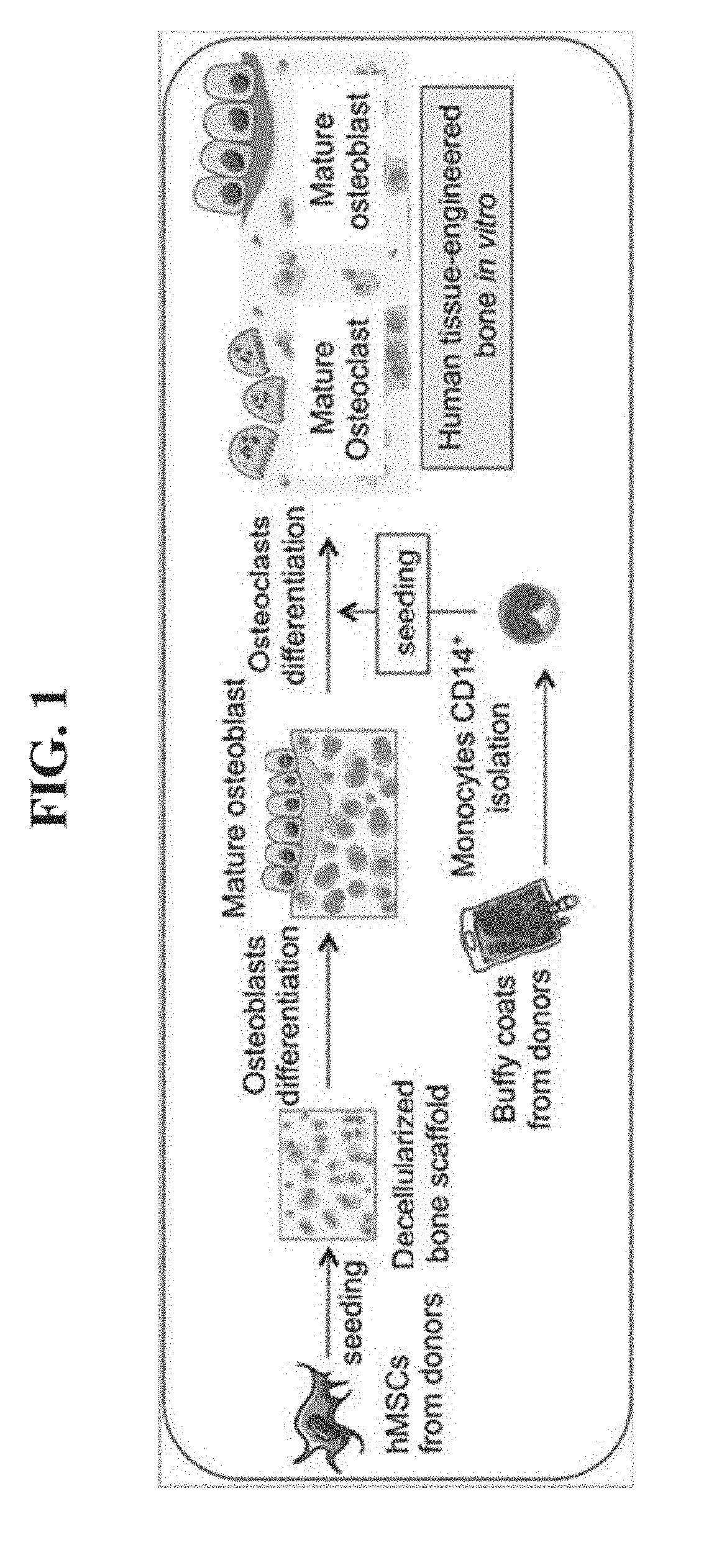
A 3D decellularized bone scaffold seeded with cancer cells, such as prostate cancer cells or Ewing's sarcoma is provided. The three-dimensional includes Ewing's sarcoma (ES) tumor cells; and an engineered human bone scaffold. The engineered human bone scaffold further includes osteoblasts that secrete substance of the human bone, and osteoclasts that absorb bone tissue during growth and healing. The engineered human bone scaffold includes the tissue engineered three-dimensional model which recapitulates the osteolytic process. The engineered human bone scaffold is engineered by co-culturing of osteoblasts and osteoclasts. The osteoblast is produced by cell differentiation process from mesenchymal stem cells. The osteoclast is produced by cell differentiation from human monocytes, wherein the human monocytes are isolated from buffy coats. The scaffold can be used with cancer cell lines to identify therapeutic targets to slow, stop, and reverse tumor growth and progression as well as to predict the efficacy of potential therapeutics.

Description

CROSS-REFERENCE TO RELATED APPLICATIONS[0001]This application is a continuation of International Application No. PCT / US2016 / 068478, filed Dec. 23, 2016, which claims priority to U.S. Provisional Application No. 62 / 387,121 filed Dec. 23, 2015, and a continuation-in-part of U.S. application Ser. No. 14 / 908870, which is a continuation of International Application No. PCT / US14 / 49416, filed Aug. 1, 2014, which claims the benefit of U.S. Provisional Application No. 61 / 861,957, filed Aug. 2, 2013 and claims the benefit of U.S. Provisional Application No. 61 / 862,447, filed Aug. 5, 2013, all of which are incorporated by reference in their entirety herein.STATEMENT REGARDING FEDERALLY SPONSORED RESEARCH OR DEVELOPMENT[0002]This invention was made with government support under grants EB002520 and EB17103 awarded by the NIH. The government has certain rights in the invention.BACKGROUNDTechnical Field[0003]The disclosed subject matter relates to providing a three-dimensional decellularized bone ...

Claims


Application Information

Patent Timeline
IPC IPC(8): C12N5/077C12N5/09C12N5/071
CPCC12N5/0654C12N5/0655C12N5/0693C12N5/0697C12N2502/1311C12N2502/1317C12N2502/30C12N2506/115C12N2506/1353C12N2533/54C12N2533/90C12N5/0643C12N5/0663C12N2502/1142C12N2503/04C12N2513/00C12N2533/80
Inventor VUNJAK-NOVAKOVIC, GORDANAVILLASANTE, ARANZAZU
Owner THE TRUSTEES OF COLUMBIA UNIV IN THE CITY OF NEW YORK
Why Eureka
Try Eureka