
Coated implants for long-term controlled release of antibody therapeutics

Pending Publication Date: 2019-05-23
THE RGT OF THE UNIV OF MICHIGAN
View PDF2 Cites 0 Cited by
  • Summary
  • Abstract
  • Description
  • Claims
  • Application Information

AI Technical Summary

Benefits of technology

The patent text describes a new way to treat diseases such as wet age-related macular degeneration (AMD) using a special implant that can release a therapeutic antibody over a period of several weeks. This implant is designed to be stable and safe, with a controlled release of the antibody that maintains its effectiveness. Compared to other methods, this approach reduces the risk of infection and inflammation, and provides a more convenient and effective treatment for patients.

Problems solved by technology

This leads to displacing the macula from its normal position, finally resulting in rapid central vision loss.
AMD is the major cause of vision loss in the developed countries, especially in people aged 60 or older as expected from its name.
The first FDA-approved anti-VEGF agent was Macugen® (pegaptanib, anti-VEGF aptamer) which slowed vision loss compared with conventional treatment such as laser coagulation or photodynamic therapy, however it did not improve visual acuity in patients.
However, monthly intravitreal injection is very inconvenient for patients and repeated injections introduce risk of infection, inflammation and hemorrhage.
A drawback of PLGA formulations for protein encapsulation is formation of an acidic microenvironment during release due to the degraded acid byproducts in the polymer, which can destabilize the encapsulated proteins and result in incomplete release.
Drying (e.g., lyophilization) and milling processes, which also can damage proteins, are needed to prepare protein powder for encapsulation.
When too high amount of these osmotically active excipients are necessary in the formulation a problem arises that the biologically protein is released too fast.
In various aspects, it was not expected that one could accomplish both polypeptide / protein stability and slow and continuous release from such a preparation.

Method used


Image

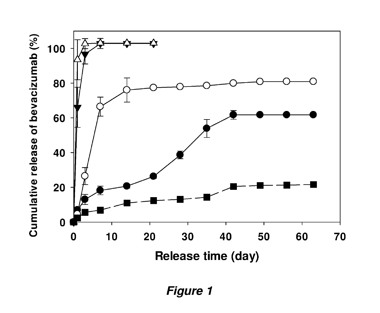
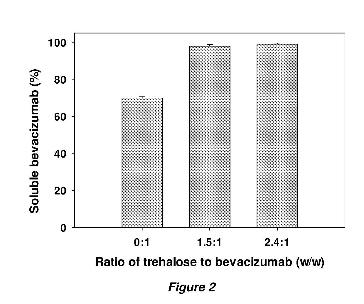
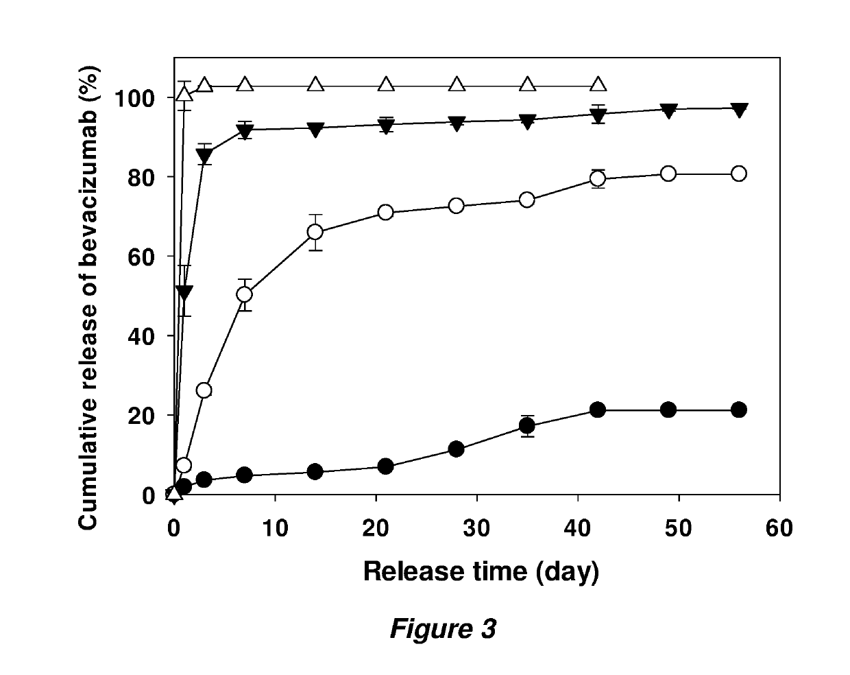
  • Coated implants for long-term controlled release of antibody therapeutics
  • Coated implants for long-term controlled release of antibody therapeutics
  • Coated implants for long-term controlled release of antibody therapeutics

Examples


example 1

Studies of Anti-VEGF Antibodies

Materials

[0050]The Avastin®, commercial solution of bevacizumab was purchased from the pharmacy and used within its shelf-life period. PLGA 50:50 (inherent viscosity=0.64 dL / g and Mw=54.3 kDa, ester terminated) was purchased from LACTEL Absorbable Polymers (Birmingham, Ala.). Trehalose dihydrate (trehalose), MgCO3, guanidine hydrochloride, DL-dithiothreitol (DTT), ethylenediamine-tetraacetic acid (EDTA), Na2HPO4, NaH2PO4, anti-human IgG-alkaline phosphatase antibody produced in goat and p-nitrophenyl phosphate liquid substrate system (pNPP) were purchased from Sigma-Aldrich Chemicals (St. Louis, Mo.). Tween 80 (10%), acetone, KH2PO4, K2HPO4, KCl, phosphate buffered saline (PBS), Amicon Ultra-15 Centrifugal Filter Units (10,000 MWCO), silicone rubber tubing, and Coomassie plus reagent assay kit were purchased from Fisher Scientific (Hanover Park, Ill.). Recombinant human vascular endothelial growth factor (VEGF) was a generous gift from Genentech.

Prepar...

example 2

n in a Rabbit Retinal Leakage Model

[0067]Materials, preparation of antibody powder, and preparation of coated implants were as in Example 1.

Measurement of Antibody Loading in Implants

[0068]Implants (3-5 mg) were dissolved in 1 mL of acetone for 1 h and centrifuged to precipitate protein. PLGA dissolved in supernatant was removed and the protein pellet was washed with acetone and centrifuged three times more to remove residual PLGA. The pellet was then air-dried, reconstituted in 0.5 mL of PBST (phosphate buffered saline with 0.02% Tween 80, pH 7.4) at 37° C. overnight and analyzed by SE-HPLC. The mobile phase (0.182 M KH2PO4, 0.018 M K2HPO4, and 0.25 M KCl [pH 6.2]) was run at a flow rate of 0.5 mL / min through a gel column (TSK-GEL G3000SWx1; Tosoh Bioscience, Tokyo, Japan), and elution was monitored at 280 nm. The volume of injection was 50 μL, and the running time was 30 minutes. All samples were filtered through 0.45 am filter. Extracted loading was calculated by the following eq...

example 3

Release Study of PLGA-Encapsulated Bovine Serum Albumin (BSA)

[0071]Materials, preparation of BSA powder, and preparation of coated implants were as in Example 1 with slight modifications. For BSA powder, the same composition (trehalose:BSA=1.5:1, w / w) as applied in Example 1 was used with BSA as an additional model protein in place of bevacizumab. The resulting BSA powder was suspended into 50% (w / w) PLGA solution in acetone with 3% (w / w) MgCO3 in a 2-mL centrifuge tube, then mixed and transferred into a 3-mL syringe. The suspension was extruded into silicone rubber tubing (I.D.=0.5 mm), then dried at room temperature for 48 h followed by vacuum drying at 40° C. and −23 in. Hg vacuum for an additional 96 h. The final dried implants were obtained by removal of silicone tubing. For coated implants, the obtained core implants were put back into silicone tubing (I.D.=0.8 mm) and 70% pure PLGA solution in acetone within a 3-mL syringe was extruded over the core implants to coat the surfa...


PUM

PropertyMeasurementUnit
Lengthaaaaaaaaaa
Lengthaaaaaaaaaa
Fractionaaaaaaaaaa

Abstract

Implants for anti-VEGF therapy provide both stability and controlled release of bevacizumab and other structurally sensitive polypeptides while maintaining protein / peptide stability in the micronized powder; achieving near zero order and complete release (>80%). Cylindrical implants suitable for intravitreal injection.

Description

INTRODUCTION[0001]Wet age-related macular degeneration (AMD) is a condition in which new abnormal blood vessels grow up and leak fluid or blood behind the macula. This leads to displacing the macula from its normal position, finally resulting in rapid central vision loss. AMD is the major cause of vision loss in the developed countries, especially in people aged 60 or older as expected from its name.[0002]There are two forms of AMD: the wet and dry form. The wet form of AMD accounts for only 10% of the cases, but is responsible for 90% of vision loss, whereas the more common dry form results in relatively mild symptoms.[0003]In wet AMD, the growth of abnormal blood vessels under the retina is stimulated by overexpression of vascular endothelial growth factor (VEGF). Therefore, anti-VEGF therapies have been developed to neutralize the overexpressed VEGF.[0004]The first FDA-approved anti-VEGF agent was Macugen® (pegaptanib, anti-VEGF aptamer) which slowed vision loss compared with con...

Claims


Application Information

Patent Timeline
IPC IPC(8): A61K9/00C07K16/22A61K47/34A61K47/26
CPCA61K9/0051C07K16/22A61K9/0024A61K47/34A61K47/26C07K2317/24A61L27/18A61L27/34A61L27/54A61L27/58A61L2300/252A61L2300/256A61L2430/16A61L27/505A61L2300/622A61L2300/604A61L2400/06C08L67/04
Inventor SCHWENDEMAN, STEVEN P.CHANG, RAE SUNG
Owner THE RGT OF THE UNIV OF MICHIGAN
Learn More