
TREATMENT OF CNS INJURY WITH RNAi THERAPEUTICS

a technology of cns injury and rnai, applied in the field of medicine and neurology, can solve the problems of limited clinical studies, few approved drugs to improve functional outcome, and long life of disability, and achieve the effects of enhancing lesion contraction, promoting seclusion of cns infiltrating blood borne monocytes, and enhancing astrocyte migration

Active Publication Date: 2020-01-30
BIOAXONE BIOSCI
View PDF0 Cites 0 Cited by
  • Summary
  • Abstract
  • Description
  • Claims
  • Application Information

AI Technical Summary

Benefits of technology

The patent text describes a new discovery of a specific molecule called PTEN siRNA that can target and inhibit the expression of PTEN, a protein involved in neurological injuries such as spinal cord and optic nerve damage. This discovery has been exploited to develop a new pharmaceutical composition that can treat these injuries by using self-delivery technology. The patent text also describes a specific version of this siRNA molecule called BA-434, which has been found to be effective in promoting axon regeneration and astrocyte cell migration to the injury site. Overall, this patent provides a new tool for treating neurological injuries and promoting the survival or regeneration of mature CNS neurons.

Problems solved by technology

SCI often leads to a lifetime of disability and there are no approved drugs to improve functional outcome.
Despite a plethora of targets and compounds that show recovery of function and walking in rodent models of SCI, very few have been taken forward to clinical studies.
While neurons in the central nervous system (CNS) have the capacity to regenerate their axons after injury, they fail to do so, in part because regeneration is limited by intrinsic signaling pathways that are upregulated in mature neurons suppress axon regrowth; failure to regenerate is also due to growth inhibitory molecules that are present in the CNS environment, and receptors for growth inhibitory molecules activate neuron-intrinsic signaling pathways that prevent axon regrowth and regeneration.
One barrier to progress in the field of spinal cord injury SCI has been the failure to translate experimental treatments to clinically relevant drugs after target identification.
However, clinically relevant ways to transiently suppress PTEN without creating unwanted side effects have not been devised.
After CNS injury, oligodendrocytes frequently undergo cell death resulting in axonal demyelination and only insufficient axonal remyelination occurs.
However very few of the techniques tested in animals have proven robust for human use.
For example, intrathecal delivery of antibodies to promote regeneration caused unacceptable infection rates in one clinical trial, and the protocol was changed to intermittent, intrathecal bolus injections, a procedure which is difficult and painful for patients.
However, reliance on components made by separate manufacturers does not ensure an approved supply of components, necessitating complex manufacturing of individual kit components for drug manufacture.
In vivo delivery of siRNA has been one challenging aspect of developing a therapeutic drug using siRNA technology.
Also, unlike DNA plasmids and viral vectors, direct-delivery of siRNA does not require a nuclear localization signal and it carries a negligible risk for genomic integration.
While different delivery methods have been developed, little is known about how modifications to enhance delivery affect mRNA localization and trafficking, which ultimately influence translation, function, and specificity.
In addition, delivery to the CNS has been challenging because siRNAs do not penetrate the blood brain barrier.
However, this is not a clinically suitable delivery because in humans the cell bodies of corticospinal neurons in the cortex can be meters from injured axons in the spinal cord.

Method used


Image

  • TREATMENT OF CNS INJURY WITH RNAi THERAPEUTICS
  • TREATMENT OF CNS INJURY WITH RNAi THERAPEUTICS
  • TREATMENT OF CNS INJURY WITH RNAi THERAPEUTICS

Examples


example 1

Assay for Identification of Self-Deliverable PTEN sdRNA Compounds

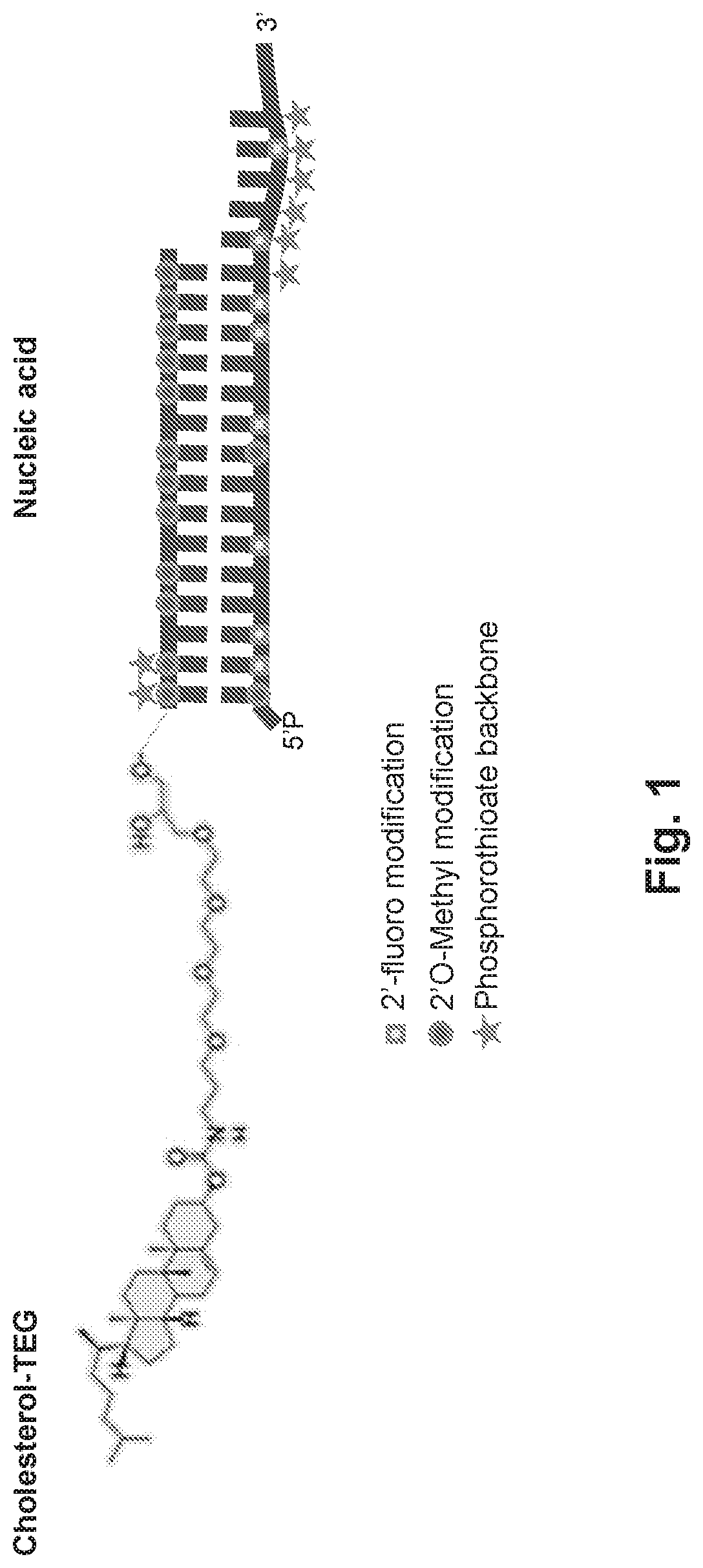
[0114]The PTEN targeting agents described herein were designed and sent for synthesis as separate modified sense (“s”) and antisense (“as”) RNA strands. These strands were then annealed to form the sdRNA prior to testing (FIG. 36).

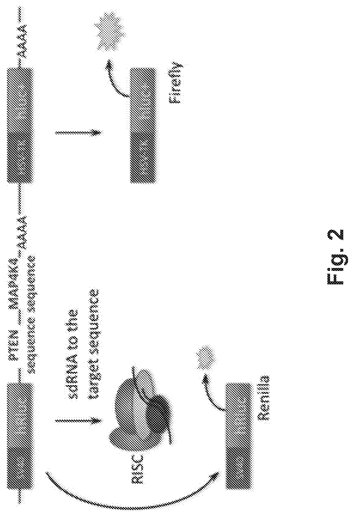
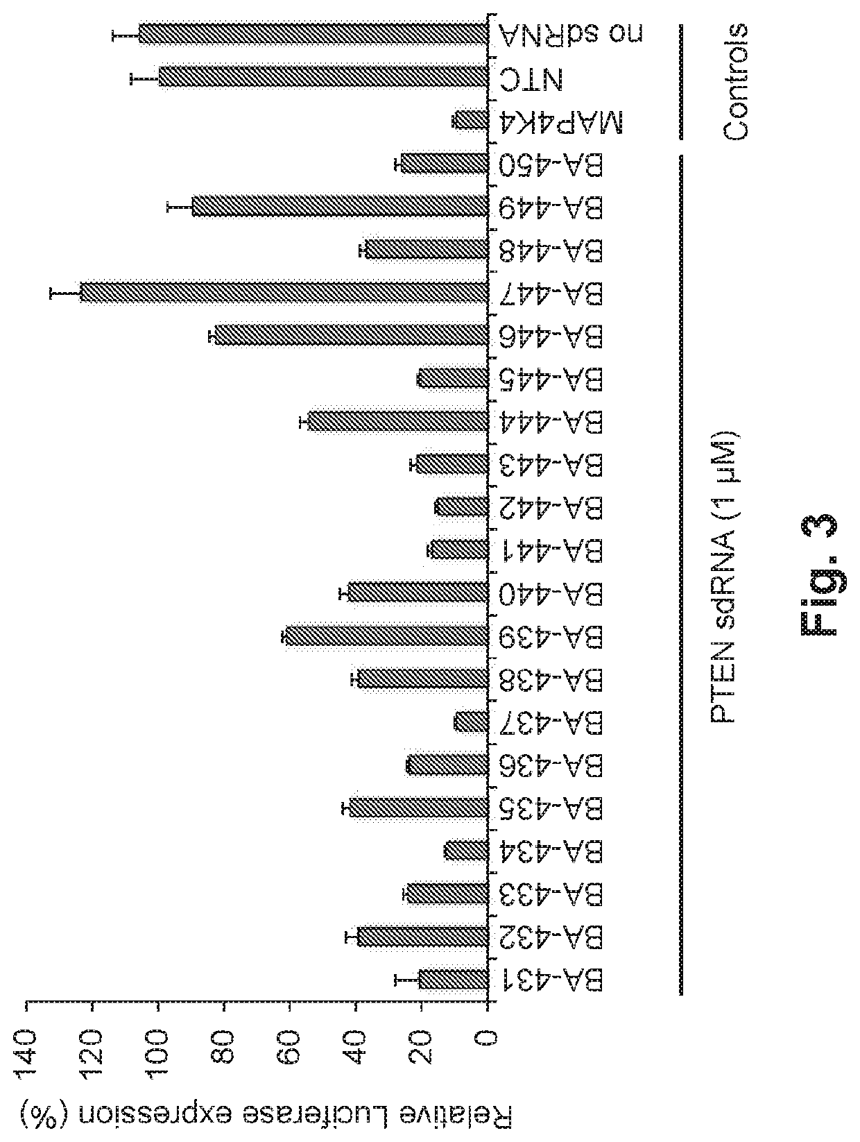
[0115]The PTEN targeting agents were identified by treating HeLa cells carrying rat PTEN sequence with designed sdRNA compounds. For that, a luciferase reporter plasmid (FIG. 2) was constructed. Renilla luciferase sequence was followed by PTEN sequence and previously validated MAP4K4 sdRNA targeting sequences, as a positive control. Firefly luciferase sequence under separate promoter was used for relative luminescence signal normalization.

[0116]HeLa cells were transfected with the PTEN plasmid using Fugene HD (Promega, Madison, Wis.) according to the manufacturer instructions. Briefly, cells were seeded at 2.5×10⁶ cells / 10 cm2 dish in the EMEM (ATCC, Manassas, Va.) medium without antibiotics....

example 2

Delivery of sdRNA Into Rat Pheochromaocytoma PC-12 Cells

[0121]PC-12 cells (ATCC) were cultured on collagen I coated vessels in the presence of 100 ng / ml Nerve Growth Factor (NGF) (Sigma-Aldrich, St. Louis, Mo.). For transfection, cells were collected by trypsinization and diluted with RPMI medium containing 1% FBS and 100 ng / ml NGF and seeded into 96-well plate at 10,000 cells / well.

[0122]MAP4K4 sdRNA conjugated with Cy3 fluorophore was added to the cells at 1 μM final concentration. Images were taken on EVOS FL Imaging System (Life Technologies, Carlsbad, Calif.) at various incubation times between 10 min and 24 hrs.

[0123]FIG. 4 demonstrates that Cy3-fluorescence is clearly visible inside the cells, which proves passive, cellular uptake of MAP4K4 sdRNA.

example 3

Validation of sdRNA Gene Silencing Efficacy in PC-12 Cells

[0124]PC-12 cells were cultured as described in EXAMPLE 1. The efficacy of MAP4K4 sdRNA was tested by qRT-PCR. 2× solutions of MAP4K4 and NTC (non-targeting control) sdRNAs were prepared in serum-free EMEM medium, by diluting 100 μM oligonucleotides to 0.08 μM-4 μM final concentration in the total volume of medium for each oligo concentration. Oligonucleotides were dispensed into a 96-well plate at 50 μl / well.

[0125]PC-12 cells were collected for transfection by trypsinization in a 50 ml tube, washed twice with medium containing 10% FBS without antibiotics, spun down at 200× g for 5 min at room temperature and resuspended in EMEM medium containing twice the required amount of FBS for the experiment (2%) and NGF (200 ng / ml). Cell concentration was adjusted to 120,000 cells / ml. Cells were dispensed into 50 μl / well into the 96-well plate with pre-diluted oligos and placed in the incubator for 48 hr.

[0126]Gene expression was analy...


PUM

PropertyMeasurementUnit
volumeaaaaaaaaaa
volumeaaaaaaaaaa
timeaaaaaaaaaa

Abstract

Self-delivering PTEN RNA and methods of reducing PTEN expression are provided herein. Also provided are methods of treating spinal cord injury (SCI) and other neurotrauma with PTEN sdRNA.

Description

CROSS-REFERENCE TO RELATED APPLICATIONS[0001]This application claims the benefit of and priority to Provisional Application No. 62 / 367,199, filed Jul. 27, 2016, the contents of which is incorporated by reference in its entirety.FIELD OF THE INVENTION[0002]The present disclosure relates to the fields of medicine and neurology, and more specifically, to compositions and treatments for promoting axon regeneration with specific RNA interference (RNAi) drugs.BACKGROUND[0003]In America, 12,000 people suffer SCI each year, most frequently from motor vehicle accidents or falls; in children SCI are mostly due to sports or water recreational activities. SCI often leads to a lifetime of disability and there are no approved drugs to improve functional outcome. Approximately 70% of patients suffer injuries to the cervical spinal cord which leads to quadriplegia and dependence on care for daily living.[0004]The current treatment for SCI is decompression surgery and rehabilitation. Despite a pleth...

Claims


Application Information

Patent Timeline
Patent Type & Authority Applications(United States)
IPC IPC(8): C12N15/113A61P25/00A61P27/02
CPCC12N2310/3515C12N2310/14C12N2310/346C12Y301/03067C12N2310/315A61P25/00C12N15/1137C12N2310/344A61P27/02A61K31/713A61P43/00C12N2310/321C12N2310/3521C12N2310/322C12N2310/3533
Inventor RUSCHEL, JOERGMCKERRACHER, LISANIEDERST, EMILYROSEN, KENNETH M.
Owner BIOAXONE BIOSCI
Learn More