
Three-dimensional culture of primary cancer cells using tumor tissue

a tumor tissue and culture technology, applied in the field of three-dimensional culture of primary cancer cells using tumor tissue, can solve the problems of limited number of cancer cell lines that cannot fully explain the pathological conditions of tumors, can not achieve cancer cell proliferation with high probability, and can lose the original tumor nature of cancer cell lines, etc., to achieve high-throughput performance

Active Publication Date: 2020-03-05
MITSUBISHI CHEM MEDIENCE
View PDF0 Cites 8 Cited by
  • Summary
  • Abstract
  • Description
  • Claims
  • Application Information

AI Technical Summary

Benefits of technology

The invention provides a way to create a cell mass using primary cancer cells that can be easily handled and perform well in high-throughput experiments. The method involves starting with tumor tissue and then inhibiting the growth of other cells while keeping the cancer cells alive. This results in a pure cell mass with cancer cells as the main component.

Problems solved by technology

However, it has been pointed out that cancer cell lines have lost the nature of their original tumor due to the homogenization of population and the accumulation of gene mutations as a result of adaptation to an ex vivo environment with long time cell culture.
In addition, it has been also pointed out that the limited number of cancer cell lines cannot fully explain the pathological conditions of tumor composed of cancer cells with diverse genetic heterogeneity.
However, even with such creative techniques, it is difficult to achieve cancer cell proliferation with high probability, and a more reliable culture method has been desired.
However, this method requires embedding cells in gel at low temperatures, and thus, lacks high-throughput performance, and is not sufficiently versatile in drug development and the like.
This method also requires embedding cells in gel at low temperatures as in the first method, and thus, lacks high-throughput performance.
However, as the drug sensitivity test requires a complicated step of selecting and arranging CTOSs of a uniform size, high-throughput performance is poor and cancer cells have poor proliferative ability in the method as compared to the method in which cells are embedded in an extracellular matrix.
Therefore, the method is insufficient in terms of practical use.
In particular, in the xenograft tumor of breast cancer, proliferation of cell mass could not be clearly confirmed, verification of versatility has remained unsatisfactory, and there is a question about practical use.
However, each method is problematic in terms of proliferative ability, handleability, high-throughput performance, versatility, etc. of cancer cells.

Method used


Image

  • Three-dimensional culture of primary cancer cells using tumor tissue
  • Three-dimensional culture of primary cancer cells using tumor tissue
  • Three-dimensional culture of primary cancer cells using tumor tissue

Examples


example 1

n of Tumor Tissue and Cancer Cell Dispersion Treatment

[0078]A human cancer patient-derived xenograft (hereinafter referred to as “PDX”) tumor, which was subcutaneously grown in an immunodeficient mouse [super SCID mouse (strain name: C3H / HeJ / NOs-scid; LPS-nonresponder)], was aseptically extracted in a safety cabinet according to an ordinary method, and every necrotic area of the tumor was removed with surgical scissors. The tumor was immediately immersed in a Japanese Pharmacopoeia saline solution and preserved on ice. Next, the Japanese Pharmacopoeia saline solution was removed from the tumor, and the tumor was washed three times repeatedly with specimen treatment solution (included in a Cancer Organoid Culture Kit, ORGANOGENIX, Inc.).

[0079]Cancer cell dispersion treatment was performed as described below as a preparation step for three-dimensional culture. The washed tumor was placed in a 10-cm petri dish on ice, shredded to a size of about 1 mm square with surgical scissors, and ...

example 2

nd Culture of Cells in Three-Dimensional Culture Plate

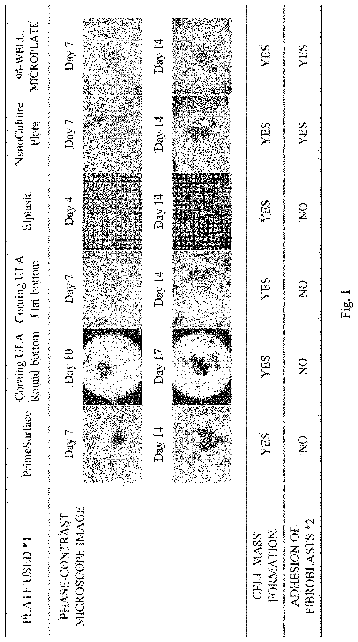
[0080]At first, it was examined whether it would be possible to perform three-dimensional culture using a PDX tumor by means of a Cancer Organoid Culture Kit (ORGANOGENIX, Inc.) described as capable of three-dimensional culture of primary cancer cells. A Cancer Organoid Culture Kit is for a method of performing culture with a plate prepared by allowing a low-adhesive plate to have an uneven scaffolding structure for forming a cell mass of cancer cells and a medium containing serum at 1% by volume or more.

[0081]Primary cancer cells were prepared using PDX tumor of pancreatic cancer (1) (procured from National institutes of Biomedical Innovation, Health and Nutrition) in accordance with Example 1. The necessary amount of cells counted after dispersion treatment were collected in a 15-mL tube, and the supernatant was removed by centrifugation at 300×g for 5 min. Thereafter, a cell suspension was prepared using NanoCulture Medium P t...

example 3

on of Concentration of Extracellular Matrix to Be Added to Medium

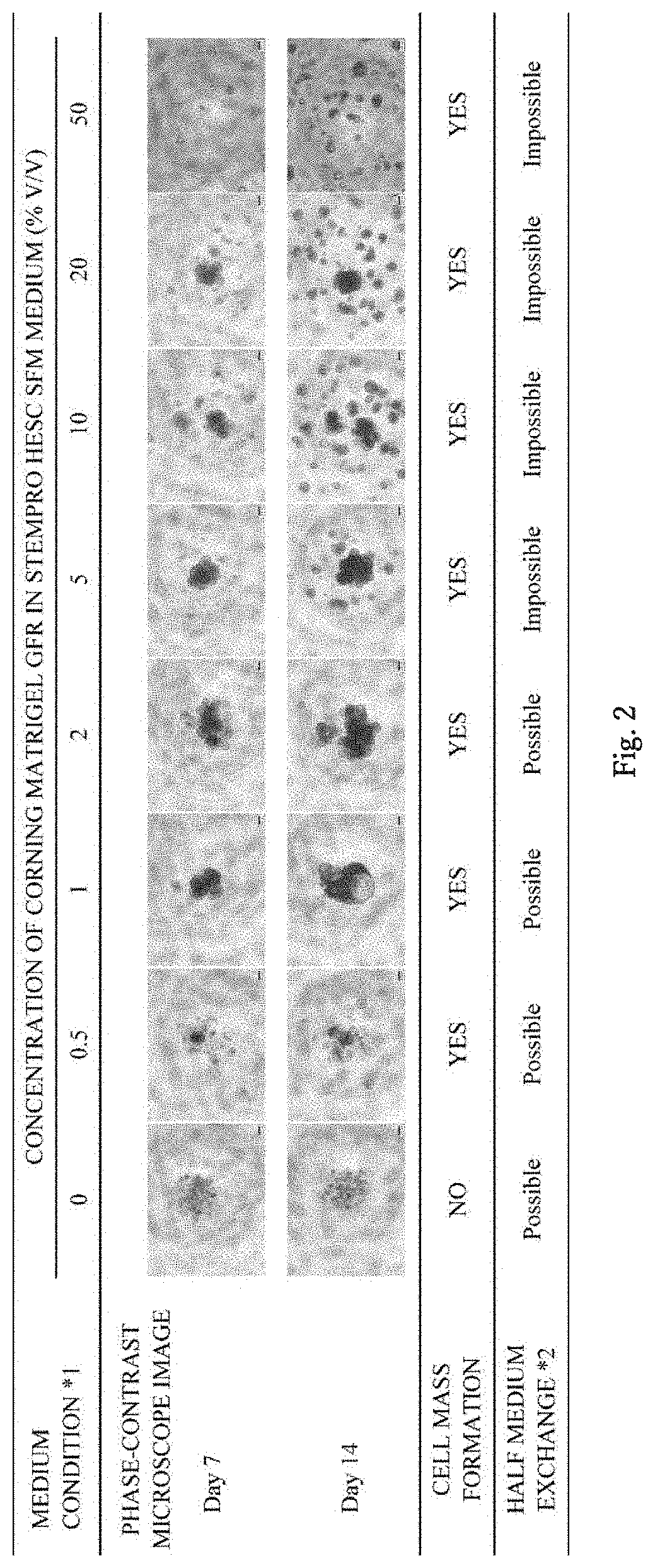
[0085]Primary cancer cells were prepared using pancreatic cancer (1) PDX tumor (procured from National Institutes of Biomedical Innovation, Health and Nutrition) in accordance with Example 1. The necessary amount of cells counted after dispersion treatment were collected in a 15-mL tube, and the supernatant was removed by centrifugation at 300×g for 5 min. Thereafter, a medium prepared by adding Coming Matrigel GFR (Corning Incorporated) to StemPro hESC SFM (Thermo Fisher Scientific K.K.) to yield a final concentration of 0%, 0.5%, 1%, 2%, 5%, 10%, 20%, or 50% v / v was used for preparing cell suspension so that the cell count was 5×10⁴ cells / mL. The cell suspension in an amount of 200 μL was seeded on PrimeSurface (Sumitomo Bakelite Co., Ltd.), and static culture was initiated in a CO2 incubator set to 37° C., and 5% CO2. The seeded cell count was 1×10⁴ cells / 200 μL / well, and the day of seeding was determined to be Day ...


PUM

PropertyMeasurementUnit
diameteraaaaaaaaaa
diameteraaaaaaaaaa
pHaaaaaaaaaa

Abstract

A method of producing a cell mass by three-dimensional culture of primary cancer cells having proliferative ability and properties of handleability, versatility, and high-throughput performance, in which a tumor tissue is used as a starting material, proliferation of cells such as fibroblasts other than cancer cells is inhibited, and the cell mass includes primary cancer cells as a main component. The object is achieved by providing a method of producing a cell mass by three-dimensional culture of primary cancer cells using a tumor tissue, including: a three-dimensional culture step of culturing cells obtained from the tumor tissue in a medium containing a 5% v / v or less extracellular matrix on a substantially low-adhesive cell culture substrate.

Description

TECHNICAL FIELD[0001]The present invention relates to a method of producing a cell mass by three-dimensional culture of primary cancer cells using a tumor tissue, and a cell mass, a screening method, a determination method, and a kit.BACKGROUND ART[0002]Established cancer cells (cancer cell lines) have been conventionally used for a method of evaluating tumor ex vivo. However, it has been pointed out that cancer cell lines have lost the nature of their original tumor due to the homogenization of population and the accumulation of gene mutations as a result of adaptation to an ex vivo environment with long time cell culture. In addition, it has been also pointed out that the limited number of cancer cell lines cannot fully explain the pathological conditions of tumor composed of cancer cells with diverse genetic heterogeneity. Therefore, in order to understand a tumor more correctly, a culture system using primary cells of the tumor has been gaining attention. In addition to a tumor ...

Claims


Application Information

Patent Timeline
Patent Type & Authority Applications(United States)
IPC IPC(8): C12N5/09G01N33/50
CPCC12N5/0693C12N2533/10G01N33/5011C12N2533/90C12N2513/00C12N2533/54C12N2533/52G01N33/15G01N33/5082
Inventor KAKINUMA, HIDEAKISHIMADA, YUKIKOINOUE, HIROAKIMORIKAWA, TAKASHI
Owner MITSUBISHI CHEM MEDIENCE
Learn More