Red fluorescent probe for use in detection of peptidase activity

a fluorescent probe and activity technology, applied in the field of fluorescent probes for detection of peptidase activity, can solve the problems of difficult complete removal, inability to apply probes to cancer cells present in living tissues or within organs such as lymph node metastases, and achieve the effect of accurately visualizing and detecting cancer cells and the lik

Pending Publication Date: 2020-03-19
THE UNIV OF TOKYO
View PDF0 Cites 0 Cited by
  • Summary
  • Abstract
  • Description
  • Claims
  • Application Information

AI Technical Summary

Benefits of technology

[0015]Also, using the red fluorescent probe of the present invention in combination with a conventional green fluorescent probe enables multicolor imaging usin

Problems solved by technology

The single most reliable cancer treatment method at the present time is the early detection and reliable surgical removal of the cancer, but it is difficult to completely remove cancer tissue that is difficult to see completely, leading to recurrence.
However, for the absorption and emission wavelengths of such conventional fluorescent p

Method used


Image

  • Red fluorescent probe for use in detection of peptidase activity
  • Red fluorescent probe for use in detection of peptidase activity
  • Red fluorescent probe for use in detection of peptidase activity

Examples


example 1

1. Synthesis of Fluorescent Probe

[0086]1-1. Synthesis of gGlu-MHM4ThPCR550

[0087]A fluorescent probe 1 (gGlu-MHM4ThPCR550) having the following structure which is a compound of formula (I) of the present invention was synthesized.

[0088]gGlu-MHM4ThPCR550 (compound 16) was synthesized according to the synthesis scheme shown below.

[Synthesis of Compound 4]

[0089]Compound 4 was synthesized according to the literature (O'Sullivan, S., Doni, E., Tuttle, T. and Murphy, J. A., Angew. Chem., 2014, 53, 474-478).

[Synthesis of Compound 5]

[0090]Vilsmeier reagent (7.4 g, 57.7 mmol) was dissolved in anhydrous DMF (40 mL), and the mixture was stirred in an Ar atmosphere at 0° C. Next, compound 4 (10.0 g, 10.9 mL, 57.7 mmol) was added, and stirring was continued for 20 hours at room temperature. Saturated NaHCO3 was added to terminate the reaction, and the mixture was extracted using CH2Cl2. The organic solution was dried using Na2SO4, filtered, and evaporated. The residue was purified by flash column...

example 2

[0146]2. Study of Red Probe Structure Based on pKcycl Calculation

[0147]The structure of suitable fluorescent probe compounds capable of exhibiting fluorescence by cleavage of an acyl residue of formula (I) by a peptidase which is the target was studied based on the calculated pKcycl values.

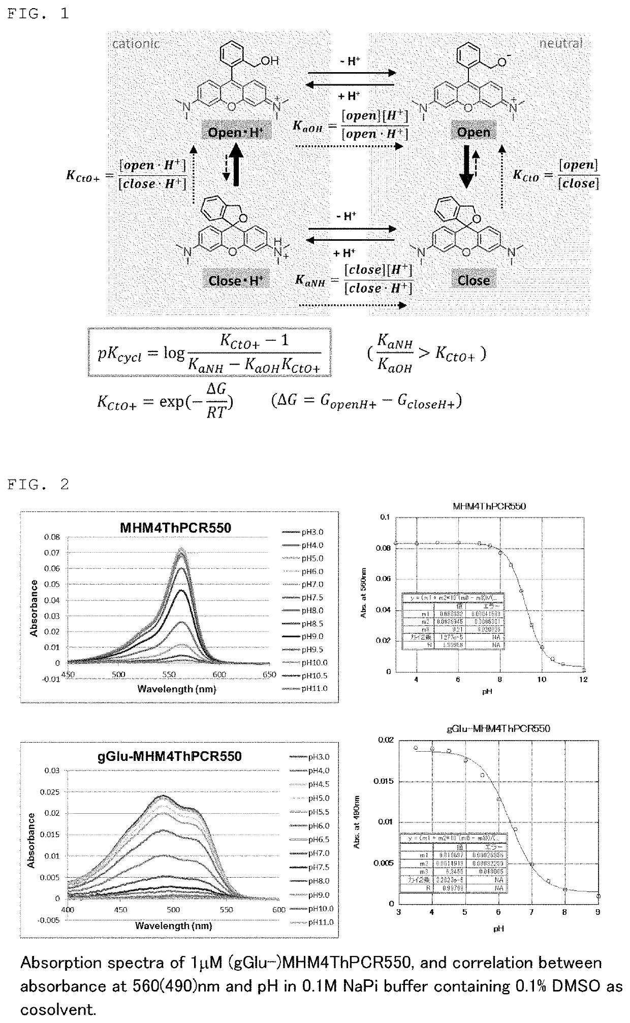
[0148]As shown in FIG. 1, an equilibrium / kinetic model consisting of only four molecular species, in consideration of protonation of amino groups and deprotonation of hydroxymethyl groups (HM), was devised as a model of intramolecular equilibrium of compounds having a rhodamine skeleton. A formula that calculates pKcycl from the free energy difference of the closed-ring form / ring-opened form by the analyzed cationic reaction was derived, assuming that acid-base equilibrium (lateral direction in the model) is reached quickly enough in the HM groups and amino groups. When the calculation results of close-1, open-1 were used as this free energy difference, it was understood that pKcycl of existing de...

example 3

3. Absorption / Fluorescence Spectrum Measurement of the Fluorescent Probe of the Present Invention

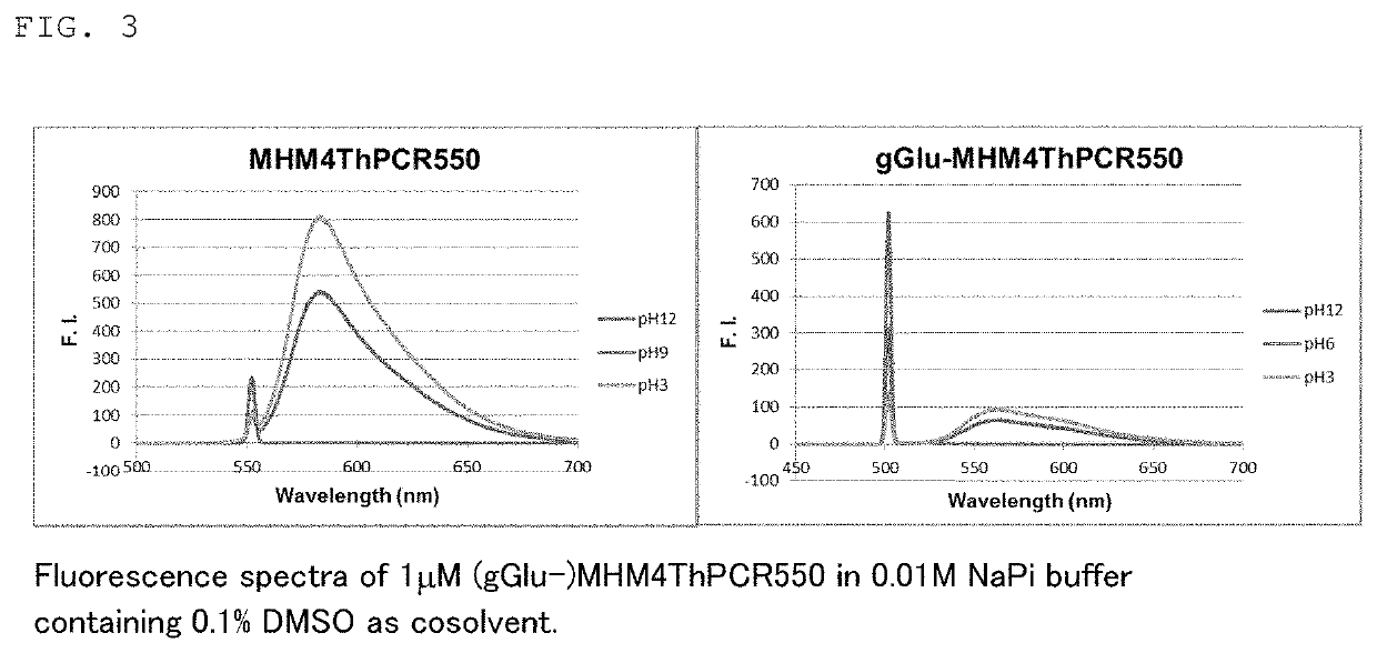
[0150]The absorption spectra and fluorescence spectra of fluorescent probe 1 (gGlu-MHM4ThPCR550) and fluorescent probe 2 (gGlu-HM3ThPSiR600) synthesized in Example 1 were each measured.

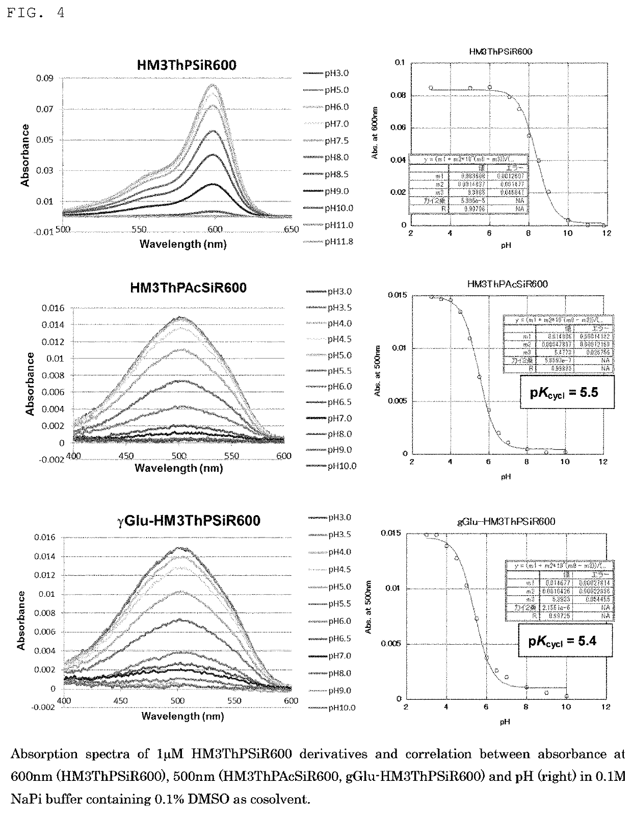
[0151]FIGS. 2 and 3 respectively show the absorption spectra and fluorescence spectra of fluorescent probe 1 (gGlu-MHM4ThPCR550) and, as a comparison, MHM4ThPCR550 having no gGlu group. The pKcycl values computed from the results in FIG. 2 are shown below in Table 3.

TABLE 3gGlu-MHM4ThPCR550MHM4ThPAcCR550MHM4ThPCR550pKcycl 9.2—6.3(Measured)pKcycl 8.75.5—(Calculated)

[0152]In addition, as shown in FIG. 3, while the fluorescent probe of the closed-ring structure exhibited virtually no fluorescence near 660 nm, MHM4ThPCR550 which took on a ring-opened structure at pH 6 was understood to exhibit strong fluorescence intensity near 660 nm.

[0153]FIG. 4 shows the absorption spectrum of fluorescent probe 2 (gGlu-HM3T...


PUM


Abstract

[Problem]
A problem addressed by the present invention is to provide a novel fluorescent probe having excellent tissue permeability that is capable of detecting the peptidase activity expressed at a high level in cancer cells and the like as a response of long-wavelength red fluorescence.
[Solution]
A compound represented by formula (I) or a salt thereof:
[In the formula, A represents a ring structure selected from the group consisting of a thiophene ring, a cyclopentene ring, a cyclopentadiene ring, and a furan ring;
  • X represents a C0-C3 alkylene group;
  • Y represents O, S, C(═O)O, or NH,
  • Z represents O, C(Ra) (Rb), Si(Ra) (Rb), Ge(Ra) (Rb), Sn(Ra) (Rb), Se, P(Rc), or P(Rc) (═O) (where Ra and Rb each independently represent a hydrogen atom or an alkyl group, and Rc represents a hydrogen atom, an alkyl group, or an aryl group);
  • R1 and R2 each independently represent from one to three of the same or different substituents selected from the group consisting of a hydrogen atom, a hydroxyl group, a halogen atom, and an alkyl group, a sulfo group, a carboxyl group, an ester group, an amide group, and an azide group each of which may be substituted;
  • R3 represents an acyl residue derived from an amino acid (where the acyl residue is a residue obtained by removing an OH group from a carboxyl group of the amino acid);
  • R4 and R5 each independently represent a hydrogen atom or an alkyl group (where when R4 or R5 is an alkyl group, the R4 or R5, together with R2, may form a ring structure comprising a nitrogen atom to which R4 and R5 are bonded).]

Description

TECHNICAL FIELD[0001]The present invention relates to a fluorescent probe for detection of peptidase activity. More specifically, the present invention relates to a novel fluorescent probe capable of detecting peptidase activity such as aminopeptidase by fluorescence in the red region, and to a detection method and device using said fluorescent probe.BACKGROUND ART[0002]With the number of cancer patients and deaths increasing year by year, the development of treatment methods continues to be anticipated. The single most reliable cancer treatment method at the present time is the early detection and reliable surgical removal of the cancer, but it is difficult to completely remove cancer tissue that is difficult to see completely, leading to recurrence.[0003]On the other hand, enhanced expression of γ-glutamyl transferase (GGT), which is a peptidase (protease), has been observed in cancer cells, and this enhanced expression is reported to be related to drug resistance. The detection o...

Claims


Application Information

Patent Timeline
IPC IPC(8): C09B11/24C12Q1/37A61K49/00
CPCC09B11/24C12Q1/37A61K49/0008A61K49/0023C07D495/10C07F7/10G01N33/533G01N33/582G01N33/57484C09B11/28C09B6/00A61K49/0021A61K49/0041
Inventor URANO, YASUTERUKAMIYA, MAKOTACHIBANA, RYO
Owner THE UNIV OF TOKYO
Why Eureka
Try Eureka