Human cell-deposited extracellular matrix coatings for textiles and fibers

a technology of extracellular matrix and textiles, which is applied in the field of biocompatibilization of biomedical materials, can solve the problems of reducing the performance and lifetime of the implant device, affecting the healing of the patient, and generating an inflammatory response of many implantable devices

Inactive Publication Date: 2020-10-08
THE SECANT GRP
View PDF1 Cites 0 Cited by
  • Summary
  • Abstract
  • Description
  • Claims
  • Application Information

AI Technical Summary

Problems solved by technology

Many implantable devices generate an inflammatory response due to a lack of human-specific signaling moieties that help the body recognize the implant as human-compatible.
The increased incidence of elevated immune system response reduces performance and lifetime of the implant while simultaneously slowing healing of the patient due to extended inflammation of the local microenvironment at the site of implantation.
While this process presents the ECM components that provide native adhesive cues and cytokines, it does not match the native underlying ECM structure a

Method used


Image

  • Human cell-deposited extracellular matrix coatings for textiles and fibers
  • Human cell-deposited extracellular matrix coatings for textiles and fibers
  • Human cell-deposited extracellular matrix coatings for textiles and fibers

Examples


example 1

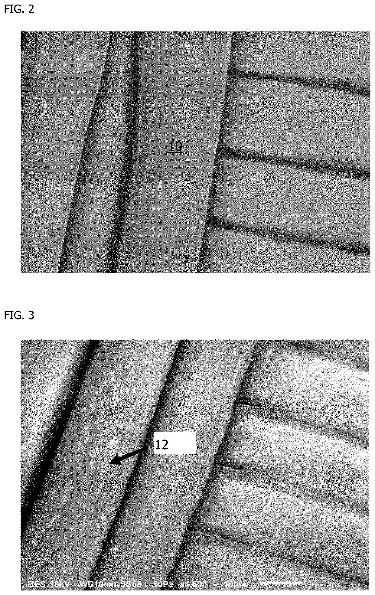
[0096]Human cardiac fibroblast cells were cultured on a woven PVDF textile for six days of cell culture followed by induction of cellular apoptosis using UV light and saline washing to remove cells. FIG. 2 shows the woven PVDF textile 10 after manufacture and prior to cell culture. The woven PVDF textile 10 does not have a coating of ECM present prior to seeding cells for culture. FIG. 3 shows the woven PVDF textile after human cardiac fibroblast cell culture and cell removal. The visible texture on the woven PVDF textile in FIG. 2 indicates the presence of human cardiac fibroblast cell-deposited ECM 12 on the textile.

example 2

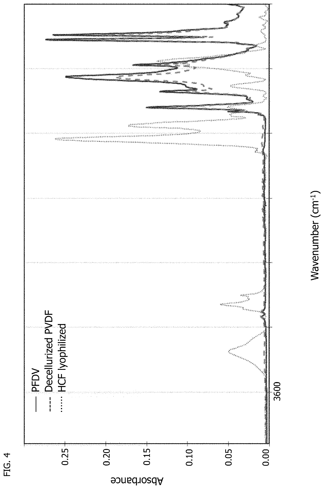
[0097]Fourier transform infrared (FTIR) spectroscopy was used to characterize both the uncoated PVDF woven textile and decellularized, coated PVDF woven textile of Example 1 along with lyophilized human cardiac fibroblast (HCF) cells to chemically identify the deposition of extracellular matrix by cells. FIG. 4 shows the full spectra of the three samples. FIG. 5 shows a magnified region of interest of wavenumber 900 cm−1 to 1100 cm−1, where a C—O stretch bond indicative of the presence of polysaccharides at wavenumber 1041 cm−1 is seen with lyophilized cells but is not present with native PVDF textile or decellularized PVDF, indicating the cells are no longer present in the decellularized textile of Example 1. FIG. 6 shows another magnified region of interest of wavenumber 1500 cm−1 to 1800 cm−1, where two amide peaks at wavenumbers 1653 cm−1 and 1544 cm−1 for the decellularized PVDF of Example 1 are indicative of the presence of proteinaceous deposited ECM by cardiac fibroblasts cu...

example 3

[0098]Similar cell cultures to those described in Example 1 were done on a PET mock leno weave, on a PET plain weave, and on a PGA textile coated with PGS. During the cell cultures on the four different samples, an alamarBlue® assay was used to calorimetrically determine cell counts by associating the cell proliferation rate measured by the assay against a standard curve of known cell dilutions using a UV-vis spectrometer to determine cell count after one and six days of culture over two rounds separate of cultures, the results of which are shown in FIG. 8. The assays demonstrated that increased levels of proliferation occur during the second-round cardiac fibroblast culture, when the cells are cultured on the decellularized textile scaffolds coated with ECM deposited by cardiac fibroblasts during the first round of cell culture. FIG. 9 shows that the proliferation factor, a ratio of the cell count at day 6 to the cell count at day 1, is notably higher in the second round, when card...


PUM

PropertyMeasurementUnit
Adhesion strengthaaaaaaaaaa

Abstract

A process of forming a coated textile includes culturing human cells on a fiber of a textile such that the human cells produce and deposit human extracellular matrix (hECM) on the textile. The process also includes removing the human cells from the hECM to provide the coated textile of the textile and a coating comprising a residual of the hECM produced and deposited by the human cells on the textile during the culturing. A coated textile includes a textile and a coating on the textile. The coating includes hECM in a cell-deposited state in the coating. A solid-state bioreactor composition includes a poly(glycerol sebacate) (PGS) adduct. The PGS adduct includes PGS and a promoting factor or a promoting factor precursor. Another method includes implanting a coated textile in a human. The coated textile is an autograft. The coating includes hECM deposited by human cells from the human.

Description

CROSS-REFERENCE TO RELATED APPLICATIONS[0001]This application claims priority to and the benefit of U.S. Provisional Application No. 62 / 828,604 filed Apr. 3, 2019, which is hereby incorporated by reference in its entirety.FIELD OF THE INVENTION[0002]The present disclosure is generally directed to bio-compatibilization of biomedical materials. More specifically, the present disclosure is directed to the usage of primary human cells or cell lines to generate human extracellular matrix (hECM)-coated woven, braided, non-woven, or knit biomedical textiles or fibers for use as or in an implantable medical device, component, or scaffold in vivo, a bioreactor scaffold for in vitro cell expansion or stem cell differentiation, or a substrate for routine cell culture applications.BACKGROUND OF THE INVENTION[0003]Many implantable devices generate an inflammatory response due to a lack of human-specific signaling moieties that help the body recognize the implant as human-compatible. The increase...

Claims


Application Information

Patent Timeline
IPC IPC(8): A61L27/36C12M3/00A61L27/58A61L27/24C12M1/12
CPCC12M21/08A61L27/58A61L27/24C12M25/14A61L27/3633A61L2420/04C12N5/0068C12N5/0656C12N2533/30C12N2533/40A61L27/34C08L67/00C12N5/0697C12M21/16D06M10/10
Inventor GINN, BRIANGABRIELE, PETER D.HARRIS, JEREMY J.
Owner THE SECANT GRP
Why Eureka
Try Eureka