Methods of Whole Genome Digital Amplification

a technology of whole genome and amplification, applied in the field of methods and compositions for amplifying trace amount of dna, can solve problems such as false negatives, and achieve the effects of reducing amplification bias, high fidelity, and high whole genome coverag

Inactive Publication Date: 2021-09-16
PRESIDENT & FELLOWS OF HARVARD COLLEGE
View PDF0 Cites 2 Cited by
  • Summary
  • Abstract
  • Description
  • Claims
  • Application Information

AI Technical Summary

Benefits of technology

[0016]Embodiments of the present disclosure are directed to a method of amplifying DNA using the methods described herein such as a small amount of genomic DNA or a limited amount of DNA such as a genomic sequence or genomic sequences obtained from a single cell or a plurality of cells of the same cell type or from a tissue, fluid or blood sample obtained from an individual or a substrate. According to certain aspects of the present disclosure, the methods described herein can be performed in a single tube to create the fragments which are then isolated within microdroplets and amplified within the microdroplets with the amplicons being collected from the microdroplets. The term droplet or microdroplet may be used interchangeably herein. The methods described herein avoid, inhibit, prevent, or reduce amplification bias associated with prior art amplification methods where many fragments are amplified together within the same reaction mixture. The methods described herein can provide substantial coverage of the entire genome of a single cell producing amplified DNA for high-throughput sequencing.
[0017]According to an additional aspect, methods are provided herein for performing whole genome amplification of single cells with high fidelity and amplification uniformity or coverage across different loci in the genome which is useful for further sequencing or analysis using high throughput sequencing platforms known to those of skill in the art. More uniform whole genome amplification normally leads to higher whole genome coverage. Coverage represents the percentage of a single cell genomic DNA that can be preserved after amplification. For example, 50% coverage means half of the genetic materials have been lost during the process of single cell whole genome amplification. Methods provided herein minimize loss and amplification bias and provide substantially complete or complete genome coverage of DNA sequencing of genomic DNA from a single cell. Methods described herein can amplify greater than 90 percent, greater than 95 percent, greater than 96 percent, greater than 97 percent, greater than 98 percent, or greater than 99 percent of genomic DNA from a single cell while greater than 70 percent or 75 percent of the genomic DNA can be sequenced with a sequencing depth of 7× or 10× or 15× or 30× with little, substantially few or no chimera sequences.
[0018]Aspects of the methods of th

Problems solved by technology

If a human single cell has a heterozygous mutation, the lack of amplification in one of the tw

Method used


Image

  • Methods of Whole Genome Digital Amplification
  • Methods of Whole Genome Digital Amplification
  • Methods of Whole Genome Digital Amplification

Examples


example i

General Protocol

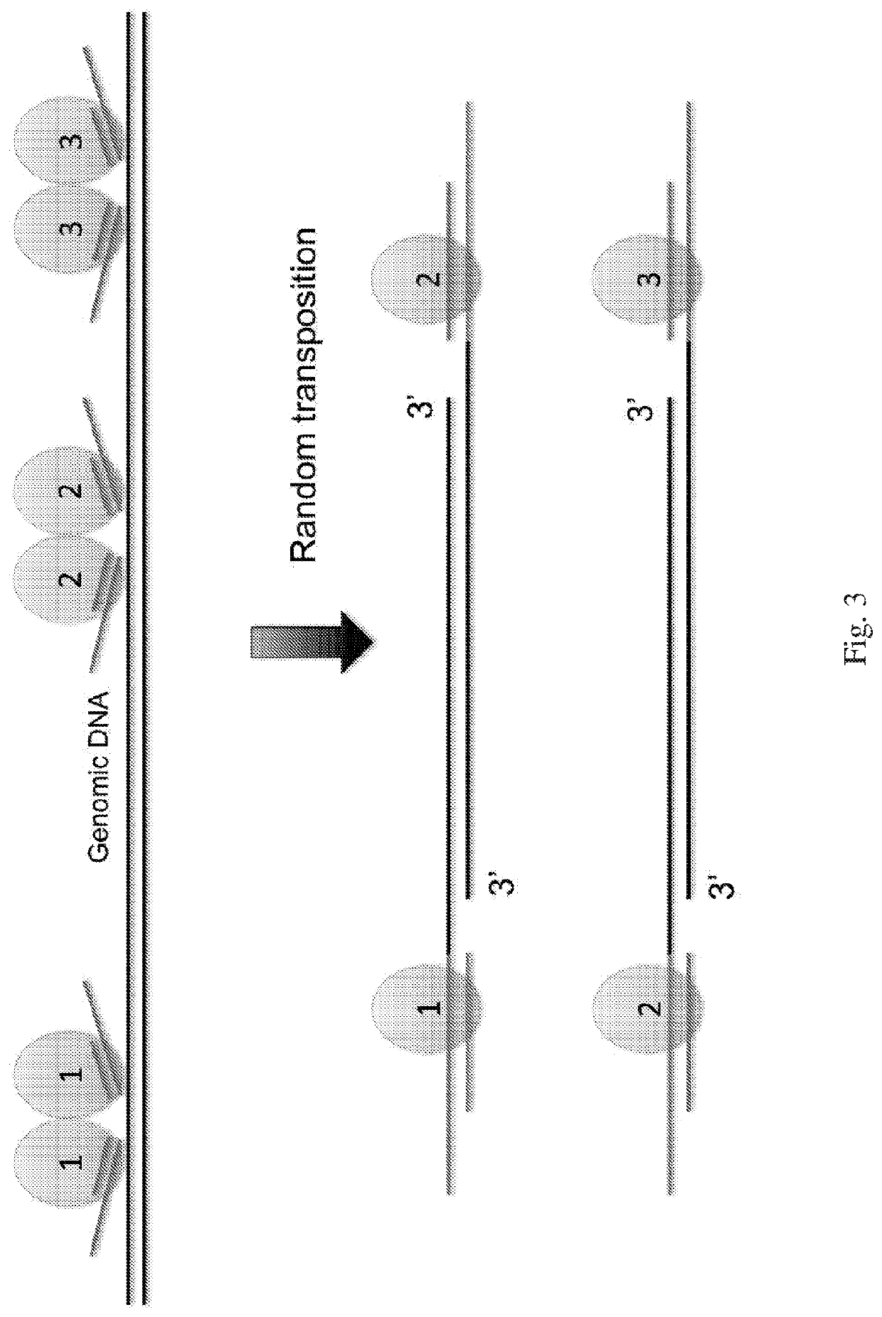
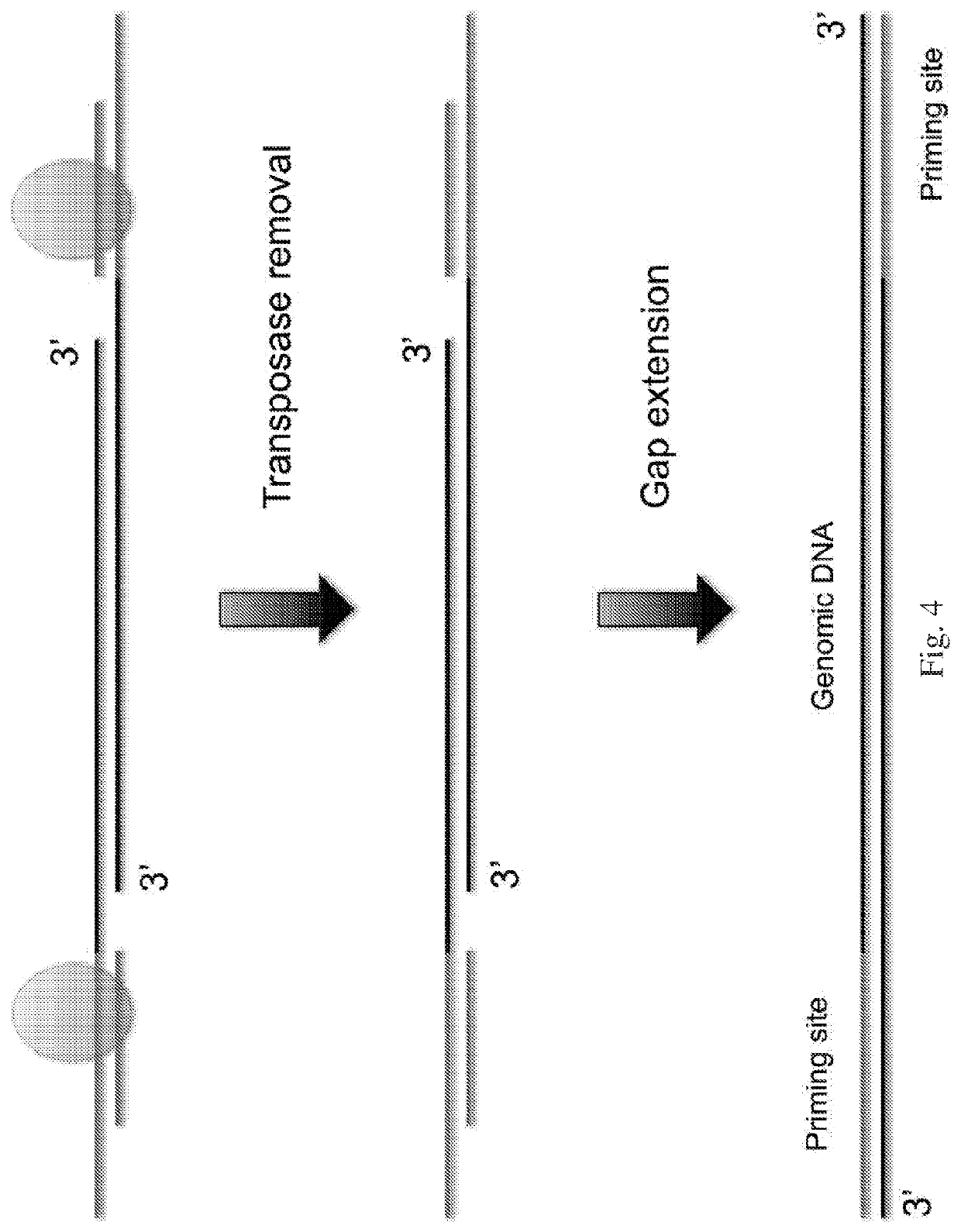
[0093]The following general protocol is useful for whole genome amplification. A single cell is lysed in lysis buffer. Transposome is formed by incubating equal molar of transposon DNA and Tn5 transposase at room temperature for 1 hour. The transposome and transposition buffer are added to the cell lysis which is mixed well and is incubated at 55° C. for 10 minutes. 1 mg / ml protease is added after the tranposition to remove the transpoase from binding to the single cell genomic DNA. Deep vent (exo-) DNA Polymerase (New England Biolabs), dNTP, PCR reaction buffer and primers are added to the reaction mixture which is heated to 72° C. for 10 min to fill in the gap generated from the transposon insertion. The reaction mixture is loaded to the microfluidic device to form micro droplets. The droplets containing single cell genomic DNA template, DNA polymerase, dNTP, reaction buffer and primer are collected into PCR tubes. 40 to 60 cycles of PCR reaction are performed to a...

example ii

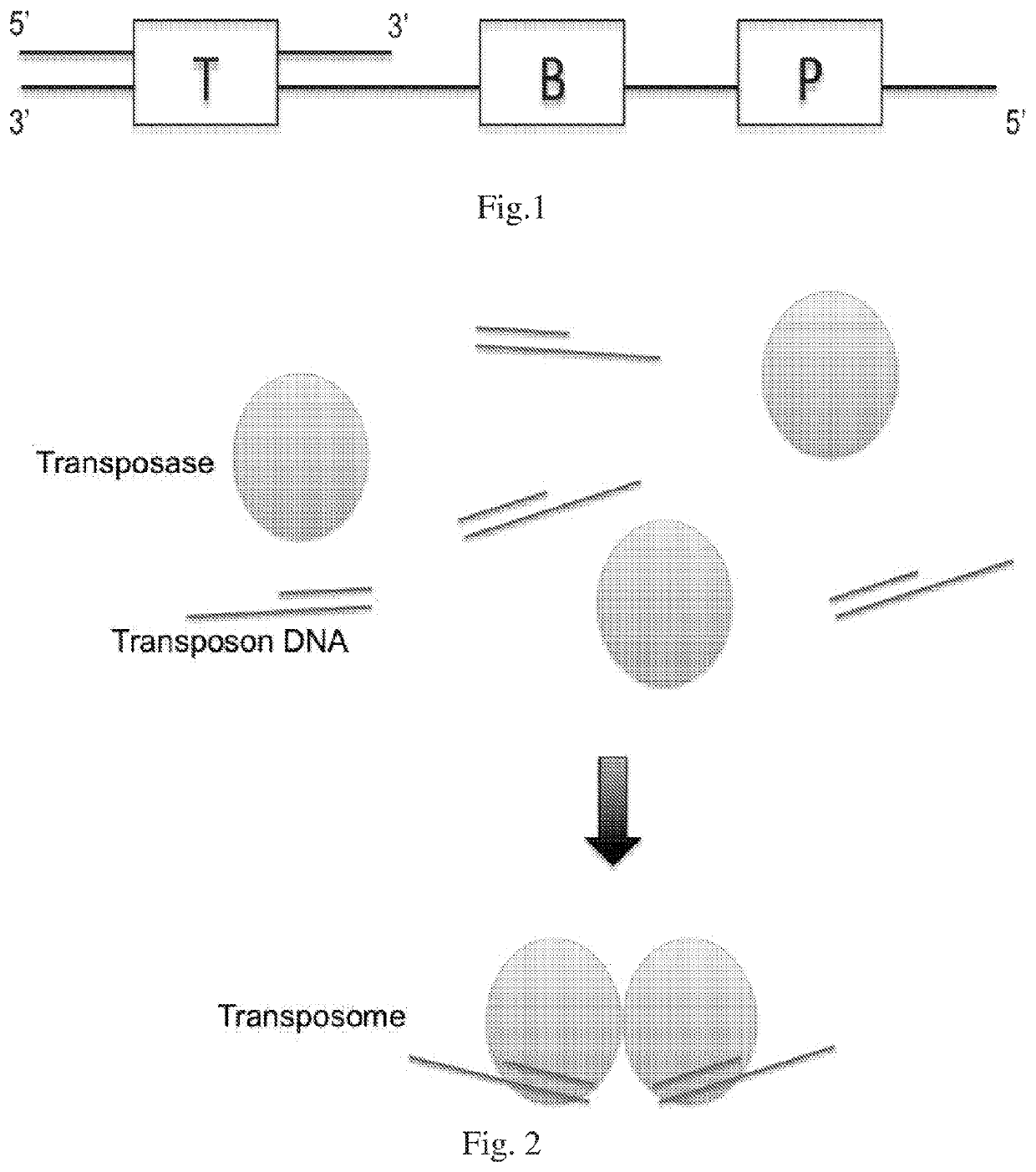
Combining Transposase with Transposon DNA

[0094]Tn5 transposase (Epicentre) is mixed with transposon DNA in equal molar number in a buffer containing EDTA and incubated at room temperature for 10-60 minutes. The final transposome concentration is 0.1-10 μM. The transposon DNA construct has a double stranded 19 bp transposase binding site on one end, and a priming site on the other end. The single stranded priming site forms a 5′ protruding end. Barcode sequences with variable length and sequence complexity could be designed as needed between the 19 bp binding site and the priming site. The transposome may be diluted by many folds in 50% Tris-EDTA and 50% glycerol solution and preserved at −20° C.

example iii

Cell Lysis

[0095]A cell is selected, cut from a culture dish, and dispensed in a tube using a laser dissection microscope (LMD-6500, Leica) as follows. The cells are plated onto a membrane-coated culture dish and observed using bright field microscopy with a 10× objective (Leica). A UV laser is then used to cut the membrane around an individually selected cell such that it falls into the cap of a PCR tube. The tube is briefly centrifuged to bring the cell down to the bottom of the tube. 3-5 μl lysis buffer (30 mM Tris-Cl PH 7.8, 2 mM EDTA, 20 mM KCl, 0.2% Triton X-100, 500 μg / ml Qiagen Protease) is added to the side of the PCR tube and span down. The captured cell is then thermally lysed using the using following temperature schedule on PCR machine: 50° C. 3 hours, 75° C. 30 minutes. Alternatively, mouth pipette a single cell into a low salt lysis buffer containing EDTA and protease such as QIAGEN protease (QIAGEN) at a concentration of 10-5000 μg / mL. The incubation condition varies ...


PUM

PropertyMeasurementUnit
Temperatureaaaaaaaaaa

Abstract

The present disclosure provides a method for genomic DNA amplification, such as whole genome amplification, including using a transposase system to make fragments of the genomic DNA including primer binding sites, isolating in oil each fragment within its own aqueous microdroplet along with PCR amplification reagents, amplifying each fragment within its own aqueous microdroplet, demulsifying the microdroplets to obtain the amplicons and sequencing the amplicons.

Description

STATEMENT OF GOVERNMENT INTERESTS[0001]This invention was made with government support under 5DP1CA186693 from the National Institutes of Health. The Government has certain rights in the invention.BACKGROUNDField of the Invention[0002]Embodiments of the present invention relate in general to methods and compositions for amplifying trace amount of DNA, such as DNA from a single cell, in order to determine its genetic sequences, particularly the entire genome.Description of Related Art[0003]The capability to perform single-cell genome sequencing is important in studies where cell-to-cell variation and population heterogeneity play a key role, such as tumor growth, stem cell reprogramming, embryonic development, etc. Single cell genome sequencing is also important when the cell samples subject to sequencing are precious or rare or in minute amounts. Important to accurate single-cell genome sequencing is the initial amplification of the genomic DNA which can be in minute amounts.[0004]M...

Claims


Application Information

Patent Timeline
IPC IPC(8): C12Q1/6853C12Q1/6806
CPCC12Q1/6853C12Q1/6806C12Q1/6844C12Q2521/301C12Q2535/122C12Q2563/159C12Q2563/179C12Q2565/629
Inventor XIE, XIAOLIANG SUNNEYXING, DONGCHANG, CHI-HAN
Owner PRESIDENT & FELLOWS OF HARVARD COLLEGE
Why Eureka
Try Eureka