Use of polymerized lipid diagnostic agents

a technology of lipid diagnostic agents and liposomes, which is applied in the field of nanoscale polymerized liposome particles, can solve the problems of insufficient ligand-bearing magnetic resonance contrast agents, prior attempts to construct bifunctional agents, and inability to achieve the versatility of chemical modification of agents, etc., and achieves the effect of enhancing polymerized liposomes, effective recirculation in the bloodstream, and well tolerated

Inactive Publication Date: 2000-07-18
NANOVALENT PHARMA +1
View PDF4 Cites 58 Cited by
  • Summary
  • Abstract
  • Description
  • Claims
  • Application Information

AI Technical Summary

Benefits of technology

This invention, in one embodiment, relates to nanoscale polymerized liposome particles based upon lipids having a polymerizable functional group and a metal chelator to attach an imaging enhancement agent, such as paramagnetic or radioactive ions, which assemble to form imaging enhancement polymerized liposomes. In preferred embodiments, the imaging enhancement polymerized liposomes are derivatized with antibodies and / or ligands for in vivo binding to cell surface receptors of targeted cells. In particular, these receptors can be located on the endothelium which eliminates the need for distribution of the active agent out of the blood pool. Paramagnetic polymerized liposomes according to this invention have been found to be well tolerated by rabbits, mice and rats, even on repeated administration, and effectively recirculate in the bloodstream, avoiding rapid endocytosis by the reticuloendothelial system.

Problems solved by technology

These agents have not achieved the versatility in chemical modification to provide for both long recirculation times and active targeting.
Prior attempts to construct bifunctional, ligand-bearing magnetic resonance contrast agents have not been satisfactory due to insufficient sensitivity, poor target specificity and lack of characterization.
Receptor-directed contrast agents for MRI have been attempted using iron oxide particles, but the chemistry and characterization of the particle has been poorly defined and thus it has been difficult to achieve control over non-specific adhesion, blood pool half life and the versatility for both T1 and T2* imaging modes.
In addition, no radioisotope imaging is possible using these iron-based agents which further limits their usefulness.
In the former case, one is limited by the amount of contrast enhancement that can be achieved by direct attachment of chelator to an antibody.
In the latter case, the iron oxide particle is not amenable to control over surface functionality needed to reduce non specific adhesion and the particle is not well characterized or well tolerated in vivo.
However, detection of micro-agglutinated products, approximately 1 .mu.m in size, is very difficult.
While polymerase chain reaction (PCR) based technologies have the technology is limited to detection of nucleic acids.
The expression of glycoproteins on a cell surface is currently detected using assays requiring multiple steps and frequently resulting in low sensitivity.
Currently, the evaluation of the pathophysiology of the cell adhesion molecules is generally limited to in vitro assays.

Method used


Image

  • Use of polymerized lipid diagnostic agents
  • Use of polymerized lipid diagnostic agents
  • Use of polymerized lipid diagnostic agents

Examples


example i

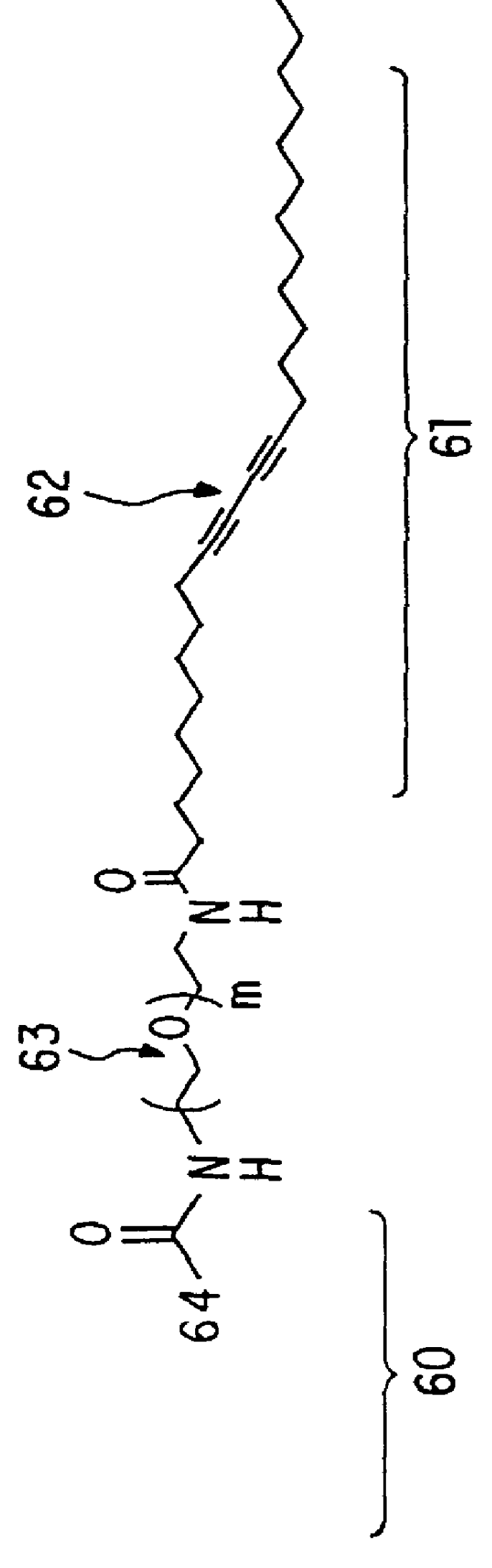
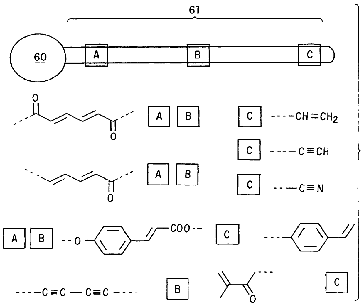
Paramagnetic polymerizable lipids having Gd.sup.+3 and PDA headgroups were synthesized by first preparing the succinimidyl ester by stirring pentacosadiynoic acid (PDA, Lancaster; 10.0 g, 26.7 mmol), N-hydroxysuccinimide (NHS, Aldrich; 5.00 g, 43.4 mmol) and 1-(3-dimethylaminopropyl)-3-ethylcarbodiimide hydrochloride (EDC, Aldrich; 6.01 g, 31.3 mmol) in 660 ml CH.sub.2 Cl.sub.2 at room temperature and shielded from light. The reaction was followed by thin layer chromatography (CHCl.sub.3 / MeOH, 8 / 1) and deemed complete after approximately 5 hours. The solution was washed with water, 1% HCl, saturated sodium bicarbonate and brine. The organic phase was then dried with MgSO.sub.4, filtered, and concentrated under reduced pressure to yield the N-succinimidyl 10,12-pentacosadiynoic acid ester as a slightly yellow solid (10.84 g; 23.0 mmol; 86%).

The succinimidyl ester was dissolved in CH.sub.2 Cl.sub.2 (250 ml) and then slowly added, in dropwise fashion, to a stirred solution of 1,11-dia...

example ii

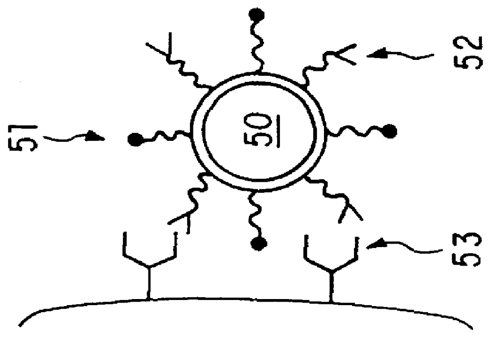
The procedures of Example I were followed except that instead of using DAPC, pentacosydiynoic acid (PDA) was used as the filler lipid. The solution turned blue over the course of one hour irradiation. The resulting polymerized liposomes had the same general properties as reported in Example I.

example iii

Two month old Lewis rats were anaesthetized either with 40-75 mg / Kg dose of sodium pentobarbitol i.p. or by 1.5% isoflurane by inhalation. Paramagnetic polymerized liposome, as prepared in Example I, was administered i.v. over 60-90 seconds through a 24G catheter in a laterial tail vein at a dose of 0.015 mmol Gd.sup.+3 / kg body weight.

Axial magnetic resonance images of the abdomen were obtained prior to paramagnetic polymerized liposome administration and periodically for up to 2 hours post administration. All magnetic resonance images were obtained using an OMEGA-CSI imager (GE, Milwaukee, Wis.) at field strengths of 2.0 or 4.7 Telsa using the standard spin-echo acquisition sequence. T1 weighted images were obtained using a repetition time (TR) of 400 ms, echo time (TE) of 18 ms, and 2 excitations (NEX) per 128 phase encoding steps, completing a 256.times.128 data matrix in under 2 minutes. Slice thickness (ST) was 2mm and the interslice gap was 2 mm. Four axial slices were acquir...


PUM

PropertyMeasurementUnit
diametersaaaaaaaaaa
sizeaaaaaaaaaa
concentrationaaaaaaaaaa

Abstract

Polymerized liposome particles which are linked to a targeting agent and may also be linked to a contrast enhancement agent and/or linked to or encapsulating a treatment agent. The targeting imaging enhancement polymerized liposome particles interact with biological targets holding the image enhancement agent to specific sites providing in vitro and in vivo study by magnetic resonance, radioactive, x-ray or optical imaging of the expression of molecules in cells and tissues during disease and pathology. Targeting polymerized liposomes may be linked to or encapsulate a treatment agent, such as, proteins, drugs or hormones for directed delivery to specific biological sites for treatment.

Description

1. Field of the InventionThis invention relates to polymerized liposomes which are linked to a targeting agent and may also be linked to at least one of an image contrast enhancement agent and a therapeutic or treatment agent to provide targeted polymerized liposome diagnostic agents and targeted polymerized liposome therapeutic agents, respectively. In one embodiment, this invention relates to liposomes which may be linked to contrast ions for magnetic resonance imaging and radioisotope imaging or optical imaging by using chromophores attached to the liposome or chromophores inherent in the particle in which the polymerization adds stability in vivo. The paramagnetic or radioactive polymerized liposomes may also be linked to antibodies and ligands for specific interaction with biological targets holding the contrast agent to specific biological sites, providing in vitro and in vivo study of the expression of molecules in or on the surface of cells and tissues during disease and pat...

Claims


Application Information

Patent Timeline
Patent Type & Authority Patents(United States)
IPC IPC(8): A61K49/18A61K49/22A61K49/06A61K51/12G01N33/58
CPCA61K49/1812A61K49/22G01N33/586A61K51/1237Y10S977/927Y10S977/795Y10S977/928Y10S977/93Y10S977/775Y10S977/918Y10S424/812Y10S977/907Y10S977/897Y10S436/829Y10S977/808
Inventor LI, KING CHUENBEDNARSKI, MARK DAVIDSTORRS, RICHARD WOODLI, HENRY Y.TROPPER, FRANCOIS DANIELSONG, CURTIS KANG HOONSIPKINS, DOROTHY ANNAKUNIYOSHI, JEREMY KENJI
Owner NANOVALENT PHARMA
Why Eureka
Try Eureka