U.S. Pat. No. 4,637,986 (to Brown, et al.) describes a
flow cytometry lysing reagent for producing a 3-part differential of leukocytes. The lysing reagent is a hypotonic
aqueous solution enabling hypotonic
lysis of red blood cells. The lysing reagent comprises a leukoprotective agent for preserving the
lymphocyte cellular integrity during analysis, and buffers to provide the correct pH environment for optimal
lysis. The preferred leukoprotective agents include methyl oxyethanol, ethyl oxyethanol and butyl oxyethanol. Brown et al. specifically teach the importance of the
pH range of the lysing reagent in order to provide rapid lysing of red blood cells, but prevent deterioration of the leukocytes. More specifically, the preferred
pH range is from approximately 8.1 to about 8.8, with optimal around 8.5. Brown et al. teach that if pH of the lysing reagent is above 9.0, a rapid deterioration of the white blood cells occurs, which results in disadvantageous effects on counting accuracy. Furthermore, Brown et al. teach that the specific
buffering agent is critical to the proper operation of the lysing agent. More specifically, it is found that mineral buffers such as
phosphate and carboxylic acids or molecules containing a carboxyl group are not effective, and the
boric acid /
TRIS buffer combination is also less effective. Brown et al. specifically teach the optimal buffer
system of an organic,
nitrogen containing buffer balanced with an ethane
sulfonic acid buffer to maintain pH of the lysing reagent from 8.1 to 8.8.
on counting accuracy. Furthermore, Brown et al. teach that the specific
buffering agent is critical to the proper operation of the lysing agent. More specifically, it is found that mineral buffers such as
phosphate and carboxylic acids or molecules containing a carboxyl group are not effective, and the
boric acid /
TRIS buffer combination is also less effective. Brown et al. specifically teach the optimal buffer
system of an organic,
nitrogen containing buffer balanced with an ethane
sulfonic acid buffer to maintain pH of the lysing reagent from 8.1 to 8.8.
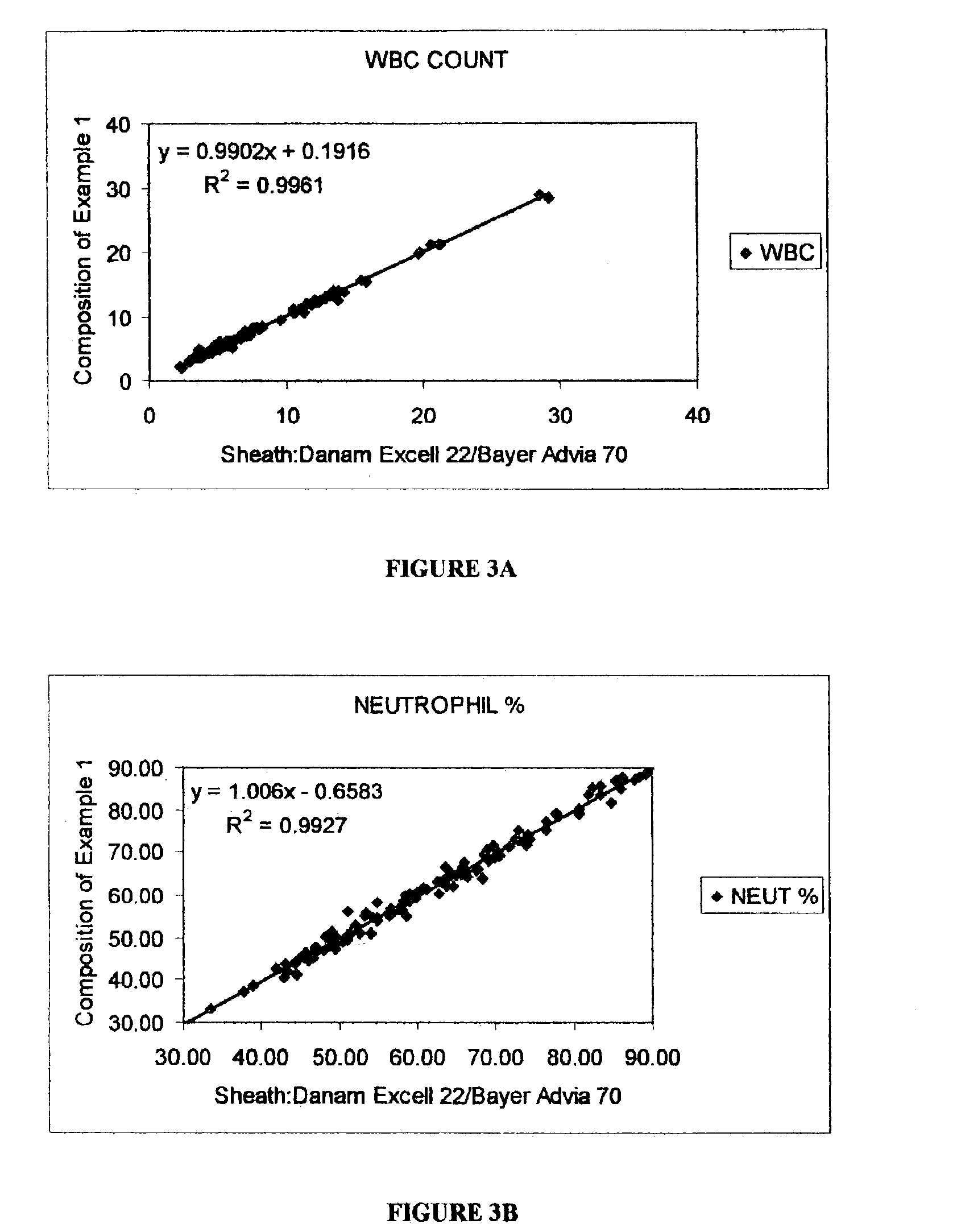
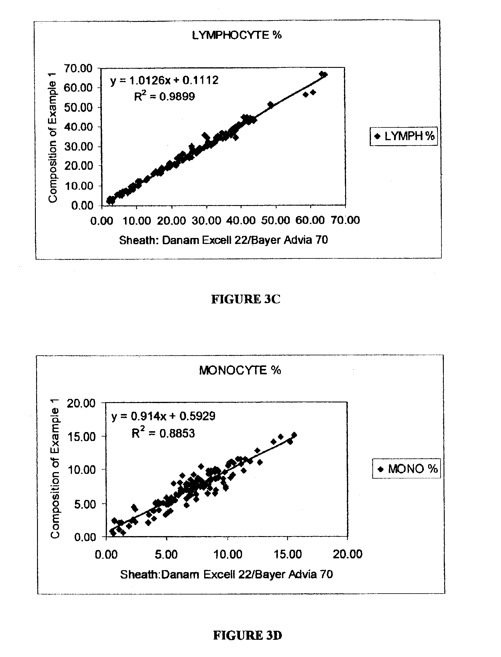
U.S. Pat. No. 5,510,267 (to Marshall) describes a
flow cytometry lytic reagent and a method for providing a 5-part differential analysis of leukocytes. The method includes diluting a blood sample with a neutral and near isotonic
diluent, mixing the diluted sample with the lytic reagent to lyse red blood cells, and analyzing the sample mixture in a
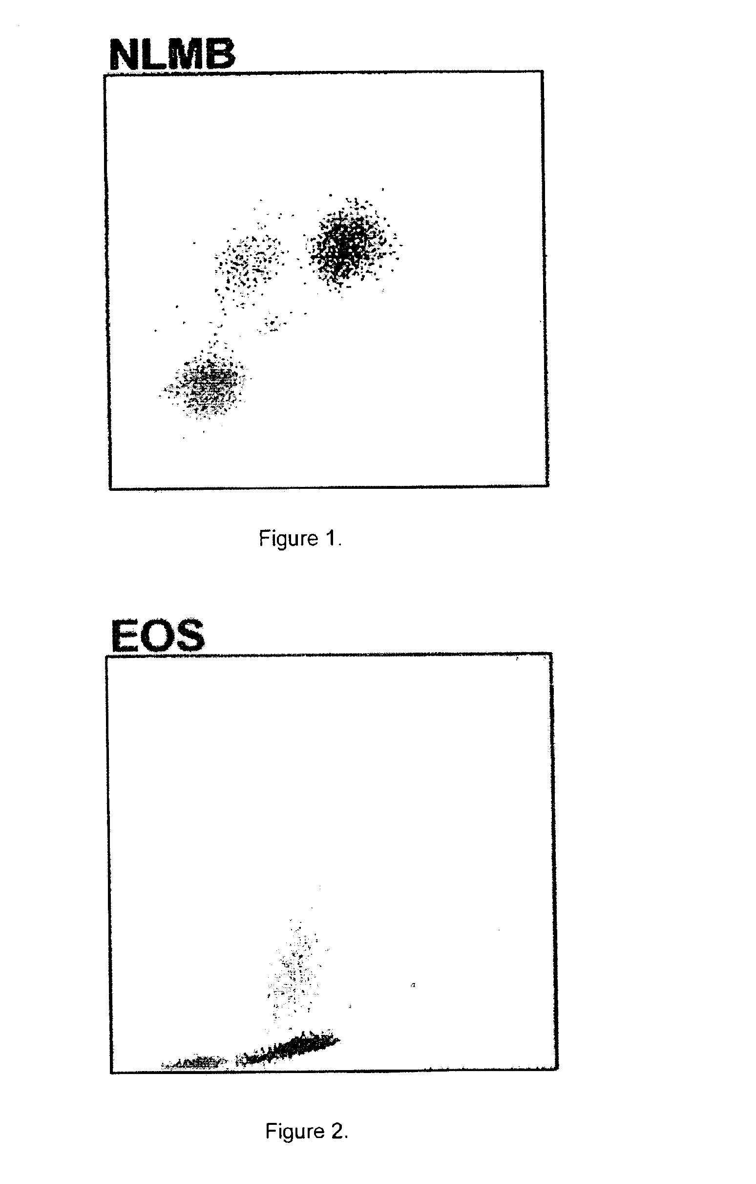
flow cell by measuring 0°, 10°, 90° and 90° depolarized light scatter signals to differentiate leukocytes into five subpopulations, including neutrophils, lymphocytes, monocytes, eosinophils and basophils. Marshall teaches that the lytic reagent includes 2-
phenoxyethanol which combines the function of leukoprotective and
antimicrobial; Triton X-100 (octylphenoxypolyethoxyethanol) a lytic and
wetting agent; and an organic buffer with pKa at or near 8.5 to maintain pH of the lysing reagent at 8.5. Furthermore, Marshall teaches the importance of pH to the function of the lytic reagent. More specifically, the optimal pH is 8.5, and with a lower range of 8.1 without significant effects on the reagent performance. However, if pH of the lytic reagent increases to 9.0, partial destruction of white blood cells can occur.
Both U.S. Pat. No. 4,637,986 and U.S. Pat. No. 5,510,267 teach that
red blood cell lysis under alkaline condition is more rapid, which is desirable for automated
hematology analyzers. However, at pH above 9,
white blood cell damage occurs, which impacts the accuracy of the leukocyte counting and differential analysis.
Therefore, it is apparent that there is a need for an improved lytic reagent composition which enables rapid
red blood cell lysis under alkaline condition, but enables preserving the leukocytes for leukocyte differential analysis using automated
hematology analyzers.
In one aspect, the present invention provides a lytic reagent composition for differential analysis of leukocytes using
optical measurements. The lytic reagent composition comprises a short chain
alkyl oxyethanol selected from the group consisting of 2-methoxyethanol, 2-ethoxyethanol, 2-propoxyethanol, and 2-isopropoxyethanol in a sufficient amount to preserve leukocytes; a non-lysing
nonionic surfactant as a debris solublizer; and an inorganic buffer to maintain pH of the lytic reagent composition in a range from about 9.1 to about 10.7.

