Controlled ocular lens regeneration

a technology of ocular lens and control, which is applied in the field of ocular tissue regeneration, can solve the problems of less than ideal procedures

Active Publication Date: 2005-09-20
GWON ARLENE E
View PDF9 Cites 32 Cited by
  • Summary
  • Abstract
  • Description
  • Claims
  • Application Information

AI Technical Summary

Benefits of technology

[0007]A method for treating an ocular condition in a mammal, as well as for controlling the shape of regenerating lens tissue that generally includes the removal of an original lens to leave an empty lens capsule and thereafter disposing a scaffold within the empty lens capsule. The scaffold enables proliferation and differentiation of lens fibers, or tissue, thereafter the scaffold is selectively removed by laser photoablation.

Problems solved by technology

This procedure is less than ideal because the current synthetic intraocular lenses are unable to accommodate appreciably, and secondary opacification of the posterior capsule is a common occurrence.
While intraocular implantation of multifocal or accommodating intraocular lenses (IOLs) attempt to address the need for far and near vision in the cataract patient, they are complicated by the development of posterior capsule opacification and visual dysphotopsias.

Method used


Image

  • Controlled ocular lens regeneration
  • Controlled ocular lens regeneration
  • Controlled ocular lens regeneration

Examples


example 1

Preparation of Animals

[0081]Endocapsular lens extraction by phacoemulsification and irrigation / aspiration of the lens through a 2–3 mm capsulorhexis was performed in rabbits. Following removal of the original lens, a high viscosity hyaluronic acid was injected into the capsule bag, and a collagen patch was placed inside the capsulorhexis and brought up against the capsulotomy with an air bubble in the bag. The animals were followed postoperatively by slit lamp biomicroscopy. Following euthanasia, eyes were enucleated, paraffin embedded and H & E slides prepared for routine histology.

example 2

Insertion of Collagen Patch in Lens Capsule

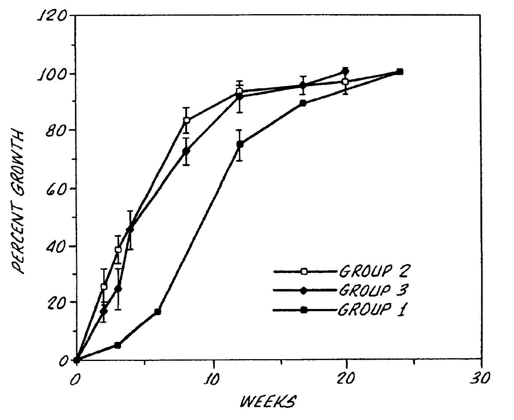
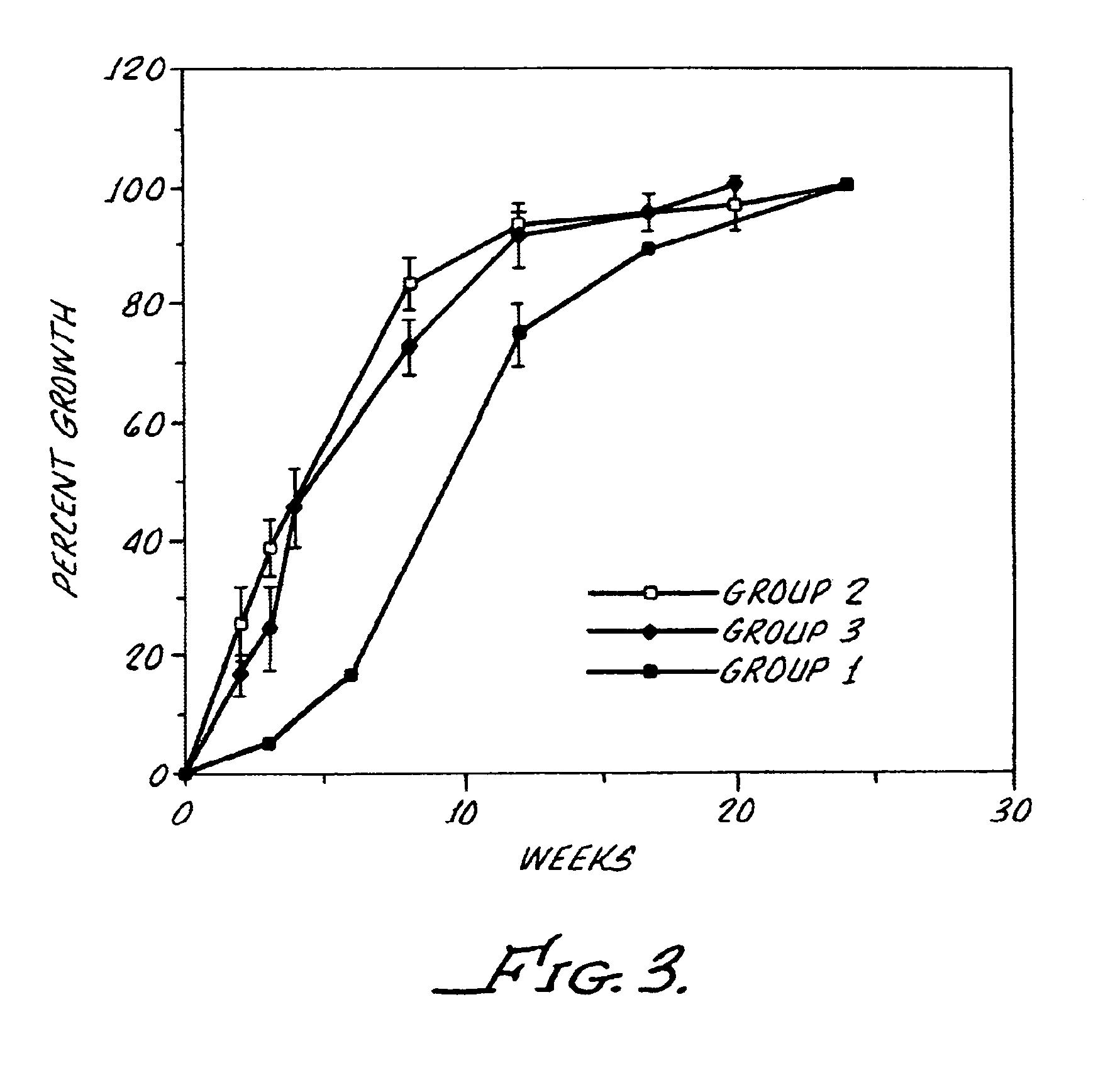
[0082]The inventor restored the lens capsule integrity by inserting a collagen patch at the time of endocapsular lens extraction surgery to seal the anterior capsulotomy and to improve the shape and structure of the regenerated lenses. Lens regeneration was first noted as early as one to two weeks following surgery. Regenerated lens filled approximately 50% of the capsule bag at two weeks and 100% by five weeks. Subsequent growth was in the anterior-posterior direction. Lens thickness increased by 0.3 mm per month. The regenerated lenses were spherical with normal cortical structure and a nuclear opacity. Restoration of lens capsular integrity with a collagen patch following endocapsular lens extraction enhanced the shape, structure, and growth rate of the regenerated lenses. In addition, lens regeneration was shown to occur in two cats.

example 3

Filling of Capsule Bag with Air, Sodium Hyaluronate, or Perfluoropropane

[0083]After insertion of a collagen patch the inventor then filled the capsule bag with air, sodium hyaluronate (Healon®), or perfluoropropane gas to prevent adhesions between the anterior and posterior capsules and to maintain capsule tautness and shape. Lens thickness measurements at one and two months represent the filled capsule bag containing lens regrowth as well as most probably a mixture of balanced salt solution (BSS), Healon®, aqueous, and collagen degradation products and injured lens epithelial cells. From 4 to 12 months, lens thickness increased gradually but in progressively smaller increments.

[0084]The collagen patch occluded the anterior capsulotomy for up to two weeks before dissolution resulting in a linear scar at the capsulotomy site. The capsule bag was distended and maintained taut without surface wrinkles for one, five, and eight weeks in the Healon®, air, and perfluoro-propane groups, res...


PUM


Abstract

The present invention addresses the treatment of ocular conditions by the enhancement of lens regeneration. This is accomplished by the administration of a high viscosity composition including a hyaluronic acid compound and laser photoablation. Excess high viscosity composition may be removed by focal laser photophacoablation. Additionally, a collagen product may be injected within the lens capsule to improve lens cell proliferation and differentiation, and to improve the configuration, shape and structure of regenerated lenses. Various embodiments involving the enhancement of lens regeneration are described. For example, lens regeneration may be enhanced by filling the lens capsule bag with the inventive hyaluronic acid compound; by inserting at least one collagen patch in the lens capsule; and/or by injecting a collagen-based product into the lens capsule.

Description

[0001]The present invention generally relates to the regeneration of ocular tissue and more particularly relates to the treatment of conditions, as for example, lenticular disorders and controlled regeneration of lens tissue.[0002]Regeneration and repair are the fundamental features of the healing response. The ability to regenerate (i.e., to replace damaged tissue with healthy cells of similar type) varies among tissues and may be seen in the corneal epithelium and conjunctiva. While the healing response in and around the eye occurs primarily because of tissue repair mechanisms (i.e., damaged tissue is replaced by a newly generated fibrous connective tissue) rather than regeneration, there is substantial data suggesting that regeneration of the natural lens is possible. Ideally, the regenerated lens, with or without a suitably flexible and biocompatible polymeric lens, would have all the properties of the natural lens including clarity, protein content, histology, focusing power an...

Claims


Application Information

Patent Timeline
Patent Type & Authority Patents(United States)
IPC IPC(8): A61F9/007A61F9/008A61F9/011A61K9/00
CPCA61F9/00802A61K9/0051A61F9/00736A61F9/008A61F2009/0087A61F2009/00887
Inventor GWON, ARLENE E.
Owner GWON ARLENE E
Why Eureka
Try Eureka