Transdifferentiation of epidermal basal cells into neural progenitor cells, neuronal cells and/or glial cells

a technology of epidermal basal cells and neural progenitor cells, applied in the field of medical arts, can solve the problems of preventing the regeneration of neural tissues, hindering the development of neurological injury therapies, and limited structural self-repair potential of the central nervous system of the adul

Inactive Publication Date: 2005-09-27
CEDARS SINAI MEDICAL CENT
View PDF16 Cites 17 Cited by
  • Summary
  • Abstract
  • Description
  • Claims
  • Application Information

AI Technical Summary

Benefits of technology

[0075]The present invention also relates to a method of identifying a novel nerve growth (or neurotrophic) factor that employs transdifferentiated cells of the invention. The methods involve transdifferentiating a population of proliferating epidermal basal cells into neuronal progenitor cells, neuronal cells, or glial cells; culturing the transdifferentiated cells; exposing the cultured cells, in vitro, to a potential nerve growth factor; and detecting the presence or absence of an effect of the potential nerve growth factor on the survival of the cells or on a morphological or electrophysiological characteristic and/or molecular biological property of the cells. The transdifferentiated cells are assayed in vitro to determine whether there is an effect of a potential nerve growth factor on a physiological or molecular biological property of the transdifferentiated cells. For example, which, if any, neuronal or glial cell types develop from neural progenitors, the maturation of particular cell types, and the continued support of cell survival (e.g., effect on cell numbers) can be determined. In addition, experimental techniques, based on an electrophysiological characteristic (patch clamp, different types of intracellular recording, etc.) or molecular biological properties (gene expression profiles, organization of cytoskeleton, organization of ion channels and receptors etc.) can be used to detect the effects of potential nerve growth/neurotrophic factors on particular cell types. The potential factor can be, but need not be an isolated compound; the inventive transdifferentiated cells can be used to test, or assay, the effect, or lack thereof, of potential growth factor sources (tissue homogenates, expression cDNA library products, etc.) on the survival and functional characteristics of the cells to detect candidates for further isolation.
[0076]The use of transdifferentiated epidermal basal cells bypasses the difficulties in isolating and culturing neuronal cell types from the brain, and, therefore, the inventive method of identifying a novel nerve growth factor is a benefit to research in this area.
[0077]This same advantage pertains to the inventive method of using cells transdifferentiated from epidermal basal cells to identify a potential chemotherapeutic agent by transdifferentiating a population of epidermal basal cells into neuronal progenitor, neuronal, or glial cells by the inventive method described above; culturing the transdifferentiated cells; exposing the cultured cells, in vitro, to a potential chemotherapeutic agent; and detecting the presence or absence of an effect of the potential chemotherapeutic agent on the survival of the cells or on a morphological or electrophysiological characteristic and/or molecular biological property of said cells. An effect altering cell survival, a morphological or electrophysiological characteristic and/or a molecular biological property of the cells indicates the activity of the chemotherapeutic agent. The potential chemotherapeutic agent can be an agent intended to treat a nervous system disorder, or the method can be used to test an agent intended or proposed for treating any other type of disorder for its effects on cells possessing neural progenitor, neuronal or glial cell features. Experimental assay techniques, based on an electrophysiological c...

Problems solved by technology

Once damaged, the central nervous system of the adult has limited potential for structural self-repair.
The general inability of the adult to generate new neurons (excitable cells specialized for the transmission of electrical signals from one part of the body to another) typically prevents the regeneration of neural tissues.
This limitation has hindered the development of therapies for neurological injury, for example from stroke or physical trauma, or for degenerative diseases, such as Huntington disease, Alzheimer disease, and Parkinsonism.
Because the brain is protected by a blood-brain barrier that effectively blocks the flow of large molecules into the brain, peripheral injection of growth factor drugs, or other potentially therapeutic gene products,...

Method used


Image

  • Transdifferentiation of epidermal basal cells into neural progenitor cells, neuronal cells and/or glial cells
  • Transdifferentiation of epidermal basal cells into neural progenitor cells, neuronal cells and/or glial cells

Examples


example 1

Methods

Preparation of Epidermal Cell Culture and Dedifferentiation of Cell Population.

[0084]Human adult skin was obtained from surgery procedures or skin biopsy. Before cultivation, as much as possible of the subepidermal tissue was removed by gentle scraping. Primary cultures were initiated by culturing 4-10 2×2 mm explants / 35 mm tissue culture dish in Dulbecco's modified Eagle medium (DMEM; GIBCO-BRL, Life Technologies, Inc.) with 15% fetal calf serum (GIBCO-BRL, Life Technologies, Inc.), 0.4 μg / mL hydrocortisone, and 10 ng / mL epidermal growth factor (EGF; Collaborative Research, Inc.). The medium was changed every three days. Thirty to thirty-five day old cultures were used for subsequent experimentation. Differentiated keratinized cell layers were stripped off by incubating the cultures in Ca2+-free minimal essential medium (SMEM; GIBCO-BRL, Life Technologies, Inc.). After 72 hours in calcium-free medium, suprabasal layers were detached and removed by aseptic aspiration after sh...

example 2

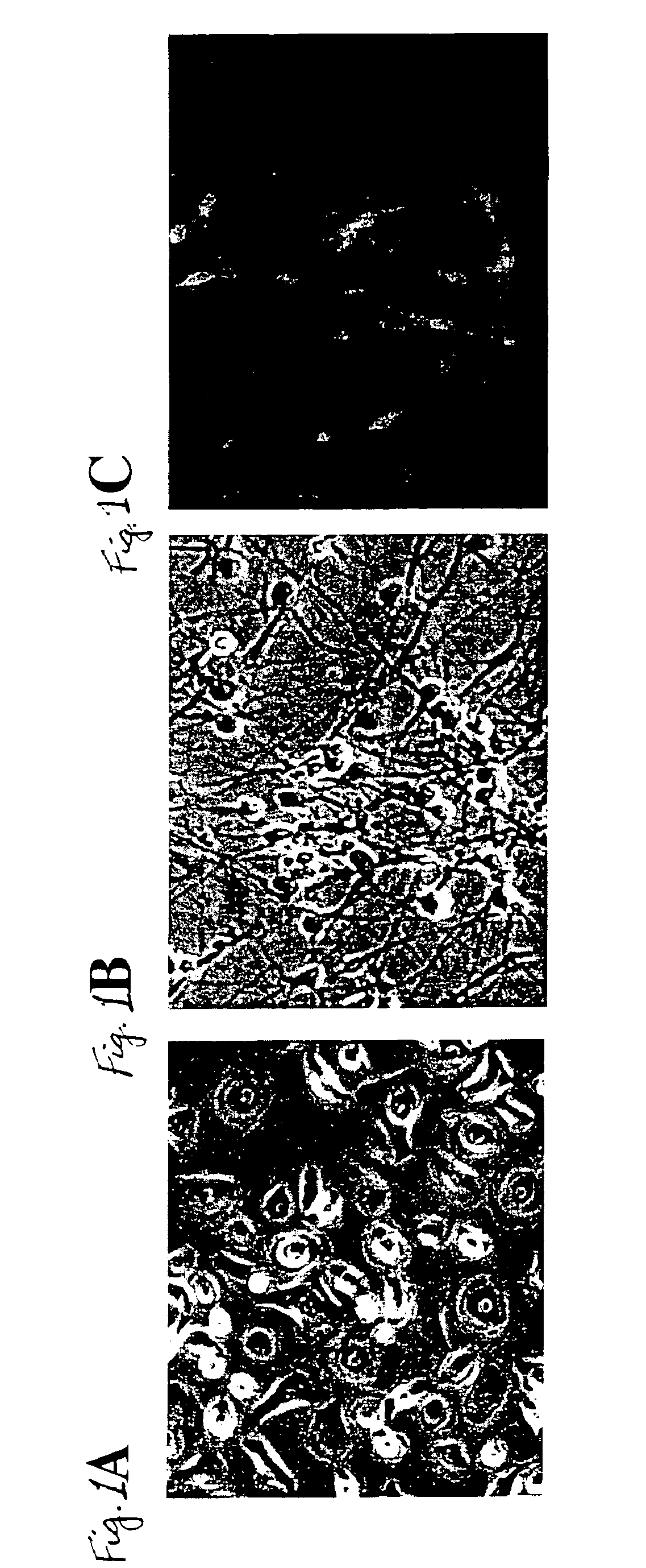
Transdifferentiation of Epidermal Basal Cells

[0094]Different treatment combinations were tested including fetuin (to antagonize or inhibit endogenous BMP signal transduction activity) with or without MSX1 and / or HES1 antisense oligonucleoddes (to suppress MSX1 and HES1 expression) to demonstrate transdifferentiation of epidermal basal cells into neuronal or neuron-like cells. After a 3-day treatment with the various combinations of fetuin and / or antisense oligonucleotides, cells in all treatment groups were grown in the presence of all-trans retinoic acid (10−7 M) and BDNF (20 ng / mL) for 7 additional days before immunostaining for the neural-specific antigen neurofilament M. The values in Table 1 show the relative effectiveness of the various treatments.

[0095]The results in Table 1 demonstrate that transdifferentiation of epidermal basal cells into Neurofilament M positive cells requires antagonism of BMP signal transduction by fetuin in combination with suppression of MSX1 and / or H...

example 3

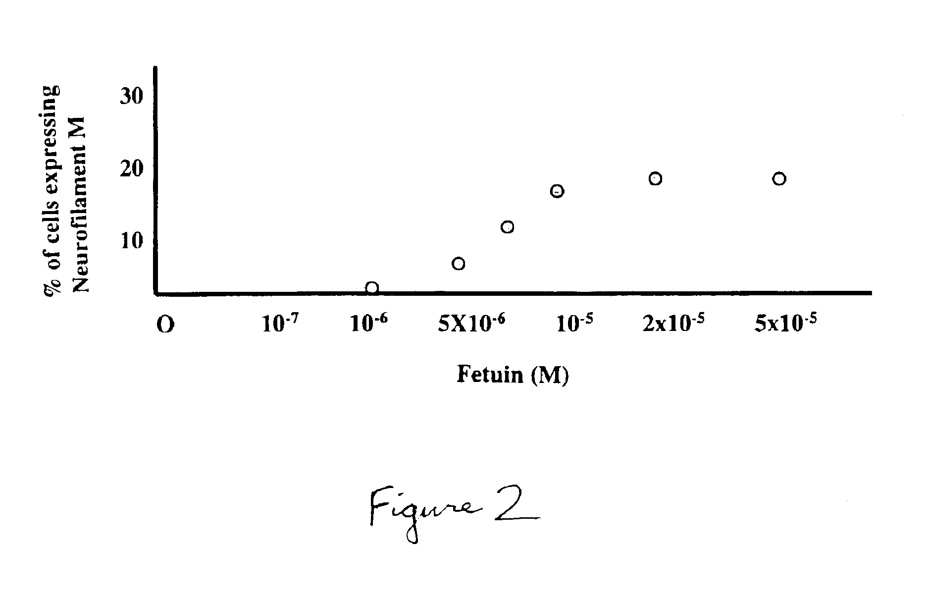
Efficiency of Transdifferention Depends on the Concentration of BMP-antagonist

[0098]FIG. 2 illustrates that suppression of BMP signal transduction to allow expression of neuronal characteristics is concentration dependent with respect to the BMP antagonist. At a concentration in the medium of 1×10⁻⁵ to 5×10⁻⁵M, fetuin had maximal effect on the expression of neurofilament M by transdifferentiated cells (FIG. 2).


PUM

PropertyMeasurementUnit
Lengthaaaaaaaaaa
Molar densityaaaaaaaaaa

Abstract

Disclosed is a method of transdifferentiating an epidermal basal cell into a cell having one or more morphological, physiological and/or immunological features of a neural progenitor, neuronal, or glial cell by culturing a proliferating epidermal basal cell population derived from the skin of a mammalian subject; exposing the epidermal basal cell(s) to an antagonist of bone morphogenetic protein (BMP), such as fetuin, noggin, chordin, gremlin, or follistatin; and growing the cell(s) in the presence of at least one antisense oligonucleotide comprising a segment of a human MSX1 gene and/or a segment of a human HES1 gene, or homologous non-human counterpart of either of these. Also disclosed is a transdifferentiated cell of epidermal origin and cell cultures derived therefrom. In addition, methods of using the inventive transdifferentiated cell(s) and cell cultures to identify a novel nerve growth factor or to screen a potential chemotherapeutic agent by detecting the presence or absence of an effect, in vitro, on a morphological, physiological and/or molecular biological property of the transdifferentiated cell(s) are described, as is a method of using the transdifferentiated cell(s) and cell cultures to screen a potential chemotherapeutic agent to treat a nervous system disorder of genetic origin. A kit useful for practicing the methods is disclosed

Description

[0001]This application is a continuation-in-part of U.S. patent application Ser. No. 09 / 234,332, filed on Jan. 20, 1999, which issued as U.S. Pat. No. 6,087,168 on Jul. 11, 2000.BACKGROUND OF THE INVENTION[0002]Throughout the application various publications are referenced in parentheses. The disclosures of these publications in their entireties are hereby incorporated by reference in the application in order to more fully describe the state ofthe art to which this invention pertains.[0003]1. Field of the Invention[0004]The present invention is related to the medical arts, particularly to the field of neural tissue regeneration.[0005]2. Discussion of the Related Art[0006]The human nervous system comprises highly diverse cell types that make specific interconnections with one another. The nervous system includes the peripheral nerves and the central nervous system. The central nervous system includes the brain, cranial nerves, and spinal cord. Once damaged, the central nervous system...

Claims


Application Information

Patent Timeline
IPC IPC(8): C12N5/06A61K35/12A61K48/00G01N33/50A61K35/30A61P25/00A61P25/16A61P25/28A61P43/00C07K14/52C12N5/079C12N5/10C12N15/02C12N15/09C12P21/02C12Q1/02C12R1/91G01N33/15
CPCC12N5/0618A61K35/12A61K48/00C12N2501/13C12N2501/135C12N2501/155C12N2501/23C12N2501/385C12N2501/41C12N2501/60C12N2503/02C12N2506/09C12N2510/00A61P25/00A61P25/16A61P25/28A61P43/00
Inventor LÉVESQUE, MICHEL F.NEUMAN, TOOMAS
Owner CEDARS SINAI MEDICAL CENT
Why Eureka
Try Eureka