Decellularized tissue engineered constructs and tissues

a technology of applied in the field of decellularized tissue engineered constructs and tissues, can solve the problems of limited number of available vessels, damage to tendons, ligaments and joints, and complex harvesting of organs, and achieve the effects of reducing immunogenicity of decellularized constructs, and reducing immune and inflammatory responses

Inactive Publication Date: 2005-11-08
DUKE UNIV
View PDF33 Cites 134 Cited by
  • Summary
  • Abstract
  • Description
  • Claims
  • Application Information

AI Technical Summary

Benefits of technology

[0086]Various methods may be used to assess the effects of a particular decellularization protocol in regards to the extent of decellularization achieved and/or in regards to the alterations in the non-cellular structural components (e.g., the extracellular matrix). Samples of the tissue may be stained, e.g., with hematoxylin and eosin, and examined by light microscopy. When hematoxylin and eosin staining is employed extracellular matrix components appear pink, and nuclei appear as purple spots as shown in FIGS. 2, 3, and 4. Staining procedures and stains that differentiate between cells and extracellular matrix, and stains that differentiate between various extracellular matrix components (e.g. collagen and elastin) are well known in the art. The number of cells present in the tissue can be determined by visual inspection at about 20× to 100× magnification. To assess the percent decellularization achieved, the number of cells present in a given area of decellularized tissue is compared with the number of cells present in an equivalent area of control tissue that has not been subjected to decellularization. The integrity of the non-cellular structural components can also be assessed by visual inspection. For example, deterioration in these components may be evidenced by fragmentation or separation between fibrils of extracellular matrix material.
[0087]Other techniques for assessing the extent of decellularization include immunohistochemistry and electron microscopy. Immunohistochemistry may be used to detect specific cellular components including components that may be particularly immunogenic such as histocompatibility antigens. Details regarding processing of tissues for light and electron microscopy and for immunohistochemistry may be found in the references cited at the beginning of this section, in particular Bader, et al. Other appropriate techniques are known to those of ordinary skill in the art. An estimate of the density of cells remaining after decellularization may also be obtained by determining the DNA content of the tissue, e.g., by measuring the fluorescence intensity of a dye such as Hoechst 33258 upon binding to DNA as described in pending application “Tissue-Engineered Constructs”, Ser. No. 09/109,427.
[0088]In certain embodiments of the invention the removal of cells and cellular components results in reduced immunogenicity of the decellularized construct as compared with the construct before decellularization. A variety of approaches may be used to demonstrate the reduced immunogenicity of the decellularized construct. For example, the humoral immune response to extracts made from decellularized constructs may be compared with the humoral immune response to extracts made from control constructs that have not been decellularized (See Example 4 in U.S. Pat. No. 5,613,982). Briefly, rabbits are immunized with NaCl extracts of either decellularized or control constructs, and immune sera are obtained. The immune sera are screened for the presence of IgG and IgM antibodies against antigens present in extracts made from non-decellularized (control) constructs. Another approach to assessing the reduction in immune and inflammatory responses to decellularized constructs compared with control constructs involves implanting samples of the constructs into rabbits, removing the implants and surrounding tissue after a period of time such as two weeks, and subjecting the removed implants and tissue to histopathologic analysis (See Example 5 in U.S. Pat. No. 5,613,982). The presence of inflammatory and immune system cells in the samples serves as an indicator of the degree of the inflammatory and immunologic response triggered by the implants. A variety of other methods known to those skilled in the art may be employed to assess the reduction in immunogenicity ...

Problems solved by technology

Accidents frequently result in damage to tendons, ligaments, and joints.
However, the severe shortage of donors, the complexity of harvesting organs and delivering them to the recipient, and the potential for transmission of infectious agents are significant shortcomings of this approach.
However, the number of available vessels is limited, and those available may not be optimal in terms of strength or other parameters.
However, use of synthetic materials has a number of disadvantages.
Use of animal tissues also poses problems of immunogenicity as well as the potential to transmit diseases.
In addition, harvested animal tissues may be suboptimal in terms of size, shape, or other properties, thus limiting the utility and flexibility of this approach.
However, these materials are essentially non-biodegradable and thus represent a nidus for infection or inflammation following implantation into the body.
However, fragments of degradable polyesters can trigger significant and undesirable inflammato...

Method used


Image

  • Decellularized tissue engineered constructs and tissues
  • Decellularized tissue engineered constructs and tissues
  • Decellularized tissue engineered constructs and tissues

Examples


example 1

Preparation of a Primary Cell-Seeded Construct

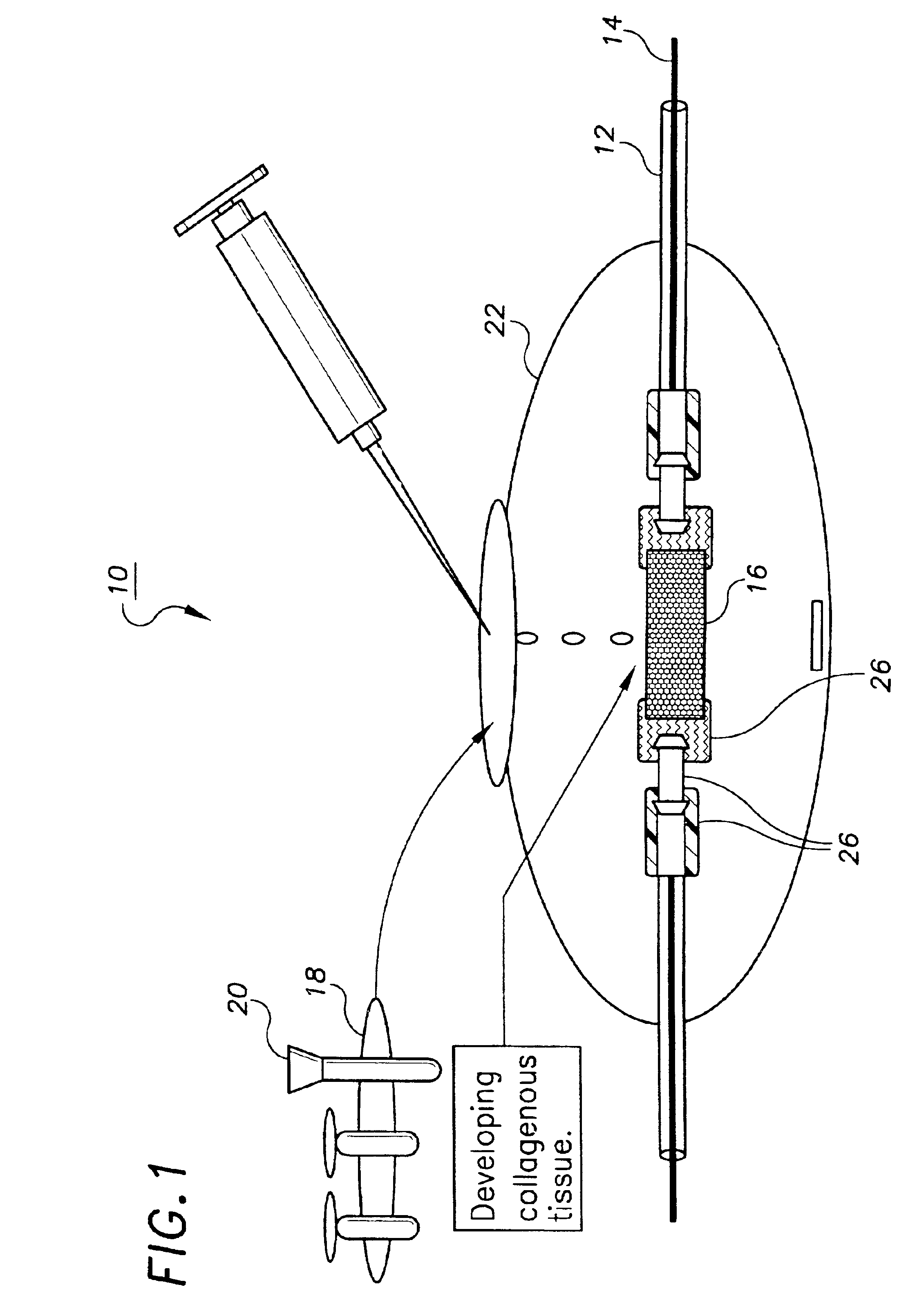
[0104]This example describes the preparation of a tissue engineered construct suitable for decellularization, in this case a small caliber artery, using a bioreactor system. A more detailed description of many aspects of this process is found in the pending application referenced above. As an initial step, a non-woven mesh made of fine polyglycolic acid (PGA) fibers (Albany International Research Co., Mansfield, Mass.) was produced and further processed to yield a porous substrate with a hydrophilic surface. The processing enhances wettability and increases the number of cells which are deposited on the surface during seeding. Briefly, the treatment began with three successive 30 minute washes in hexane, dichloromethane, and diethyl ether followed by lyophilization overnight. The PGA mesh was then placed briefly in ethanol, removed to distilled water, and placed in a 1.0 normal solution of NaOH for 1 minute, during which the solution was...

example 2

Decellularization of a Tissue Engineered Bovine Artery Construct Using Ionic Detergent Solutions

Materials and Methods

[0109]Small caliber arteries were engineered using bovine aortic smooth muscle cells as described in Example 1. After an 8 week culture period a vessel was removed from the bioreactor, washed with PBS, and sliced into segments 2 mm thick. The slices were immersed in 50 ml of a decellularization solution containing 1 M NaCl, 25 mM EDTA, 8 mM CHAPS in sterile PBS at pH 7.2. The samples were incubated with continuous stirring for 1 hour at room temperature and were then washed three times in PBS. The samples were then placed in 50 ml of a second decellularization solution containing 1 M NaCl, 25 mM EDTA, 1.8 mM SDS in sterile PBS at pH 7.2 and incubated for 1 hour at room temperature with continuous stirring. After removal from the decellularization solution the segments were washed twice with PBS for 5 minutes to remove residual solution.

[0110]The decellularized artery ...

example 3

Decellularization of a Tissue Engineered Bovine Artery Construct Using a Nonionic Detergent Solution

Materials and Methods

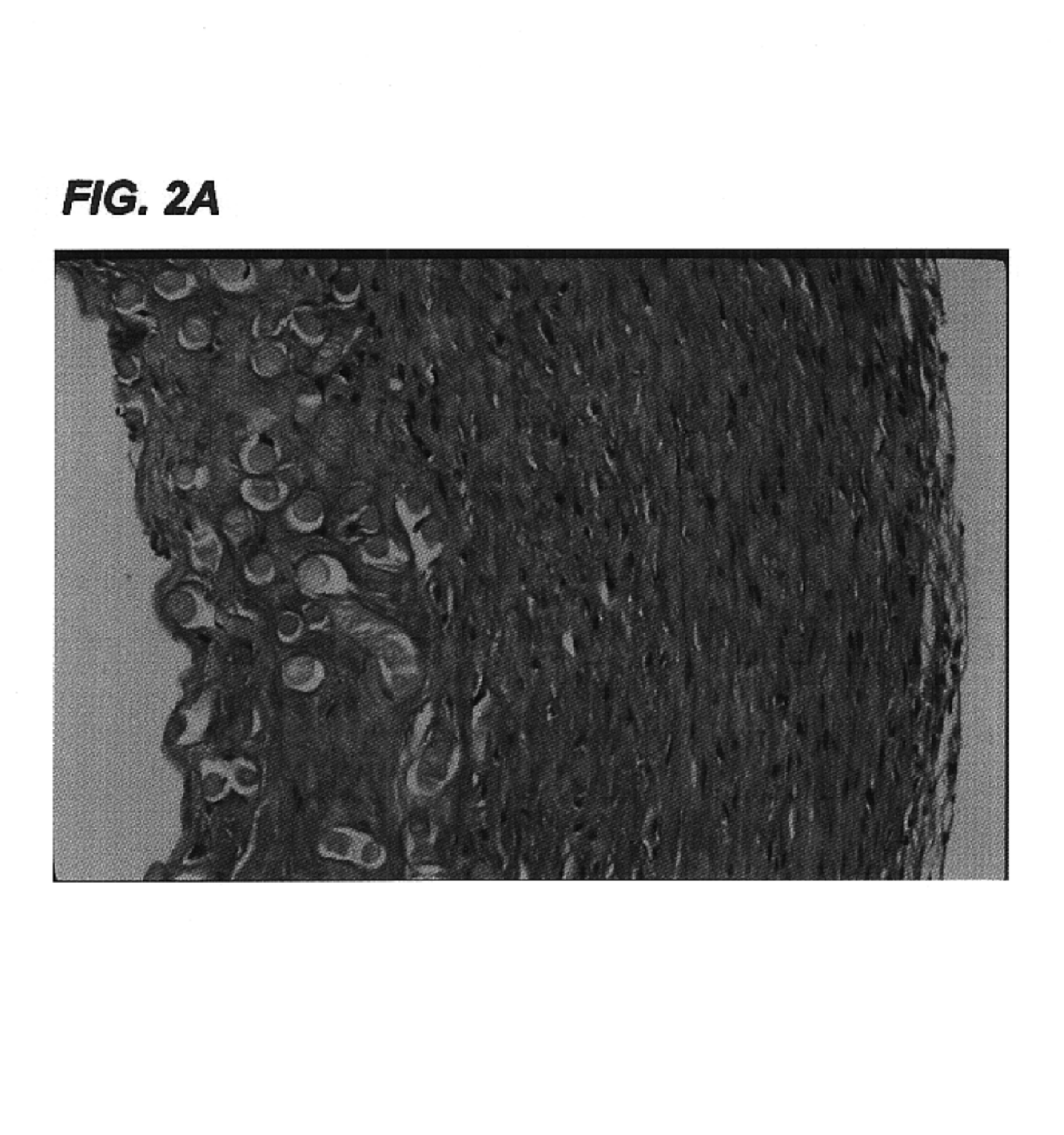
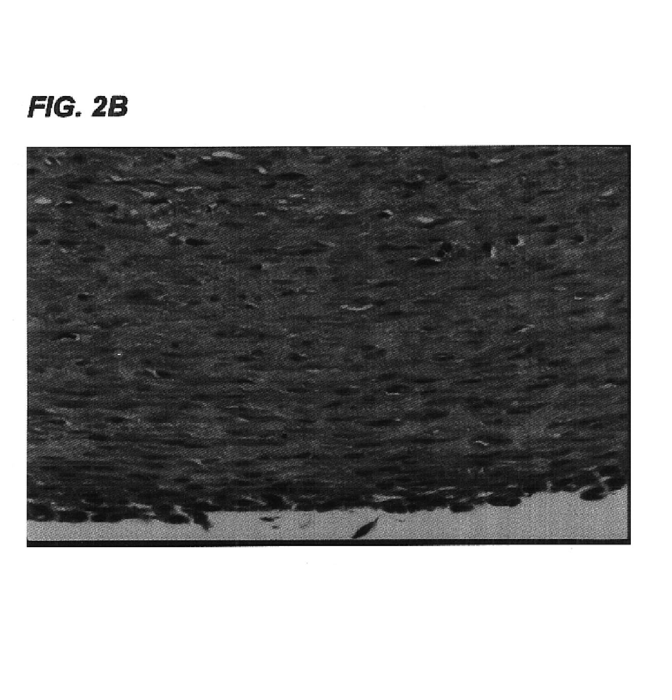
[0113]Small caliber arteries were engineered using bovine aortic smooth muscle cells as described in Example 1. After an 8 week culture period a vessel was removed from the bioreactor, rinsed with PBS, and sliced into segments 2 mm thick. The slices were immersed in 50 ml of a decellularization solution containing 1% Triton X-100® (Sigma), 0.02% EDTA (Sigma), 20 μg / ml RNAse A (Sigma), and 0.2 mg / ml DNAse (Sigma) in sterile PBS without Ca2+ or Mg2+ and incubated for 24 hours in a 10% CO2 atmosphere at 37° C. with continuous stirring. After removal from the decellularization solution, the segments were washed several times with PBS to remove residual solution.

[0114]The decellularized artery and a control artery that had been produced under identical conditions but not subjected to decellularization were fixed in formalin, embedded in paraffin, sectioned, and stained...


PUM


Abstract

New methods for producing tissue engineered constructs and engineered native tissues are disclosed. The methods include producing a tissue engineered construct by growing cells in vitro on a substrate and then decellularizing the construct to produce a decellularized construct consisting largely of extracellular matrix components. The construct can be used immediately or stored until needed. The decellularized construct can be used for further tissue engineering, which may include seeding the construct with cells obtained from the intended recipient of the construct. During any of the growth phases required for production of the construct, the developing construct may be subjected to various tissue engineering steps such as application of mechanical stimuli including pulsatile forces. The methods also include producing an engineered native tissue by harvesting tissue from an animal or human, performing one or more tissue engineering steps on the tissue, and subjecting the tissue to decellularization. The decellularized, engineered native tissue may then be subjected to further tissue engineering steps.

Description

CROSS-REFERENCE TO RELATED APPLICATION[0001]This application claims priority to U.S. Provisional Patent Application 60 / 225,698, filed Aug. 16, 2000, the contents of which are hereby incorporated by reference in their entirety.GOVERNMENT SUPPORT[0002]The U.S. government has a paid-up license in this invention and the right under certain circumstances to require the patent owner to license others on reasonable terms as provided for by the terms of grant number HL-03492 awarded by the NHLBI.BACKGROUND OF THE INVENTION[0003]Tissue damage, dysfunction, or loss is a feature of a wide variety of medical conditions. Atherosclerosis, in which formation of fatty plaques in blood vessel walls leads to narrowing of the vessels, is one well-known example. Accidents frequently result in damage to tendons, ligaments, and joints. Degenerative diseases such as arthritis represent another source of injury to such tissues. Systemic diseases such as diabetes, cancer, and cirrhosis are yet another cause...

Claims


Application Information

Patent Timeline
IPC IPC(8): A61L27/00A61L27/38C12N5/00
CPCA61L27/3691A61L27/3839C12N5/0068C12N2533/90C12N2533/30C12N2533/40A61P19/00
Inventor MITCHELL, SHANNONKOH, JENNIFERPRABHAKAR, VIKASNIKLASON, LAURA
Owner DUKE UNIV
Why Eureka
Try Eureka