Mass spectrometric immunoassay

a mass spectrometry and immunoassay technology, applied in the field of mass spectrometry immunoassays, can solve the problems of limited immunoassay screening possibility, restricted detection limit of technique, and traditional immunoassays that do not operate at that level of detection, and achieve rapid ease and equal reliability.

Inactive Publication Date: 2005-12-13
INTRINSIC BIOPROBES
View PDF7 Cites 64 Cited by
  • Summary
  • Abstract
  • Description
  • Claims
  • Application Information

AI Technical Summary

Benefits of technology

[0015]Because detection using mass spectrometry lends an added dimension of unequivocal specificity, the mass spectrometric immunoassay under the present invention is an improvement over existing immunoassays in several ways. First, the present invention does not require monoclonal antibodies as a reagent, polyclonal antibodies produce equally reliable results. Second, radioactive materials are not required at all. Third, the presence of other substances not the subject of the immunoassay do not interfere with either detecting or quantifying the targeted antigen or antibody. Fourth, an accurate multiplex immunoassay clearly detecting and / or quantifying virtually any combination of different antigens or antibodies is possible.
[0016]In addition, the rapid ease by which the mass spectrometer can detect an antigen or antibody regardless of whether the antigen-antibody binding has reached equilibrium grants to the present invention an immunoassay that produces results in a short period of time, minutes rather than hours.

Problems solved by technology
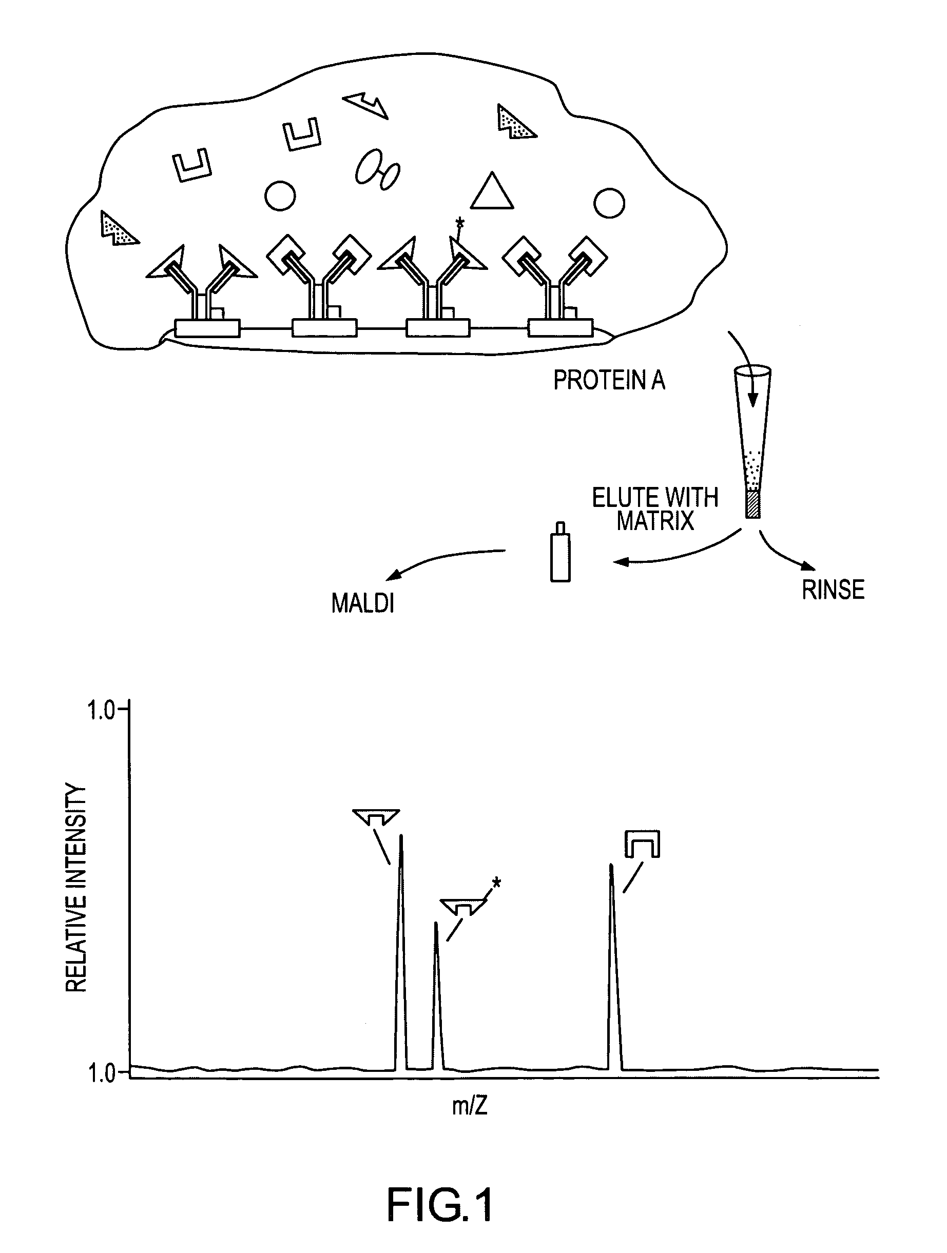
Although detection of attomole levels of an enzyme has been demonstrated, so that it was, in principle, possible to detect attomole levels of an antigen, traditional immunoassays did not operate at that level of detection because all were limited by non-specific binding of the enzyme-linked antibody to surfaces in the reaction well or vial.
This produced a background response which restricted the detection limit of the technique which could not be discriminated against because detection was indirect.
A further limitation of the traditional immunoassays employing optical detection was caused by the limited number of clearly resolvable colored enzyme products, at most two or three, which limited the possibility for an immunoassay to screen for multiple antigens in a single sample.
Multiple antigen immunoassays usually focused on a number of separate immunoassays in an array of well plates each requiring its own sample which clearly reduced the utility of this approach.
Polyclonal antibodies whose specificity is low could be used in theory but were not a practical choice for a reagent because polyclonal antibodies bind with several species of antigens making the detection of the resulting antibody-antigen complex less specific for a single given antigen species.
Yet another restriction to EIA and ELISA is that they required the antigen-antibody binding to reach an equilibrium for quantification, making the immunoassay take several hours to perform.
Until about 1988, mass spectrometry of proteins and peptides was thought difficult or impossible.
While the new MALDI techniques opened the field of biomolecular mass spectrometry, the mass spectrometric analysis of complex biological materials was not possible because of matrix overloading.
Presently, there is no mass spectrometric immunoassay which is capable of qualitatively and quantitatively determining the presence of single or multiple antigen or antibody species in a specimen.

Method used


Image

  • Mass spectrometric immunoassay
  • Mass spectrometric immunoassay
  • Mass spectrometric immunoassay

Examples


example 1

[0121]In one practice of the present invention, a single analyte, myotoxin a, was detected in human whole blood using the affinant, anti-myotoxin a, as a constituent of the affinity reagent, and then quantified using a working curve strategy. Myotoxin a is one toxin found in the venom of the prairie rattlesnake, Crotalus viridis viridis (C. v. viridis).

[0122]The antibody, anti-myotoxin a immunoglobulin IgG affinity purified from rabbit antiserum, was used to prepare the affinity reagent as follows. One milliliter of antibody solution (the solution contained the antibody at 5 mg / mL in 0.01 M tris[Hydroxymethyl] aminomethane, pH 8.2 (tris)) was incubated for two hours (gentle agitation at room temperature) with 1 mL of slurried 6% agarose beads on which protein A was supported. Following incubation, the beads were washed (3×1 mL tris) and allowed to react at room temperature with 500 μL of 0.02 M dimethylpimelimidate dihydrochloride prepared in 0.2 M triethanolamine (pH 8.2). The reac...

example 2

[0133]Myotoxin a present in a blood sample containing 0.002 mg / mL of C.v. viridis venom was quantified employing the bargraph quantification strategy. Introduced into the sample were multiple internal reference species of myotoxin a which had been iodinated (at the tyrosine residues) using the following procedure: Iodobeads (Pierce) were washed two times with 0.1 M sodium phosphate at pH 7.0, then incubated for five minutes with 0.4 M sodium iodide in a 0.1 M sodium phosphate solution at pH 7.0. An equal volume of myotoxin a, 2 mg / mL in buffer, was added to the sodium iodide / Iodobead solution. Two Iodobeads were used for each milligram of myotoxin a. The mixture was incubated overnight (˜10 hours) at room temperature, after which the reaction was stopped by removal of the Iodobeads. The modified protein was desalted and stored as described above. The reaction resulted in the production of four distinct iodinated species, with each addition incrementing the molecular weight of myotox...

example 3

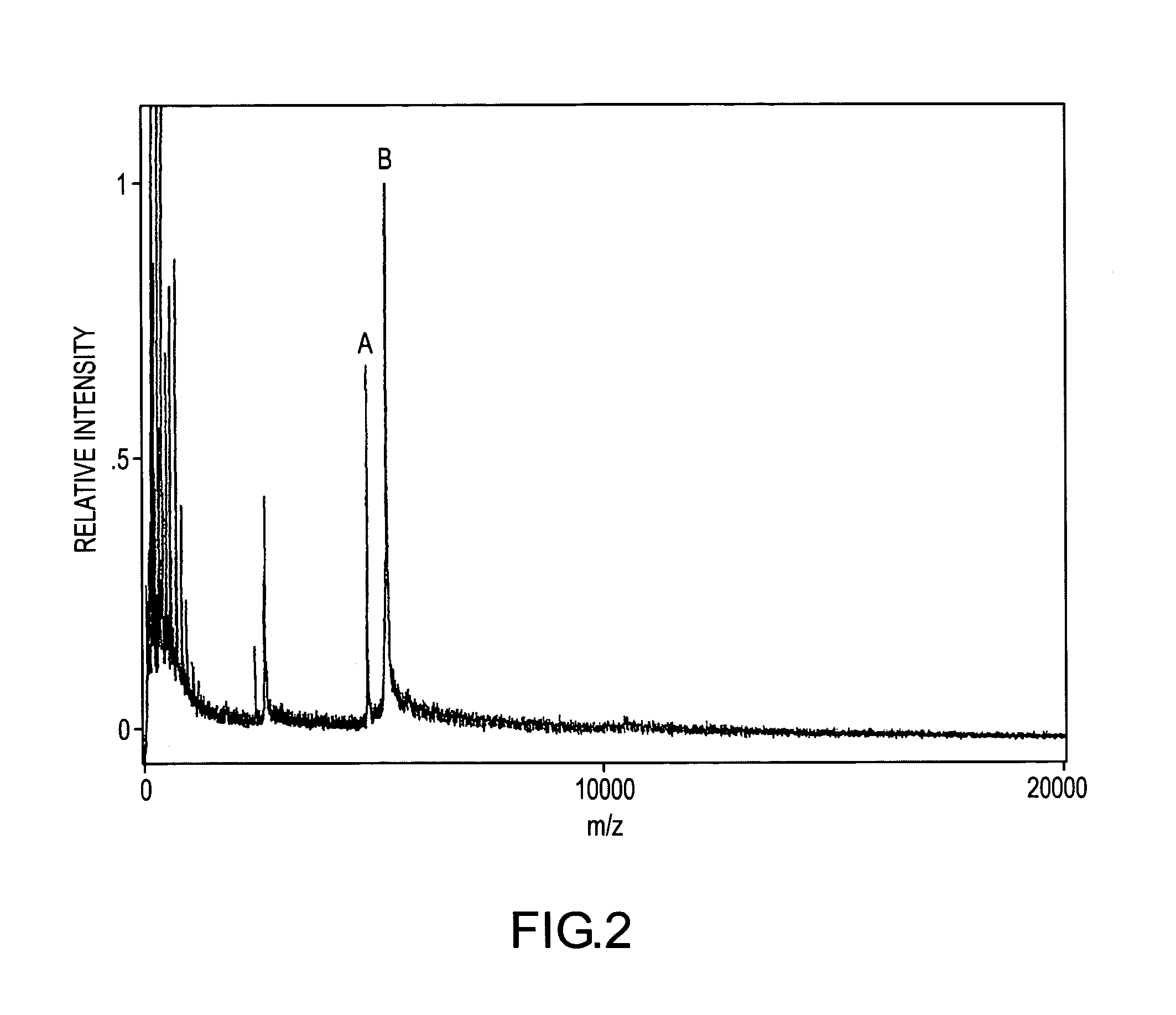
[0135]FIG. 6 demonstrates a single-point relative signal strategy for quantifying and detecting myotoxin a in human blood. A human blood sample containing an unknown concentration of myotoxin a and a 30 nM concentration of H-myotoxin a was prepared then mass spectrometrically immunoassayed according to the protocols of Example 1 above. The resulting mass spectrum shows mass spectral signals for myotoxin a, -I-, and H-myotoxin a, -J-, at their respective molecular weights. The relative mass spectrometric responses of myotoxin a and H-myotoxin a may be determined from the mass spectrum of FIG. 2 in which the concentrations of both species are known (25 nM myotoxin a, from the working curve calibration, and 40 nM H-myotoxin a). From this calibration it may be determined that equal concentrations of the two analytes give a 10% lower signal for H-myotoxin a. The myotoxin a signal is 3.5 times the H-myotoxin a signal for a known 30 nM concentration of H-myotoxin a. Therefore the myotoxin ...


PUM

PropertyMeasurementUnit
molecular weightaaaaaaaaaa
molecular weightsaaaaaaaaaa

Abstract

Rapid mass spectrometric immunoassay methods for detecting and / or quantifying antibody and antigen analytes utilizing affinity capture to isolate the analytes and internal reference species (for quantification) followed by mass spectrometric analysis of the isolated analyte / internal reference species. Quantification is obtained by normalizing and calibrating obtained mass spectrum against the mass spectrum obtained for an antibody / antigen of known concentration.

Description

[0001]This application is a continuation of application Ser. No. 09 / 024,988 filed on Feb. 17, 1998, abandoned which was a continuation of original application Ser. No. 08 / 449,903, which was filed on May 23, 1995, now abandoned.[0002]Financial Assistance for some of the work reported herein was provided by the United States Department of Energy under Grant number DEFG02-91ER61127. The United States government may have certain rights to this invention.INTRODUCTION[0003]The present invention relates to a new and useful immunoassay and more specifically to new and improved mass spectrometric immunoassay processes for the detection and / or quantification of one or more antigens or antibodies in a single determining immunoassay. Hereinafter, singular terms are intended to include plural.BACKGROUND OF THE INVENTION[0004]Immunoassay techniques first came into wide usage with the development of radioimmunoassay (RIA) in which the specificity of antigen-antibody binding was coupled with the hi...

Claims


Application Information

Patent Timeline
Patent Type & Authority Patents(United States)
IPC IPC(8): C12Q1/68G01N33/53G01N33/543
CPCC12Q1/6816C12Q1/6872G01N33/5302G01N33/6851G01N24/00G01N33/54373Y10T436/24
Inventor NELSON, RANDALL W.WILLIAMS, PETERKRONE, JENNIFER REEVE
Owner INTRINSIC BIOPROBES
Why Eureka
Try Eureka