
DNA sequences comprising dystrophin minigenes and methods of use thereof

a dystrophin and minigene technology, applied in the field of dna sequences comprising dystrophin minigenes, can solve the problems of loss of dap complex, degenerative pathology, chronic muscle damage, etc., and achieve the effects of reducing size, efficient and stable correction of both biochemical and physiological defects, and convenient and less time-consuming

Inactive Publication Date: 2006-02-21
ASKLEPIOS BIOPHARMACEUTICAL INC
View PDF11 Cites 56 Cited by
  • Summary
  • Abstract
  • Description
  • Claims
  • Application Information

AI Technical Summary

Benefits of technology

[0023]The present invention provides dystrophin minigenes that are significantly reduced in size without compromising essential functions in protecting muscles from dystrophic phenotypes. The present invention also provides viral vectors carrying the dystrophin minigenes that are capable of mediating efficient and stable correction of both biochemical and physiological defects in a mammalian subject. Furthermore, the present invention provides a method that is more convenient and less time-consuming to discern the dystrophin functional domains in vivo and to optimize the minigenes for DMD gene therapy. Finally, the present invention provides a method for treatment of muscular dystrophy.

Problems solved by technology

The progressive muscle degeneration and weakness usually confine the patients to wheelchairs by their early teens, and lead to death by their early twenties.
The absence of a functional dystrophin results in the loss of DAP complexes and causes instability of myofiber plasma membrane.
These deficiencies in turn lead to chronic muscle damage and degenerative pathology.
However, clinical trials of myoblast transplantation have met with little success owing to the poor survival of the transplanted cells (Gussoni et al., Nature Med 3, 970-977 ).
Nonetheless, the immunogenicity and inefficiency of infecting mature muscle cells remain major hurdles to overcome before the adenovirus vectors can be safely used in humans.
2. Herpes simplex virus (HSV) vectors: The main feature of an HSV vector is that it has very large packaging capacity, is usually replication defective and does not integrate into the host genome.
However, these modifications also result in a lower virus yield.
However, Recombinant retroviral vectors can only accommodate about 8 kb to 10 kb of foreign DNA.
This packaging capacity also limits its use in the genetic treatment of DMD.
Furthermore, there seems to be no potent antibody response.
However, a major obstacle for AAV vectors is the limited packaging size that only allows for genes smaller than 4.7 kb (Song et al.
Transgene expression was achieved with only very limited efficiency.
Previous attempts to generate dystrophin minigenes that were shorter than ½ of the full-length dystrophin failed to preserve the essential protective functions.
However, expression of Dp71 failed to alleviate symptoms of muscle degeneration [Cox et al.
Hum Gene Ther 11, 669-80 ) demonstrated that expression of dystrophin minigenes with both intact N- and C-terminal domains and 1 to 3 central rod repeats in mouse skeletal muscle was sufficient to restore DAP complexes but insufficient to restore myofiber morphology and to prevent dystrophic pathology.

Method used


Image

  • DNA sequences comprising dystrophin minigenes and methods of use thereof
  • DNA sequences comprising dystrophin minigenes and methods of use thereof
  • DNA sequences comprising dystrophin minigenes and methods of use thereof

Examples


example 1

Dystrophin Minigenes and AAV Vectors Carrying the Minigenes

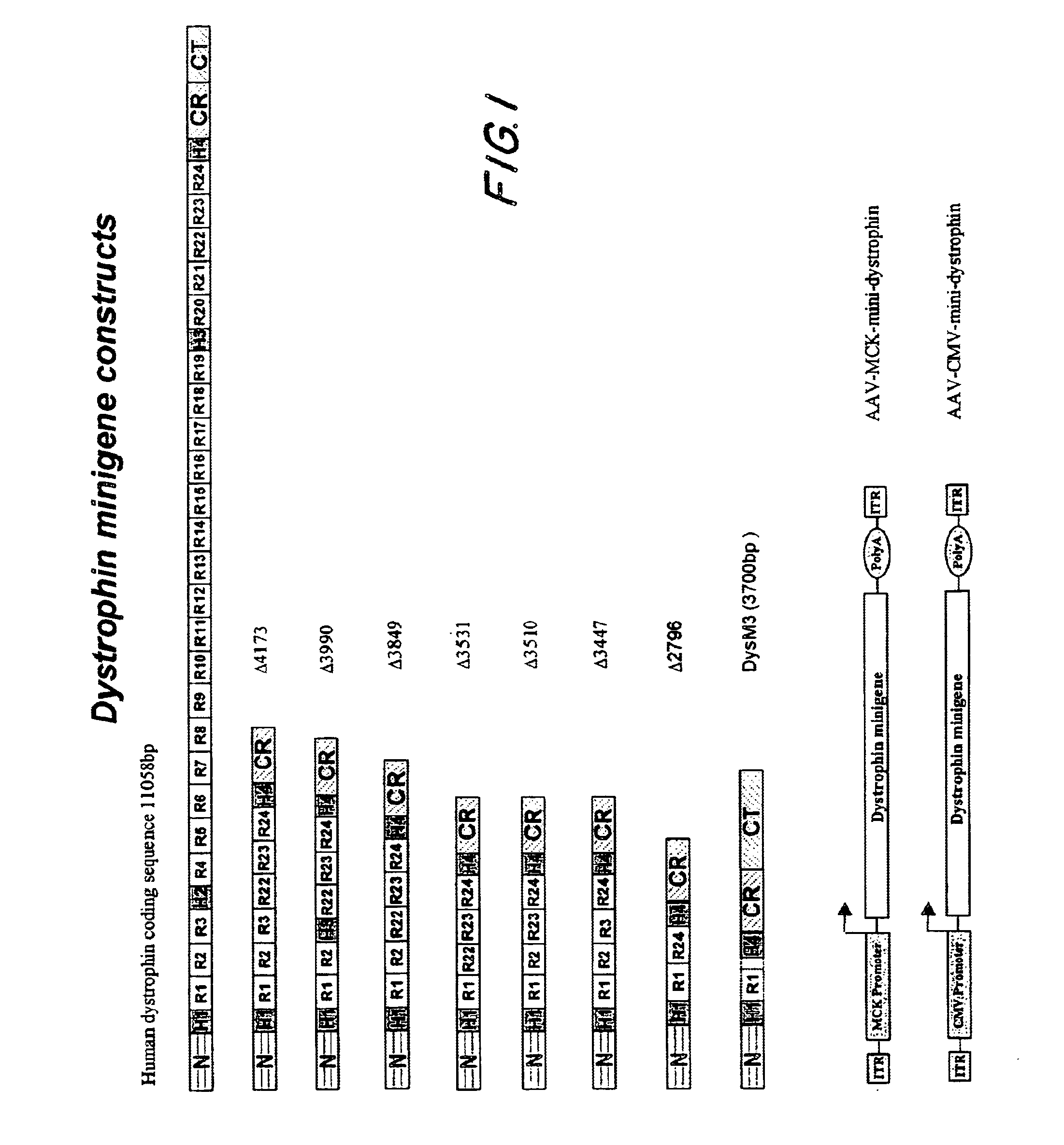
[0068]This example describes the construction of highly truncated dystrophin minigenes and AAV vectors carrying the minigenes. The dystrophin minigene constructs were made mainly by PCR cloning method using Pfu polymerase (Stratagene, CA) and human dystrophin cDNA (GenBank # NM 004006) as the template. For consistency, the numbering of the nucleotide only includes the 11,058 bp dystrophin protein coding sequence (SEQ ID NO:1).

[0069]As depicted in FIG. 1, dystrophin minigene Δ4173 (SEQ ID NO:2) contains nucleotides 1-1992 (N-terminus, hinge H1 and rods R1, R2 & R3, SEQ ID NO:3) and 8059-10227 (rods R22, R23 & R24, hinge H4 and CR domain, SEQ ID NO:4) and 11047-11058 (the last 3 amino acids of dystrophin, SEQ ID NO:5).

[0070]Dystrophin minigene Δ3990 (SEQ ID NO:6) contains nucleotides 1-1668 (N-terminus, hinge H1 and rods R1 & R2, SEQ ID NO:7), 7270-7410 (hinge H3, SEQ ID NO:8), 8059-10227 (rods R22, R23 & R24, hinge H4 and CR ...

example 2

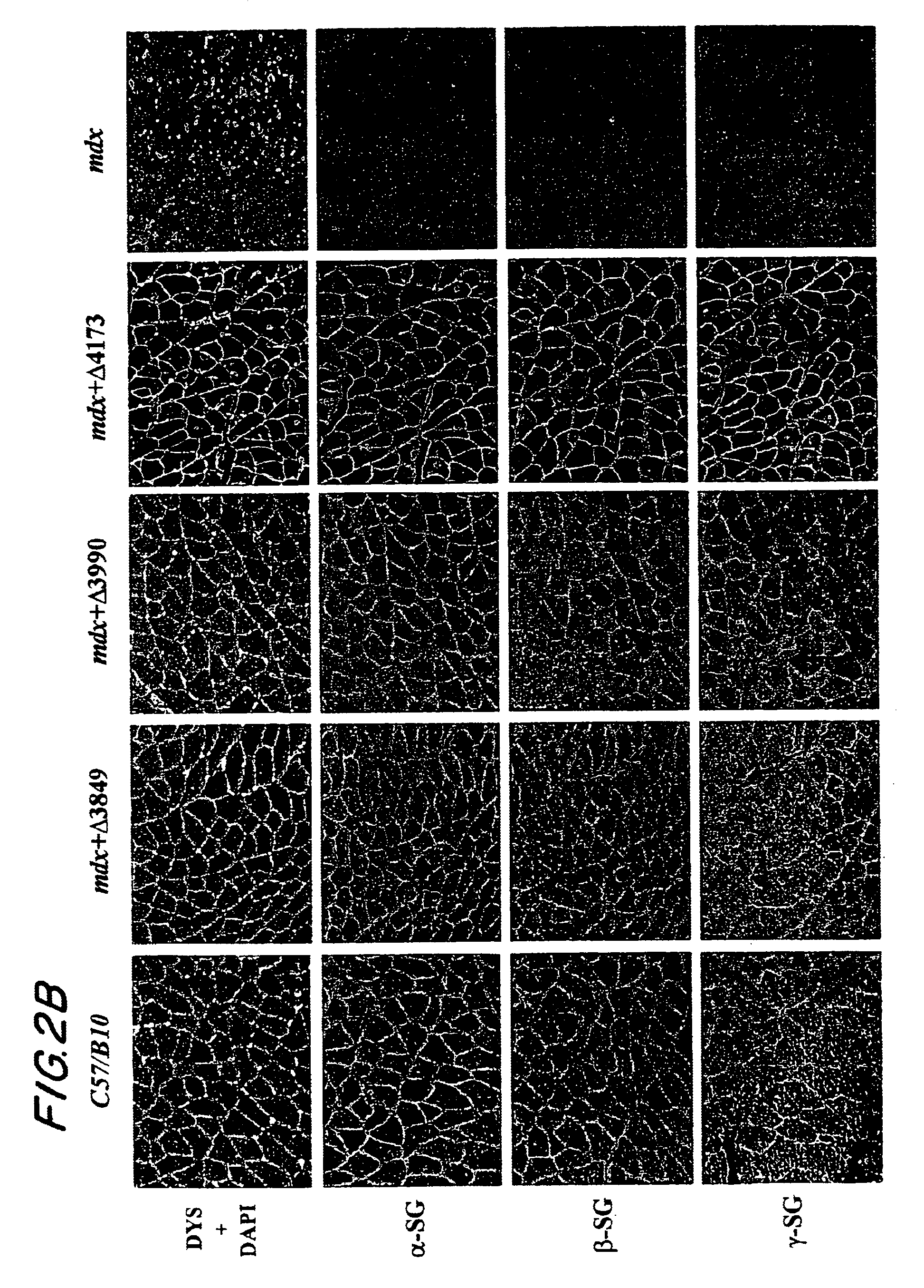
Restoration of DAP Complexes

[0080]This example describes whether dystrophin minigene products still retain the major biochemical functionality including submembrane localization and interaction with dystrophin associated protein (DAP) complexes. Healthy C57 / B10 mice and dystrophic mdx mice were purchased from The Jackson Laboratory (Bar Harbor, Me.). Ten-day old mdx pups or 50-day old mdx adult mice were injected into the hindleg gastrocnemius muscle with 50 μl (5×10¹⁰ GC) of different AAV mini-dystrophin vectors.

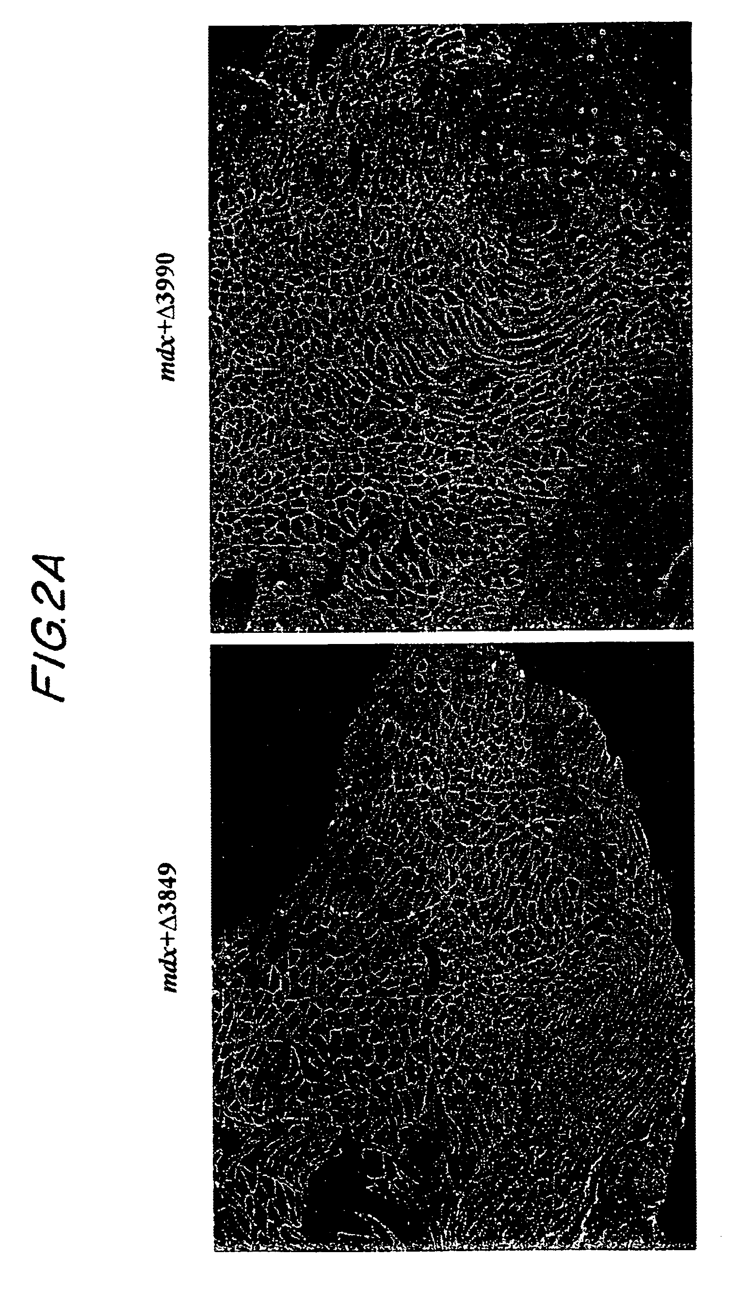
[0081]At three months and six months after vector injection, the muscles were collected for evaluation of mini-dystrophin expression and biochemical restoration of the DAP complexes, which were absent due to the primary deficiency of dystrophin. IF staining on thin sections of AAV treated muscles, using an antibody (Dys3) specific to human dystrophin, revealed widespread vector transduction and correct submembrane location of the mini-dystrophins in a majority of the myofib...

example 3

Amelioration of Dystrophic Pathology

[0083]This example demonstrates that dystrophin minigene products can protect muscle from the pathological phenotypes. The onset of the pathology in mdx mice starts at around three weeks of age with massive waves of myofiber degeneration / regeneration. This process is characterized by the presence of central nuclei in myofibers, a primary pathological sign of muscular dystrophies. The absence or reduction of central nucleation after gene therapy would suggest that the therapy is successful. Therefore, we initially chose to test the AAV mini-dystrophin constructs in young mdx mice (10-day old) before the onset of central nucleation, to see whether muscle degeneration / regeneration can be prevented.

[0084]Histological examination of the mdx muscles at 3 and 6 months after AAV mini-dystrophin (containing more than 2 rod domains) treatment, which was prior to the onset of central nucleation, showed nearly exclusive (˜98%) peripheral nucleation in the min...


PUM

PropertyMeasurementUnit
thicknessaaaaaaaaaa
frequencyaaaaaaaaaa
particle sizeaaaaaaaaaa

Abstract

The present invention provides a series of novel dystrophin minigenes that retain the essential biological functions. The expression of the dystrophin minigenes may be controlled by a regulatory element along with a small polyadenylation signal. The entire gene expression cassettes may be readily packaged into a viral vector, preferably an AAV vector. The present invention further defines the minimal functional domains of dystrophin and provides ways to optimize and create new versions of dystrophin minigenes. Finally, the present invention provides a method of treatment for Duchenne muscular dystrophy (DMD) and Becker muscular dystrophy (BMD).

Description

[0001]This application claims the priority of U.S. Provisional Patent application No. 60 / 200,777, filed Apr. 28, 2000.TECHNICAL FIELD[0002]The present invention relates to novel dystrophin minigenes that retain the essential biological functions of a full length dystrophin gene, and methods of treatment for Duchenne muscular dystrophy (DMD) and Becker muscular dystrophy (BMD) in a mammalian subject using the dystrophin minigenes.BACKGROUND OF INVENTION[0003]Duchenne muscular dystrophy (DMD) is an X-linked genetic muscle disease affecting 1 of every 3,500 newborn males (Kunkel et al. Nature (London) 322, 73-77 [1986]). The progressive muscle degeneration and weakness usually confine the patients to wheelchairs by their early teens, and lead to death by their early twenties. DMD is caused by recessive mutations in the dystrophin gene, the largest gene known to date, which spans nearly 3 million base-pairs on the X-chromosome with 79 exons, a coding sequence of about 11 kb, and a high ...

Claims


Application Information

Patent Timeline
Patent Type & Authority Patents(United States)
IPC IPC(8): C12N15/00C12N15/09C12N15/63C12N15/70C12N15/74A61K38/00C07K14/47
CPCC07K14/4708A61K38/00C12N2799/025
Inventor XIAO, XIAO
Owner ASKLEPIOS BIOPHARMACEUTICAL INC
Learn More