Method for transforming monocotyledons

Inactive Publication Date: 2006-06-13
METABOLIX
24 Cites 195 Cited by

AI-Extracted Technical Summary

Problems solved by technology

Although methods for transforming monocotyledons using Agrobacteri...

Method used

[0043]Since the Agrobacterium prepared as mentioned above has a highly virulent DNA originated from pTOK162, transformation of monocots can be achieved with a high efficiency.
[0122]Thus, the embryo tissues cultured in a dedifferentiation-inducing medium f...

Benefits of technology

[0019]By the present invention, introduction of foreign genes into monocots such as plants belonging to the family Gramineae, including rice, maize, wheat, barley and the like, with good reproducibility was first achieved. Although methods for transforming monocotyledons using Agrobacterium are known, they are not established methods as mentioned above. In contrast, according to the present invention, by inoculating Agrobacterium to cultured tissues which have not been employed in the conventional methods, genes can be very easily introduced. In the present invention, since a cultured tissue such as a callus which can be easily prepared is used, the sample materials c...

Abstract

The invention relates to a method for transforming a monocotyledonous plant. The time required from transformation to regeneration of a plant is shorter using the inventive method so that the frequency of emergence of mutants is smaller than the conventional methods. The inventive method may be generally applied even to the plants for which a regeneration method from a protoplast to a plant has not been established, and with which the preparation of the material to be subjected to the method is easy. That is, the present invention provides a method for transforming a monocotyledonous plant, comprising contacting a cultured tissue of said monocotyledonous plant during dedifferentiation thereof obtained by culturing an explant on a dedifferentiation-inducing medium for less than 7 days with a bacterium belonging to the genus Agrobacterium containing a super binary vector having the virulence region of a Ti plasmid, left and right border sequences of T-DNA of a Ti plasmid or an Ri plasmid of a bacterium belonging to the genus Agrobacterium, and a desired gene located between said left and right border sequences.

Application Domain

Technology Topic

Image

  • Method for transforming monocotyledons
  • Method for transforming monocotyledons

Examples

  • Experimental program(3)

Example

EXAMPLE 1
(1) Preparation of Sample Cultured Tissues
(i) Variety of Rice
[0047]Varieties Asanohikari, Tsukinohikari and Koshihikari, which are varieties of japonica rice were selected as samples.
(ii) Scutellum and Scutellum Callus
[0048]Mature seeds of rice were sterilized by being immersed in 70% ethanol for 1 minute and then in 1% sodium hypochlorite solution for 30 minutes. The seeds were then placed on 2N6 solid medium (inorganic salts and vitamins of N6 (Chu C. C., 1978; Proc. Symp. Plant Tissue Culture, Science Press Peking, pp. 43–50), 1 g/l of casamino acid, 2 mg/l of 2,4-D, 30 g/l of sucrose, 2 g/l of Gelrite). Scutella were removed from the seeds on Day 4 from the beginning of the culture on 2N6 solid medium and used as “scutellum” samples. On the other hand, after culturing the mature seeds for about 3, weeks, the formed calli originated from scutella were transferred to 2N6 medium and cultured therein for 4–7 days. The resulting calli were used as “scutellum callus” samples.
(ii) Shoot Apex Tissue
[0049]Mature seeds of rice were sterilized by the above-described method and were placed on ½ N6 solid medium (half strength of major inorganic salts and minor salts of N6, vitamins of N6, 1 g/l of casamino acid, 20 g/l of sucrose and 2 g/l of Gelrite). From seedlings on Day 3 after germination, tissues of 2–3 mm length containing apex dividing tissues were cut out and used as samples.
(iv) Root Segment and Root Callus
[0050]From the seedlings obtained by the method described in (iii), tip portions of 5–10 mm length of the seed roots were cut out and used as “root segment” samples. On the other hand, these radicles were cultured on 2N6 solid medium for about 2 weeks to obtain calli, and these calli were used as “root callus” samples.
(v) Suspended Cultured Cells
[0051]The calli originated from scutella were transferred to AA liquid medium (major inorganic salts of AA, amino acids of AA and vitamins of AA (Toriyama and Hinata 1985; Plant Science 41:179–183), MS minor salts (Murashige and Skoog 1962; Physiol. Plant. 15:473–497), 0.5 g/l of casamino acid, 1 mg/l of 2,4-D, 0.2 mg/l of kinetin, 0.1 mg/l of gibberellin and 20 g/l of sucrose) and the cells were cultured therein at 25° C. in the dark with shaking at 120 rpm to obtain suspended cultured cells. The medium was replaced with fresh medium every week. (2) Ti Plasmid (Binary Vector)
[0052]A hygromycin resistance gene (HPT) and β-D-glucuronidase (GUS) gene were inserted into the T-DNA region of a Ti plasmid to obtain the following plasmids:
(i) pIG121 Hm:
[0053]A plasmid in which the GUS gene containing the first intron of the catalase gene of castor bean (“intron GUS” gene) and a hygromycin-resistance gene were ligated (Nakamura et al., 1991; Plant Biotechnology II (Extra Issue of GENDAI KAGAKU, pp. 123–132); the construct was a gift of Dr. Nakamura of Nagoya University).
(ii) pTOK232
1. Insertion of Intron GUS and Hygromycin-Resistance Genes into Intermediate Vector pTOK229
[0054]The Cla I fragment (2.5 kb) of the spectinomycin-resistance gene originated from Tn7 were treated with Klenow fragment to blunt the ends. The resulting fragment was inserted into the Sma I site of pUC19 to obtain a plasmid pTOK107 (5.2 kb) having ampicillin-registance and spectinomycin-resistance genes. The obtained pTOK107 was treated with Eco RI and Hind III and the obtained 2.5 kb fragment containing the spectinomycin-resistance gene was ligated to a Eco RI-Hind III fragment (2.7 kb) of pGA482 to obtain pTOK170 (5.2 kb) containing the spectinomycin-resistance gene and has a Hind III site and a Hpa I site.
[0055]A vector pIG221 in which the first intron of catalase of castor bean and a GUS gene (“intron GUS gene”) are ligated to a 35S promoter (Ohta S. et al., 1990; Plant Cell Physiol. 31: 805–813, presented by Dr. Nakamura of Nagoya University), was digested with Eco RI and the resultant was treated with Klenow fragment to blunt the ends. A Hind III linker (pCAAGCTTG, code 4660P commercially available from TAKARA SHUZO) was ligated to the resultant. A fragment containing the 35S promoter and intron GUS gene was cut out by digesting the resulting vector with Hind III, and the fragment was inserted into the Hind III site of a plasmid pGL2 (J. Paszkowski, obtained from Friedrich Mieocher Institute) containing a hygromycin-resistance gene ligated to a 35S promoter, to obtain pGL2-IG (7.6 kb). The above-mentioned plasmid pGL2 was obtained by inserting a hygromycin-resistance gene (Gritz L. and Davis J. 1983; Gene 25: 179–188) into pDH51 (Pietrazak et al., 1986; Nucleic Acids Research 14: 5857–5868). The fragment obtained by treating pTOK170 with Hpa I was ligated to a Pvu II fragment (5.2 kb) of pGL2-IG to obtain pTOK229 (10.1 kb).
2) Insertion into Super Binary Vector pTOK162
[0056]A super binary vector pTOK162 was obtained by inserting virB, virC and virG genes of strongly virulent Agrobacterium tumefaciens A281. Insertion of the desired genes (hygromycin-resistance gene and intron GUS gene) into the binary vector was carried out by homologus recombination. That is, since both vectors contain a region originated from an E. coli plasmid pBR322, in the bacterial cells selected by resistances to spectinomycin and kanamycin, only the plasmid generated by recombination of both plasmids is retained. The plasmid obtained by the fact that the hygromycin-resistan gene and the intron GUS gene were inserted into the super binary vector is called pTOK232 (see FIG. 1).
[0057]An E. coli strain MC1000 harboring the plasmid pTOK162 was deposited under the terms and conditions of the Budapest Treaty on Sep. 30, 2004, at the Incorporated Administrative Agency National Institute of Technology and Evaluation Patent Microorganisms Depositary (NPMD), located at 2-5-8 Kazusakamatari Kisarazu-shi, Chiba-ken 292-0818, Japan, under the accession number Nite BP-24.
(3) Host Agrobacterium tumefaciens
[0058]Strains LBA4404 and EHA101 in which T-DNA regions were deleted were used as the host bacteria. Strain LBA4404 has a helper plasmid PAL4404 (having a complete vir region), and is available from the American Type Culture Collection (ATCC 37349). Strain EHA101 has a helper plasmid having the vir region originated from a strongly virulent Agrobacterium tumefaciens A281, and is available from Hood E. E. et al., 1986.
[0059]The various binary vectors described in (2) were introduced into these two strains of Agrobacterium tumefaciens, and the following strains described were used for introducing the genes. The plasmids were introduced into the Agrobacterium strains by a triple cross (Ditta G. et al., 1980; Proc. Natl. Acad. Sci. USA 77: 7347–7351).
[0060]LBA4404(pTOK232)
[0061]LBA4404(pIG121Hm)
[0062]EHA101(pIG121Hm)
(4) Preparation of Suspension of Agrobacterium tumefaciens
[0063]Colonies obtained by culturing the Agrobacterium strains on AB medium (Drlica K. A. and Kado C. I. 1974; Proc. Natl. Acad. Sci. USA 71:3677–3681) containing hygromycin (50 μg/ml) and kanamycin (50 μg/ml) for 3–10 days were collected with a platinum loop and suspended in modified AA medium (same as the composition of the above-described AA medium except that concentrations of sucrose and glucose were changed to 0.2 M and 0.2 M, respectively, and that 100 μM of acetosyringone was added, pH 5.2). The cell population was adjusted to 3×10⁹–5×10⁹ cells/ml and the suspensions were used for inoculation.
(5) Inoculation Conditions
[0064]The sample tissues were washed with sterilized water and immersed in the above-described suspensions of Agrobacterium strains, respectively, for 3–10 minutes. Thereafter, the shoot apex samples were placed on N6S3 solid medium (½ N6 major inorganic salts, N6 minor salts, N6 vitamins, Chu C. C., 1978, AA amino acids (Toriyama and Hinata 1985), 1 g/l of casamino acid, 0.2 mg/l of NAA, 1.0 mg/l of kinetin and 3 g/l of Gelrite) containing 100 μM of acetosyringone, 10 g/l of glucose and 20 g/l of sucrose. The other tissue samples such as scutellum callus samples were cultured on 2N6 solid medium containing acetosyringone, glucose and sucrose in the same concentrations as mentioned above. The co-cultures were carried out at 25° C. in the dark for 2–5 days. The resulting inoculated tissues were then washed with sterilized water containing 250 mg/l of cefotaxime and then the culture was continued on the respective solid media containing the same concentration of cefotaxime as mentioned above.
(6) Method for Examining GUS Activity
[0065]Immediately after the above-mentioned culture with the Agrobacterium strains, the tissues were immersed in 0.1 M phosphate buffer (pH 6.8) containing 0.1% TRITON X-100 at 37° C. for 1 hour. After washing off the Agrobacterium strains with phosphate buffer, phosphate buffer containing 0.1 mM of 5-bromo-4-chloro-3-indolyl-β-D-glucuronic acid and 20% methanol was added to the tissues. After incubation at 37° C. for 24 hours, the number of blue-colored tissues was counted under a microscope and the percentages thereof based on the number of samples are described. For assay of the GUS activities of the plants assumed to be transformants after the selection treatment, leaves were collected from the plants and GUS staining was performed in the same manner. If the entire leaf or the cut face of the leaf is uniformly colored in blue, the plant was judged to be a positive plant, and if the leaf or the cut face of the leaf is chimerically stained, the plant was judged to be a chimera plant.
(7) Selection of Transformed Cells and Tissues (i) Shoot Apex
[0066]Shoot apices cultured with the Agrobacterium strains for 5 days were cultured on N6S3 medium containing 250 mg/l of cefotaxime for 2 weeks. The grown shoot apex tissues were transplanted to N6S3 medium containing 40 mg/l of hygromycin and selection of the transformants was carried out.
(ii) Scutellum
[0067]Scutella cultured with the Agrobacterium strains for 3 days were cultured on 2N6 medium containing 250 mg/l of cefotaxime for 1 week. Selection of transformed cells were then carried out on 2N6 medium containing 50 mg/l of hygromycin.
(iii) Cultured Tissue (Scutellum Callus)
[0068]Tissues cultured with the Agrobacterium strains for 3 days were cultured on 2N6 medium containing 250 mg/l of cefotaxime for 1 week. Hygromycin-resistant cultured tissues were selected by culturing the cultured tissues on 2N6 medium containing 50 mg/l of hygromycin for 3 weeks (primary selection). The obtained resistant tissues were further cultured on N6-12 medium (N6 inorganic salts, N6 vitamins, 2 g/l of casamino acid, 0.2 mg/l of 2,4-D, 0.5 mg/l of 6BA, 5 mg/l of ABA, 30 g/l of sorbitol, 20 g/l of sucrose and 2 g/l of Gelrite) containing 50 mg/l of hygromycin for 2–3 weeks (secondary selection), and the calli grown on this medium were transferred to a plant regeneration medium N6S3 containing 0, 20 or 50 mg/l of hygromycin. In all of the media used after the culture with Agrobacterium strains, cefotaxime was added to 250 mg/l.
(iv) Suspended Cultured Cells
[0069]The cells cultured with the Agrobacterium strains for 5 days were cultured in 2N6 medium containing 250 mg/l of cefotaxime for 1 week, and then the selection of the transformed cells was carried out on 2N6 medium containing 50 mg/l of hygromycin.
(8) Expression of Introduced Genes in the Progeny of Transformants
[0070]Seeds of the progeny of the transformants were sown in aqueous 400-fold Homai hydrate (Kumiai Kagaku Inc.) solution containing 70 mg/l of hygromycin and incubated therein at 25° C. for 10 days, thereby examining the resistance to hygromycin. Twenty seeds of each plant of the progeny of the transformants were sown and cultured for about 3 weeks. Leaves were collected from the obtained seedlings and examined for the expression of the GUS gene.
(9) Analysis of Introduced Genes by Southern Blotting Method
[0071]DNAs were extracted from the primary transformants of varieties Asanohikari and Tsukinohikari by the method of Komari et al. (Komari et al., 1989; Theoretical and Applied Genetics 77: 547–552), and the DNAs were treated with the restriction enzyme Hind III. The resulting fragments were subjected to detection of the introduced genes by Southern blotting analysis using the HPT gene as a probe. The length of the Hind III fragment containing the HPT gene as a probe is about 5.5 kb and the length of the DNA region from the Hind III site in the T-DNA in this region to the L border sequence is about 5.4 kb (FIG. 1). The Southern blotting analysis was carried out as describe in “Molecular Cloning: A Laboratory Manual” (Sambrook et al., 1989; Cold Spring Harbor Laboratory Press). Two GUS positive plants, two GUS negative plants and two hygromycin-resistant plants were picked up from each of the two lines of the progeny of transformants of Tsukinohikari and were subjected to the Southern blotting analysis in the same manner as mentioned above.
(10) Differences in Efficiencies of Introducing Genes Depending on Sample Tissues (Expression of GUS after Culturing with Agrobacterium Strains)
[0072]In order to confirm that Agrobacterium can introduce genes into cells of monocots, various tissues of the rice variety Tsukinohikari were treated with Agrobacterium tumefaciens EHA101 having a super-virulent vir region, into which the binary vector (supra) containing the hygromycin-resistance gene and the GUS gene were introduced, and then the GUS activities were examined. The sample tissues were shoot apices, radicles, scutella, radicle calli, scutellum calli and suspended: cultured cells. In cases where the tissues were not treated with the Agrobacterium strain, no tissues exhibited GUS activity as indicated by blue color. On the other hand, in cases where the tissues were treated with Agrobacterium tumefaciens EHA101 (pIG121Hm), expression of GUS was confirmed in all of the tissues except for radicles. The ratio of the number of the tissues showing blue color to the number of treated tissues was the highest in scutellum calli (Table 1). Further, the size of the tissues expressing GUS was also largest in scutellum calli. The tissues exhibiting the second highest rate of introduction next to the scutellum calli were shoot apices. Further, while the scutellum calli and the suspension cells which are dedifferentiated tissues of scutella exhibited a high introduction rate, the introduction rate in scutella was apparently lower. This suggests that genes are more easily introduced into tissues having high cell-dividing activities.
[0073] TABLE 1 Differences in Efficiency of Introduction of GUS Gene Depending on Sample Material (Variety: Tsukinohikari) Number of GUS+ Tissues/ Number of Sample Tissues (%) Size of GUS-stained Non-Treated Portion Based on Sample Tissue Group Treated Group Treated Tissue Shoot Apex 0/30(0) 109/157(69) +++ Root Segment 0/20(0) 0/30(0) Root Callus 0/30(0) 24/115(21) + Scutellum 0/50(0) 8/89(9) + Scutellum Callus 0/141(0) 312/395(79) +++ Suspension Cells 0/232(0) 61/247(25) ++ +: 1% or less, ++: 1~10%, +++: 10% or more
[0074]It has been confirmed that the binary vector pIG121Hm used in this experiment does not express GUS gene in Agrobacterium cells because the intron of castor bean plant is inserted in the promoter of the GUS gene (Nakamura et al., 1991). Thus, from the results of the experiments described above in which the expression of the GUS gene after the culturing with Agrobacterium is used as an index, it was confirmed that genes can be introduced into rice cells by Agrobacterium.
(11) Differences in Rate of Emergence of Transformed Tissues and Cells Depending on Sample Tissues
[0075]Selection of transformed tissues and transformed cells was carried out according to the resistance to hygromycin, using shoot apices, scutella, scutellum calli and suspension cells after the culture with the Agrobacterium strain. As a result, growth of transformants exhibiting resistance to hygromycin were observed in scutellum calli and the suspension cells (Table 2). Further, all of the selected cells expressed the GUS gene. Although the shoot apex tissues exhibited high rate of introduction of GUS gene after the culture with the Agrobacterium strain, after the selection by hygromycin, all tissues died and no tissues resistant to hygromycin were obtained. Although shoot apex is a tissue containing the apical meristem, in order that cells expressing a hygromycin-resistance continue to grow after the treatment for introducing the gene, it is necessary that the gene be introduced into the limited apical meristem. The reason why no resistant tissues were obtained in spite of the fact that a number of genes were introduced into the shoot apices after the culture with the Agrobacterium strain is thought to be that the probability that the gene is introduced in the vicinity of the apical meristem is low. Further, it is easily assumed that even if a gene is introduced in the vicinity of the apical meristem so that transformed cells are obtained, the possibility that the obtained plants exhibit chimeric properties is high. Therefore, it is thought that the transformation method utilizing shoot apex, which was reported by Gould et al (1991), has more technical difficulties and less reproducibility than the method utilizing a dedifferentiated tissue such as callus.
[0076]While transformed cells were obtained from the scutellum calli and the suspension cells which were originated from scutella of mature seeds, resistant cells were not obtained from the scutellum samples. Although the introduction of the genes was tried by using injured scutella in accordance with the teachings by Raineri et al. (1990), the efficiency of introducing genes was not increased and no transformed cells were obtained. In contrast, in cases where scutellum calli were used as the samples, transformed cells were obtained with good reproducibility and high frequency, without a treatment such as injuring the samples. From these observations, it is thought that cultured tissues which were dedifferentiated or which are in the process of dedifferentiation are suited as tissues to be subjected to transformation by Agrobacterium.
[0077] TABLE 2 Differences in Rate of Energence of Transformed Tissues and Cells Depending on Sample Material (Variety: Tsukinohikari) Number of Hygromycin-resistant Tissues/ Number of Treated Tissues (%) Sample Tissue Non-Treated Group Treated Group Shoot Apex 0/20(0) 0/77(0) Scutellum 0/30(0) 0/128(0) Scutellum Callus 0/50(0) 169/743(23) Suspension Cells 0/250(0) 22/254(9)
(12) Differences in Efficiencies of Introducing Genes Depending on Varieties of Rice (Expression of GUS after Culturing with Agrobacterium Strains)
[0078]There are large differences among varieties in the conditions for establishing cultured cells and for regenerating plants from the cultured cells (Mikami and Kinoshita 1988; Plant Cell Tissue Organ Cult. 12:311–314). It is said that, among the Japonica rices, Koshihikari is difficult to culture. On the other hand, Tsukinohikari employed in the preceding section is relatively easy to culture. When using the transformation method utilizing Agrobacterium, it is practically inconvenient if such differences among varieties exist. In order to clarify this point, the differences in the efficiencies of gene introduction between Koshihikari and Tsukinohikari which have different ease of culture were examined. The sample tissues employed were scutellum calli and the Agrobacterium tumefaciens strains employed were EHA101(pIG121Hm) and LBA4404(pIG121Hm).
[0079]While GUS activity was observed in not less than 90% of calli of Tsukinohikari in each experiment, GUS activity was observed in Koshihikari at lower rates (Table 3). Thus, in cases where EHA101(pIG121Hm) or LBA4404(pIG121Hm) is used, there is a difference in the introduction efficiency between the varieties.
[0080] TABLE 3 Differences in Rate of Introduction of GUS Gene Depending on Agrobacterium Strain and Rice Variety Number of GUS+ Tissues/Number of Treated Tissues (%) Strain LBA4404 EHA101 LBA4404 Variety Experiment (pIG121Hm) (pIG121Hm) (pTOK232) Tsukinohikari 1 67/70(96) 78/87(90) 64/66(97) Tsukinohikari 2 72/86(84) 68/73(93) 82/82(100) Koshihikari 1 46/135(34) 43/116(37) 124/131(95) Koshihikari 2 28/107(26) 81/143(57) 102/103(99)
(13) Differences in Efficiencies of Introducing Genes Depending on Agrobacterium Strains (Expression of GUS Gene after Culturing with Agrobacterium Strains)
[0081]EHA101(pIG121Hm) has a helper plasmid containing the vir region of super-virulent Agrobacterium tumefaciens A281. LBA4404(pIG121Hm) has an ordinary vir region. On the other hand, although the vir region of the helper plasmid in LBA4404(pTOK232) is ordinary, a gene which is a part of the vir region of the super-virulent Agrobacterium tumefaciens A281 is contained in the binary vector. This binary vector is originated from pTOK162 and made it possible to transform at a very high rate dicotyledonous species which are difficult to transform (Saito Y. et al., 1992; Theor. Appl. Genet. 83:679–683). Thus, there is a possibility that the transformation efficiency is largely influenced by the existence a super-virulent vir region or by the manner of existence thereof. Thus, using the above-described three Agrobacterium strains whose vir regions are different, the efficiencies of introducing GUS gene were compared. The samples used were scutellum calli of Koshihikari and Tsukinohikari.
[0082]Even with LBA4404(pIG121Hm) which does not have a super-virulent vir region, tissues exhibiting GUS activities were obtained in both varieties. However, in Koshihikari, the rate was as low as about 30%. With EHA101(pIG121Hm) having the super-virulent vir region in the helper plasmid, the introduction efficiency in Koshihikari was somewhat higher. With LBA4404(pTOK232) having the super-virulent vir region in the binary vector, GUS activities were observed in not less than 95% tissues even with Koshihikari as with Tsukinohikari (Table 3). Further, as for the area of blue-colored regions in each tissue, the area was the largest with LBA4404(pTOK232), which indicates a high introduction efficiency.
(14) Differences in Selection Efficiency Depending on Strains (Hygromycin-Resistant Callus)
[0083]Using the above-mentioned 3 strains, the selection rates of hygromycin-resistant calli after culturing scutellum calli of Tsukinohikari and Koshihikari with the Agrobacterium strains were compared. As for the rate of emergence of the resistant calli, LBA4404(pTOK232) exhibited the highest rate. No differences about the rate of selection were observed between the varieties (Table 4). With the strains LBA4404(pIG121Hm) and EHA101(pIG121Hm), the rates of selection were low. Especially, with Koshihikari which is difficult to culture, the rate of emergence of hygromycin-resistant calli was as low as about 2%. Thus, it is thought that LBA4404(pTOK232) having a part of the super-virulent vir gene in the binary vector is the best as the Agrobacterium strain used for transforming rice.
[0084] TABLE 4 Differences in Transformation efficiency Depending on Agrobacterium Strain (Scutellum Callus) Number of Hygromycin-resistant Callus/ Number of Treated Callus (%) Strain LBA4404 EHA101 LBA4404 Variety Experiment (pIG121Hm) (pIG121Hm) (pTOK232) Tsukinohikari 1 91/338(27) 139/301(46) 169/305(55) Tsukinohikari 2 59/421(14) 66/425(16) 110/360(31) Tsukinohikari 3 10/521(2) 174/644(27) Tsukinohikari 4 20/349(6) 100/349(29) Koshihikari 1 6/269(2) 65/283(23)
(15) Manner of GUS Gene Expression in Hygromycin-Resistant Transformants
[0085]The thus obtained resistant calli were subjected to secondary selection, and plants were regenerated from the selected resistant calli. A group in which hygromycin was not added to the N6S3 medium for regeneration was provided. In this group, a number of plants which did not exhibit GUS activity or which exhibited GUS activity chimerally emerged. However, in cases where hygromycin was added to the regeneration medium, the number of these plants greatly decreased and the number of plants, each of which exhibits GUS activity in the whole plant, was increased (Table 5, Table 6 and Table 7). In cases where the tissue was not treated with Agrobacterium, no plants which exhibited resistance to hygromycin or GUS activity were obtained. Therefore, the plants each of which exhibited GUS activity in the whole plant and which was regenerated from hygromycin-resistant callus, are considered as transformants.
[0086] TABLE 5 Expression of GUS Gene in Plants Regenerated from Hygromycin-resistant Calli (Variety: Asanohikari) Number of Expression of GUS Gene Resistant Regenerated Stably Callus Plants Positive Chimera Negative 1 26 25 1 0 2 8 7 1 0 (Hygronycin was added to culture medium until regeneration of plants.)
Table 6 and Table 7
[0087] Expression of GUS Gene in Plants Regenerated from Hygromycin-resistant Calli (Variety: Tsukinohikari) Number of Lines Sample Calli Yielded GUS Positive Hygromycin- Regenerated Regenerated Sample Strain resistant Calli Plants Plants LBA4404(pIG121Hm) 3 1 1 EHA101(pIG121Hm) 20 17 10 LBA4404(pTOK232) 20 15 12 (Hygromycin was added to culture medium until regeneration of plants.)
[0088] Expression of GUS Gene in Plants Regenerated from Hygromycin-resistant Calli (Variety: Asanohikari) Number of Lines Sample Calli Yielded GUS Positive Hygromycin- Regenerated Regenerated Sample Strain resistant Calli Plants Plants LBA4404(pIG121Hm) 19 5 3 EHA101(pIG121Hm) 11 4 1 LBA4404(pTOK232) 19 11 11 (Hygromycin was added to culture medium until regeneration of plants.)
(16) Ploidy and Fertility of Seeds of Transformants
[0089]When cultivated in a green house, the thus obtained transformants exhibited normal growth and morphology and no plants exhibited characteristics of tetraploidy or malformation. As for the fertility of the seeds, although some plants exhibited partial infertility or complete infertility, most plants exhibited substantially normal fertility.
(17) Expression and Analysis of Introduced Genes in the Primary Transformants and in the Progeny
[0090]Fragments obtained by Hind III digesting the whole DNAs in the primary transformants were subjected to detection of the introduced gene by Southern blotting analysis using the HPT gene as a probe. In all of the tested plants, the existence of the introduced gene in a number of one to several copies was confirmed (Table 8 and Table 9). While the size of the Hind III fragment containing the HPT gene in plasmid pTOK232 is 5.5 kb, in all of the tested transformants, a band having a size larger than about 6 kb was observed. This demonstrates that the T-DNA was incorporated into the plant chromosomes. The fact that the size of the detected DNA fragment differed from plant to plant indicates that the site in which the HPT gene was introduced was different from transformant to transformant. Therefore, it was confirmed that the fact that the introduced gene was detected was not due to the Agrobacterium bacteria remaining in the plants.
[0091]The resistance to hygromycin of the progeny of the transformants was examined. With the seeds of control plants, substantially no germination was observed or the growth after germination was severely inhibited. In contrast, many of the seeds obtained from the transformants exhibited normal germination and growth (Tables 8 and 9). These hygromycin-resistant plants also expressed the GUS gene. In many lines, expression of the hygromycin resistance and the GUS genes, genetically segregated substantially in accordance with one factor segregation. As for transformant lines 1–2 and 3–2 of Asanohikari in Table 8, existence of 2 or more factors of introduced genes is expected from the segregation ratio. The results of Southern blotting analysis were also compatible with two factor segregation. In the transformant 2-1 in Table 8, existence of two copies of the introduced genes was confirmed. One of these bands represents a fragment shorter than 5 kb, so that it is assumed that a T-DNA of an incomplete form was incorporated. It is assumed that because of this, this transformant exhibited segregation like one factor segregation of the hygromycin resistance in the progeny.
[0092]As shown in Table 9, many of the transformants of Tsukinohikari exhibited one factor segregation of the hygromycin resistance and of the GUS gene in the progeny. However, Southern blotting analysis of the primary transformant revealed that most of the transformants contained a plurality of copies of the gene although some of them contained a single copy of the gene. The progeny of the transformant 18a which contained one copy of the introduced gene and the progeny of the transformant 16c which contained two copies of the introduced gene were subjected to Southern blotting analysis. Two plants each of the GUS positive, GUS negative and hygromycin-resistant plants of each line were subjected to the analysis. As a result, in all of the plants except for the GUS negative plants, the same bands as detected in the plants of the primary transformants were detected. Therefore, it was shown that the introduced genes were inherited to the subsequent generations. As for the line 16c having two copies of the introduced gene, the fact that both the progeny of GUS positive plants and hygromycin-resistant plants had the same two copies of the introduced genes suggests that a plurality of genes were incorporated into the same chromosome or the same locus.
[0093]These results indicate that the genes introduced into rice plants by Agrobacterium were incorporated in the nuclei of the plant cells and were inherited to the subsequent generations in accordance with Mendel's laws.
[0094] TABLE 8 Number of Copies of Introduced Genes in Transformants Determined by Southern Blotting Analysis and Expression of Introduced Gene in the Progeny of Transformants (Variety: Asanohikari) Number of Plants Number of of the Progeny of Transformants Copies of Resistance to Introduced Hygromycin Expression of GUS Transformants Gene Resitant Sensitive Positive Negative Control — 0 60 0 20 1–2 2 30 0 19 1 2–1 2* 64 26 13 5 3–2 2 59 1 19 1 *In one of the two copies of the introduced genes, the restriction fragment was short, so that the introduced gene was incomplete.
[0095] TABLE 9 Number of Copies of Introduced Genes in Transformants Determined by Southern Blotting Analysis and Expression of Introduced Gene in the Progeny of Transformants (Variety: Tsukinohikari) Number of Plants Number of of the Progeny of Transformants Copies of Resistance to Introduced Hygromycin Expression of GUS Transformants Gene Resitant Sensitive Positive Negative Control — 0 60 0 20 1 1 46 26 15 5 2a 2 33 18 13 5 2b 2 31 9 15 5 3 2 29 10 16 3 4a 3 22 21 13 7 4b 3 48 11 16 3 5a 3 26 13 17 3 5b 3 36 14 17 3 5c 3 24 9 17 2 6 2 47 13 — — 7 1 56 20 14 5 8 4 45 22 — — 9 1 52 18 18 2 10 4 53 10 — — 11 2 75 15 18 2 12 3 44 7 14 6 13a 2 33 18 15 5 13b 2 32 8 13 7 14a 1 72 20 15 5 14b 1 26 14 10 10 15 1~2 22 7 12 8 16a 2 31 10 15 2 16b 2 32 8 14 3 16c* 2 69 24 13 7 17 6 89 41 — — 18a* 1 35 5 15 5 18b 1 70 20 10 10 19 2 47 13 — — *Introduced genes in plants of the next generation were analyzed by Southern blotting analysis.

Example

EXAMPLE 2
(1) Maize Varieties
[0096]Maize varieties A188, F1 (A188×Black Mexican Sweet), F1 (A188×B73Ht), F1 (B73Ht×A188) and F1 P3732 were selected as the sample materials. The varieties of A188, Black Mexican Sweet and B73Ht were obtained from the National Institute of Agrobiological Resources, Ministry of Agriculture, Forestry & Fisheries, and P3732 was obtained from IWATA RAKUNOU KYODOKUMIAI.
(2) Preparation of Tissues in the Vicinity of the Growth Point
[0097]Mature seeds were immersed in 70% ethanol for 1 minute and in 1% sodium hypochlorite for 5 minutes. The seeds were then washed three times with sterilized water and were placed on LS solid medium (inorganic salts and vitamins of Linsmaier and Skoog; Linsmaier, E. and Skoog, F. 1965; Physiol. Plant 18: 100–127, 100 mg/l of casamino acid, 700 mg/l of proline, 20 g/l of sucrose and 2.3 g/l of Gelrite). After culturing the seeds at 25° C. in the dark for 4 days, tissues with a length of about 0.1×0.3 mm containing the apex dividing tissues were cut out and used as samples.
(3) Preparation of Calli Originated from Immature Embryos
[0098]Immature embryos were placed on LSD1.5 solid medium (inorganic salts and vitamins of Linsmaier and Skoog, 100 mg/l of casamino acid, 700 mg/l of proline, 1.5 mg/l of 2,4-D, 20 g/l of sucrose and 2.3 g/l of Gelrite). After culturing the embryos for 3 weeks, the formed calli originated from scutella were collected and used in the subsequent experiments.
(4) Strains of Agrobacterium
[0099]Among the strains of Agrobacterium described in Example 1, LBA4404(pTOK232) and EHA101(pIG121Hm) were used.
(5) Preparation of Suspensions of Agrobacterium Strains
[0100]Colonies of the Agrobacterium strains obtained by culturing the Agrobacterium strains on AB medium containing hygromycin (50 mg/l) and kanamycin (50 mg/l) for 3–10 days were collected using a platinum loop and the cells were suspended in the modified AA medium described in Example 1. The cell population was adjusted to 3×10⁹–5×10⁹ cells/ml and the resultants were used for inoculation.
(6) Conditions for Inoculation to Tissues in the Vicinity of the Growth Point and for Culturing
[0101]After piercing the cut out tissues with a glass needle, the sample tissues were immersed in the above-described suspensions of the Agrobacterium strains for 3–10 minutes. The tissues were then transferred to modified LS solid medium (inorganic salts of Linsmaier and Skoog, vitamins of Murashige and Skoog; Murashige, T. and Skoog, F. 1962; Physiol. Plant. 15:473–497, 0.1 mg/l of kinetin, 1.0 mg/l of casamino acid and 2.3 g/l of Gelrite) and were cultured at 25° C. under illumination for 2–3 days. Thereafter, the tissues were washed with sterilized water containing 250 mg/l of cefotaxime and then the culture was continued on LS solid medium containing the same concentration of cefotaxime.
(7) Conditions for Inoculation to Calli and for Culturing
[0102]The calli were immersed in the above-described Agrobacterium suspensions for about 5 minutes and the resulting calli were transferred to 2N6 solid medium containing acetosyringone described in Example 1 at 25° C. in the dark for 3 days to carry out the culture with the Agrobacterium strains. The calli were washed with sterilized water containing 250 mg/l of cefotaxime and then the culture was continued on LSD1.5 solid medium containing the same concentration of cefotaxime and 30 mg/l of hygromycin, thereby carrying out the selection of transformed calli.
(8) Method for Examining GUS Activity
[0103]The shoot apex tissues and the calli immediately after the culture with the Agrobacterium strains and the shoot apex tissues and the calli which were continuously cultured after the culture with the Agrobacterium strains were examined for their GUS activities by the method described in Example 1.
(9) Introduction of Genes into Shoot Apex Tissues
[0104]In order to confirm that transformation employing the growth point tissues (shoot apex tissues) reported by Gould et al (Gould J. et al., 1991; Plant Physiol. 95:426–434) can be achieved, isolated shoot apex tissues were treated with the above-described Agrobacterium tumefaciens strain EHA101(pIG121Hm), and the GUS activities of the grown plants were examined. While expression of the GUS gene was not observed in the tissues which were not treated with the Agrobacterium strain, expression of the GUS gene was observed in the spots pierced with the needle in the tissues which were treated with the Agrobacterium strain. The plants obtained by culturing the tissues were tested for GUS activities. However, no plants exhibited GUS activity. The vicinity of the growth point is a very small tissue, so that it is not easy to pierce the needle into the very small tissue to infect Agrobacterium. The results of this experiment show that the transformation by infecting the vicinity of the growth point with Agrobacterium requires high skill in cutting out and piercing the growth point and the like.
[0105] TABLE 10 Introduction of Gene into Maize Shoot Apex Tissue Number of Number of Tissues whose Number of Number of Plants Sample Apices shoot Plants in which GUS Experiment Tissues Extended Obtained was Expressed 1 24 9 2 0 2 16 8 6 0 3 17 13 5 0 4 14 1 0 0 5 45 14 7 0 6 32 14 8 0 7 30 7 1 0 Sample variety was P3732 in all experiments.
(10) Differences in Efficiencies of Gene Introduction Depending on Maize Varieties and on Strains
[0106]In any of the varieties tested, expression of the GUS gene was observed at a high frequency. No differences were observed between the EHA101(pIG121Hm) and LBA4404(pTOK232) (Table 10). The size of the stained area with respect to the entire callus was not less than 10% in many calli, so that the expression of the gene was observed in wide range of cells. The binary vectors pIG121Hm and pTOK232 used in this experiment do not express the GUS gene in Agrobacterium cells because the intron of castor bean is inserted in the promoter of the GUS gene. Therefore, the expression of the GUS gene observed in the maize calli indicates that gene introduction was carried out by the Agrobacterium strains with high frequency. After the culture with the Agrobacterium strains, from some of the sample calli, compact and knot-like calli grew. Since the grown cells exhibited expression of the GUS gene, it is thought that these cells are transformed cells. These compact and knot-like transformed calli can be regenerated into plants according to the method of Lupotto et al (Lupotto, E. and Lusardi, M. C. 1988; Maydica XXXIII:163–177).
[0107] TABLE 11 Introduction of Gene into Maize Shoot Apex Tissue Number Number of Number of Tissues Number Plants in of Sample whose shoot of Plants which GUS Experiment Tissue Apices Extended Obtained was Expressed 1 24 9 2 0 2 16 8 6 0 3 17 13 5 0 4 14 1 0 0 5 45 14 7 0 6 32 14 8 0 7 30 7 1 0 Sample variety was P3732 in all experiments.

Example

EXAMPLE 3
Transformation of Rice Cultured Tissue During Dedifferentiation
(1) Variety of Rice
[0108]TSUKINOHIKARI, which is a variety of japonica rice and which was also used in Example 1 was employed.
(2) Super Binary Vector pTOK233
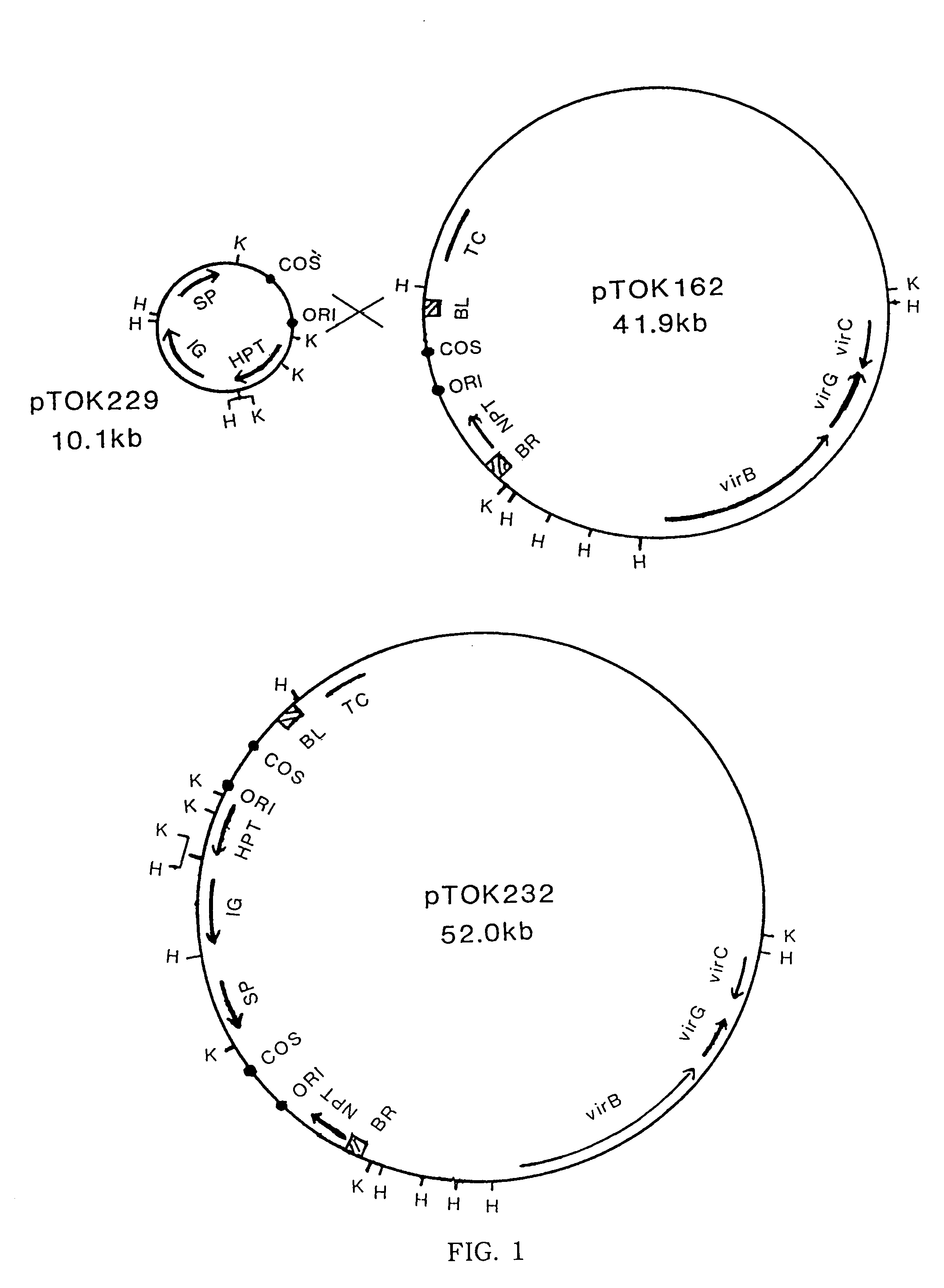
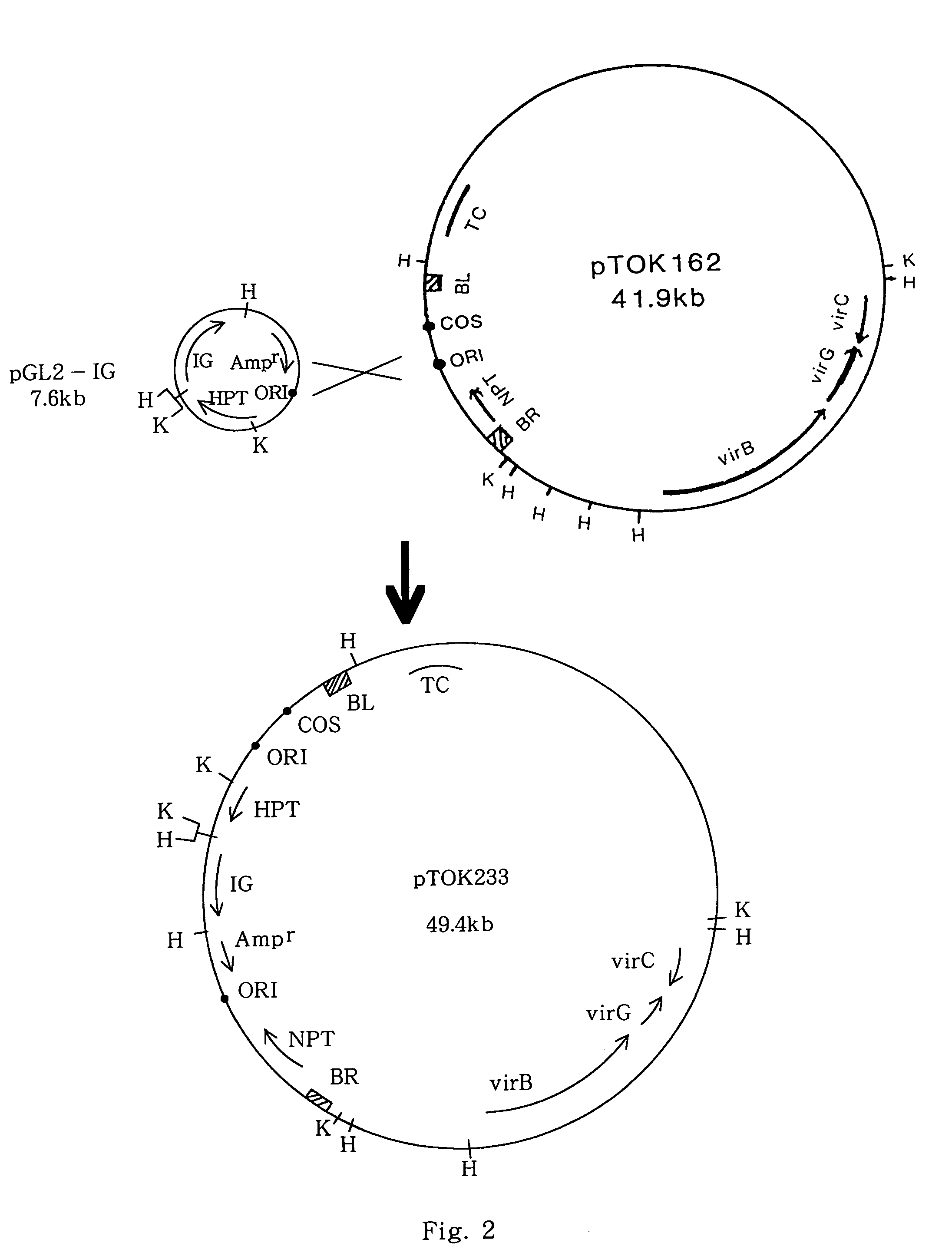
[0109]A super binary vector pTOK233 was constructed by a process similar to the construction process of pTOK232, as shown in FIG. 2, from the above-described pGL2-IG and pTOK162.
(3) The Obtained Super Binary Vector pTOK233 or the Above-Described Binary Vector pIG121Hm was Introduced into Two Strains of Agrobacterium tumefaciens by the Same Method as Used in Example 1 to Obtain the Following Three Agrobacterium tumefaciens Strains.
[0110]LBA4404(pIG121Hm)
[0111]EHA101(pIG121Hm)
[0112]LBA4404(pTOK233)
All of these strains have an intron GUS and a hygromycin-resistance gene in the T-DNA region.
(4) Embryos of Mature Seeds
[0113]From mature rice seeds cultured on LS2.5 medium (LS basal medium supplemented with 2.5 mg/l of 2,4-dichlorophenoxyacetic acid) for 5 days (4 days post-germination), embryos (3–5 mm size) containing scutella were excised. The embryos were washed with sterilized water and immersed in a cell suspension of Agrobacterium as in Example 1. The embryos were then placed on 2N6 medium containing acetosyringone and cultured at 25° C. for 3 days in the dark. Expression of GUS gene was checked using X-gluc as in Example 1. The number of embryos which exhibited GUS expression on the scutella was counted.
(5) Embryos of Immature Seeds
[0114]Immature seeds at 10–14 days after flowering were sterilized and immature embryos (1.5–2.0 mm size) were excised. The embryos were sterilized by a conventional method and cultured on 2N6 medium for 2 days. The germinated buds were excised and immersed in Agrobacterium suspension as in Example 1. The embryos were then placed on 2N6 medium containing acetosyringone and cultured at 25° C. for 3 days in the dark.
(6) Selection of Resistant Cells and Regeneration
[0115]The embryos after the culture with Agrobacterium were placed on 2N6 medium containing 50 mg/l of hygromycin and 250 mg/l of cefotaxime and cultured at 25° C. for 3 weeks under illumination. The number of the tissues from which calli exhibiting uniform GUS expression were formed was counted. The selected calli were placed on N6–7 medium containing 100 mg/l of hygromycin and 250 mg/l of cefotaxime and cultured at 25° C. for 10 days under illumination. The selected calli were placed on N6S3 medium containing 50 mg/l of hygromycin and 250 mg/l of cefotaxime and cultured at 25° C. for 40 days under illumination. Using leaves of the obtained regenerated plants, expression of GUS gene was checked using X-gluc as in Example 1.
(7) Results
[0116]The results are shown in Tables 12–14 below.
[0117] TABLE 12 GUS Expression after Culturing with Agrobacterium (embryos derived from mature seeds) Number of Sample Number of GUS+ Sample Strain Embryos Embryos EHA101 50 8 (pIG121Hm) 61 11 LBA4404 53 16 (pTOK233) 60 32
[0118] TABLE 13 Results of Transformation (embryos derived from mature seeds) Number of Lines Hygromycin- Calli Number of resistant Yielded GUS+ Sample Sample and GUS+ Regenerated Regenerated Strain Tissues Calli Plants Plants EHA101 43 0 0 0 (pIG121Hm) 78 2 0 0 LBA4404 50 1 1 1 (pTOK233) 80 8 5 5
[0119] TABLE 14 Results of Transformation (embryos derived from immature seeds) Number of Lines Hygromycin- Calli Number of resistant Yielded GUS+ Sample Sample and GUS+ Regenerated Regenerated Strain Tissues Calli Plants Plants LBA4404 65 10 4 3 (pIG121Hm) EHA101 67 15 6 6 (pIG121Hm) 41 8 3 3 LBA4404 65 41 28 25 (pTOK233) 40 28 17 13
[0120]As can be seen from Table 12, with the embryos derived from mature seeds, the number of embryos which showed GUS expression was much larger in the group treated with LBAA4404(pTOK233). As can be seen from Table 13, hygromycin-resistant calli were scarcely obtained in the group treated with EHA101(pIG121Hm) and no regenerated plants were obtained in this group. In contrast, in the group treated with LBA4404(pTOK233), regenerated plants were obtained even though the frequency was low. Most of the regenerated plants expressed GUS and so they are thought to be transformants.
[0121]As can be seen from Table 14, with the immature embryos, transformants were obtained in all groups irrespective of the strain used for transformation. However, the transformation efficiency was prominently high when LBA4404(pTOK233) was used.
[0122]Thus, the embryo tissues cultured in a dedifferentiation-inducing medium for several days were able to be transformed via Agrobacterium. For such transformation, a super binary vector such as pTOK233 contained in the strain LBA4404(pTOK233) gives prominently high transformation efficiency and so is very useful for this purpose.

PUM

PropertyMeasurementUnit
Time604800.0s
Time518400.0s
Fraction0.0fraction
tensileMPa
Particle sizePa
strength10

Description & Claims & Application Information

We can also present the details of the Description, Claims and Application information to help users get a comprehensive understanding of the technical details of the patent, such as background art, summary of invention, brief description of drawings, description of embodiments, and other original content. On the other hand, users can also determine the specific scope of protection of the technology through the list of claims; as well as understand the changes in the life cycle of the technology with the presentation of the patent timeline. Login to view more.

Similar technology patents

Nitride semiconductor laser chip

ActiveUS20100316082A1Easily obtainSufficient reliabilityOptical wave guidanceLaser detailsResonatorLaser
Owner:SHARP FUKUYAMA LASER CO LTD

Plant information display apparatus and method

InactiveUS20090228839A1Good operabilityEasily obtainProgramme controlTotal factory controlData selectionSoftware selection
Owner:YOKOGAWA ELECTRIC CORP

Classification and recommendation of technical efficacy words

  • Easily obtain
  • Decrease frequency

Methods and Compositions For Preparing Pancreatic Insulin Secreting Cells

InactiveUS20080241107A1Easily obtainBiocidePancreatic cellsTransdifferentiationPancreatic structure
Owner:BOARD OF RGT THE UNIV OF TEXAS SYST

Slave device, time synchronization method in slave device, master device, and electronic equipment system

ActiveUS20100220748A1Decrease frequencyIncreased frequencyTime-division multiplexElectric windingTime informationTime synchronization
Owner:SONY CORP

Piezoelectric filter

ActiveUS20070115079A1Reduce property degradationDecrease frequencyPiezoelectric/electrostrictive device manufacture/assemblyImpedence networksPiezoelectric resonatorsCondensed matter physics
Owner:MURATA MFG CO LTD

Generating a consensus mesh from an input set of meshes

ActiveUS20140098090A1Decrease frequency3D-image rendering3D modellingTriangulationDelaunay triangulation
Owner:AUTODESK INC
Why Eureka
Try Eureka