Optical bio-discs and fluidic circuits for analysis of cells and methods relating thereto
- Summary
- Abstract
- Description
- Claims
- Application Information
AI Technical Summary
Benefits of technology
Problems solved by technology
Method used
Image
Examples
example 1
[0319]FIG. 17A illustrates a pictorial flow chart showing the preparation of a sample, use of a bio-disc, and the provision of results which are shown in greater detail in FIGS. 52 and 53. The details of the following example such as the individual time duration of process steps, rotation rates, and other details are more particular than those described above with reference to FIGS. 17A, 52, and 53. The basic steps of the present example are, nonetheless, similar to those described above.
A. Disc Manufacturing Including Substrate Preparation and Chemistry Deposition
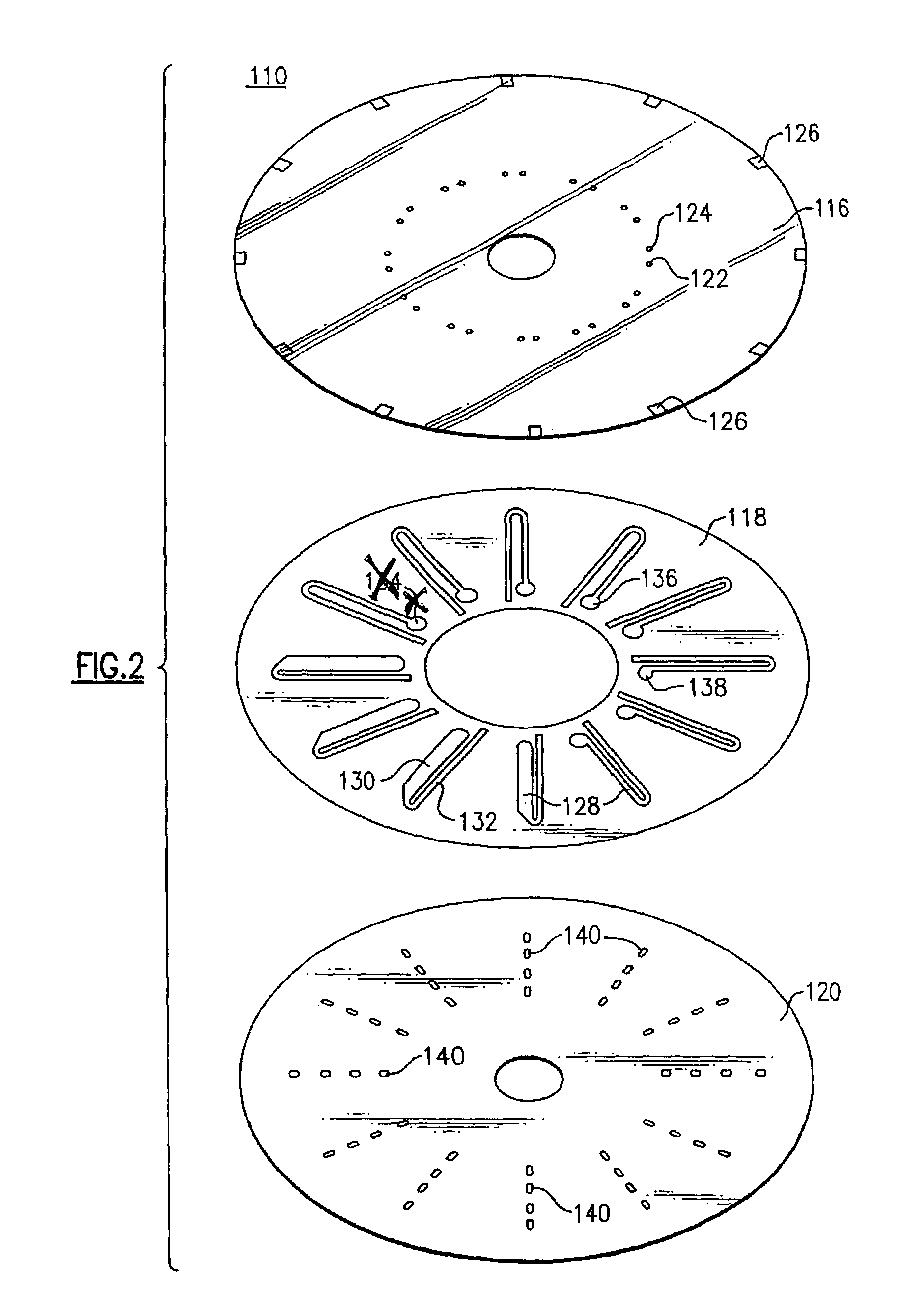
[0320]In this example, a reflective disc or transmissive disc substrate 120 (FIGS. 2 and 5, respectively) is cleaned using an air gun to remove any dust particles. The disc is rinsed twice with iso-propanol, using a spin coater. A 2% polystyrene is spin coated on the disc to give a very thick coating throughout.
[0321]The chemistry is then deposited. One embodiment includes a three step deposition protocol that incubates: s...
example 2
Mononuclear Cells Separation Procedure
[0351]Use Becton and Dickinson Vacutainer CPT (BD catalog # 362760 for 4 ml, #362761 for 8 ml) cell preparation tubes with sodium citrate. Do procedure in biohazard hood following all biohazard precautions. Steps:[0352]1. Collect blood directly into a 4 or 8 ml EDTA containing CPT Vacutainer. If the blood sample is already in an anticoagulant, pour off the EDTA in the Vacutainer first and then pour 6–8 ml of blood sample into the CPT tube.[0353]2. Centrifuge the tube at 1500 to 1800×g in a biohazard centrifuge with horizontal rotor and swing out buckets for 25 minutes at room temperature. For best results, the blood should be centrifuged within two hours of collection. However, blood older then 2 hours may be centrifuged with a decrease in MNC number and increase in RBC contamination.[0354]3. After centrifugation, remove the plasma leaving about 2 mm of plasma above the MNC layer. Collect and transfer the whitish mononuclear layer into a 15 ml c...
example 3
Isolation of MNCs from Whole Blood Using Histopaque-1077
[0363]1 ml of Histopaque-1077 was placed in a 15 ml centrifuge tube and 1 ml of whole blood is gently layered over that. Then centrifuged at 400×g for 30 min at room temperature. The mixture was aspirated carefully with a pasture pipette and the opaque interface transferred to centrifuged tube. Then 10 ml of PBS was added to the centrifuge tube. The solution was then centrifuged at 250×g for 10 min. The supernatant was decanted and the cell pellet was resuspended in 10.0 ml PBS and spun at 250×g for 10 min. The cells were then washed one more time by resuspending the pellet in 10 ml PBS and spinning at 250×g. The final cell pellet was resuspended in 0.5 ml PBS.
PUM
| Property | Measurement | Unit |
|---|---|---|
| Volume | aaaaa | aaaaa |
| Volume | aaaaa | aaaaa |
| Volume | aaaaa | aaaaa |
Abstract
Description
Claims
Application Information
 - R&D
- Intellectual Property
- Life Sciences
- Materials
- Tech Scout
- Unparalleled Data Quality
- Higher Quality Content
- 60% Fewer Hallucinations
Browse by: Latest US Patents, China's latest patents, Technical Efficacy Thesaurus, Application Domain, Technology Topic, Popular Technical Reports.
© 2025 PatSnap. All rights reserved.Legal|Privacy policy|Modern Slavery Act Transparency Statement|Sitemap|About US| Contact US: help@patsnap.com


