Wound treatment employing reduced pressure

a wound treatment and pressure reduction technology, applied in the field of wound treatment using reduced pressure, can solve the problems of large or infected wounds, inability to spontaneously close, wounds that cannot successfully fight bacterial infections, etc., to promote epithelial migration, reduce pressure, and enhance the attachment of adjacent tissu

Inactive Publication Date: 2007-04-03
WAKE FOREST UNIV HEALTH SCI INC
View PDF139 Cites 503 Cited by
  • Summary
  • Abstract
  • Description
  • Claims
  • Application Information

AI Technical Summary

Benefits of technology

[0009]In accordance with the present invention a wound treatment apparatus is provided for treating a wound by applying reduced pressure (i.e. pressure that is below ambient atmospheric pressure) to the wound in a controlled manner for a selected time period. The application of reduced pressure to a wound provides such benefits as faster healing, increased formation of granulation tissue, closure of chronic open wounds, reduction of bacterial density within wounds, inhibition of burn penetration, and enhancement of flap and graft attachment. Wounds that have exhibited positive response to treatment by the application of negative pressure include infected open wounds, decubitus ulcers, dehisced incisions, partial thickness burns, and various lesions to which flaps or grafts have been attached.
[0016]Another aspect of the invention is a method of enhancing the attachment of adjacent tissue to a wound which comprises applying negative or reduced pressure to a joined complex of the adjacent living tissue and the wound at a sufficient magnitude of reduced pressure and for a sufficient time duration to promote the migration of epithelial and subcutaneous tissue toward the complex. This method enhances attachment of adjacent tissue to tissues of the wound edges. Another use of this method is to enhance attachment of an open skin graft to the wound tissue.

Problems solved by technology

The treatment of open wounds that are too large to spontaneously close has long been a troublesome area of medical practice.
Some wounds, however, are sufficiently large or infected that they are unable to heal spontaneously.
Without sufficient blood flow, the wound is unable to successfully fight bacterial infection and is accordingly unable to close spontaneously.
Infection and poor vascularization hinder the formation of granulation tissue within wounded tissue, thereby inhibiting wound healing.
Poor blood circulation and infection at the wound may also hinder attachment of skin grafts or flaps upon wounded tissue.
Another problem encountered during the treatment of wounds is the selection of an appropriate technique for wound closure during the healing process.
However, sutures apply a closure force to only a very small percentage of the area surrounding a wound.
When there is scarring, edema, or insufficient tissue, the tension produced by the sutures can become great causing excessive pressure to be exerted by the sutures upon the tissue adjacent to each suture.
As a result, the adjacent tissue often becomes ischemic thereby rendering suturing of large wounds counterproductive.
If the quantity or size of the sutures is increased to reduce the tension required of any single suture, the quantity of foreign material within the wound is concomitantly increased and the wound is more apt to become infected.
Additionally, the size or type of a particular wound may prevent the use of sutures to promote wound closure.
Wounds resulting from ischemia, or lack of blood flow, are also often difficult to heal since decreased blood flow to a wound may inhibit normal immune reaction to fight infection.
Patients that are bedridden or otherwise non-ambulatory are susceptible to such ischemic wounds as decubitus ulcers or pressure sores.
Since the patient is often unable to feel the wound or to move sufficiently to relieve the pressure, such wounds can become self-perpetuating.
Although it is common to treat such wounds with flaps, the conditions that initially caused the wound may also work against successful flap attachment.
The progression of partial thickness burns to deeper buns is a major problem in burn therapy.
The death of tissue within the zone of stasis is caused by lack of oxygen and nutrients, reperfusion injury (re-establishment of blood flow after prolonged ischemia), and decreased migration of white blood cells to the zone resulting in bacterial proliferation.

Method used


Image

  • Wound treatment employing reduced pressure
  • Wound treatment employing reduced pressure
  • Wound treatment employing reduced pressure

Examples


example 1

Treatment of Open Wounds

[0054]In order to demonstrate the use of a negative pressure appliance in the treatment of open wounds, an animal study was conducted using pigs as subjects. Pigs are frequently used as subjects in wound healing studies since they have essentially the same skin and subcutaneous tissue structure as humans.

[0055]Five 15 kg Chester pigs were obtained and acclimated for 1 week prior to use. The animals were sedated with an intramuscular injection of ketamine (25 mg / kg):xylazine (2.5 mg / kg):acepromazine (5 mg / kg). The backs and sides of the animals were shaved and scrubbed for surgery. One percent halothane was administered by endotracheal tube for maintenance of anesthesia. Two circular wounds are created on the midline of the animals. The wounds were 2.5 cm in diameter having a depth reaching, but not including, the deep fascia over the spins (approximately 1 cm). Wounds in pigs in this site do not contract during healing. Alginate impressions were made of each ...

example 2

Reduction of Infection

[0059]During the course of the experiment described as Example 1 above, it was observed that the reduced pressure-treated wounds were much cleaner and bled more spontaneously than non-treated wounds. It was therefore undertaken to determine the relative rates of clearance of a known bacterial inoculum from treated and non-treated wounds.

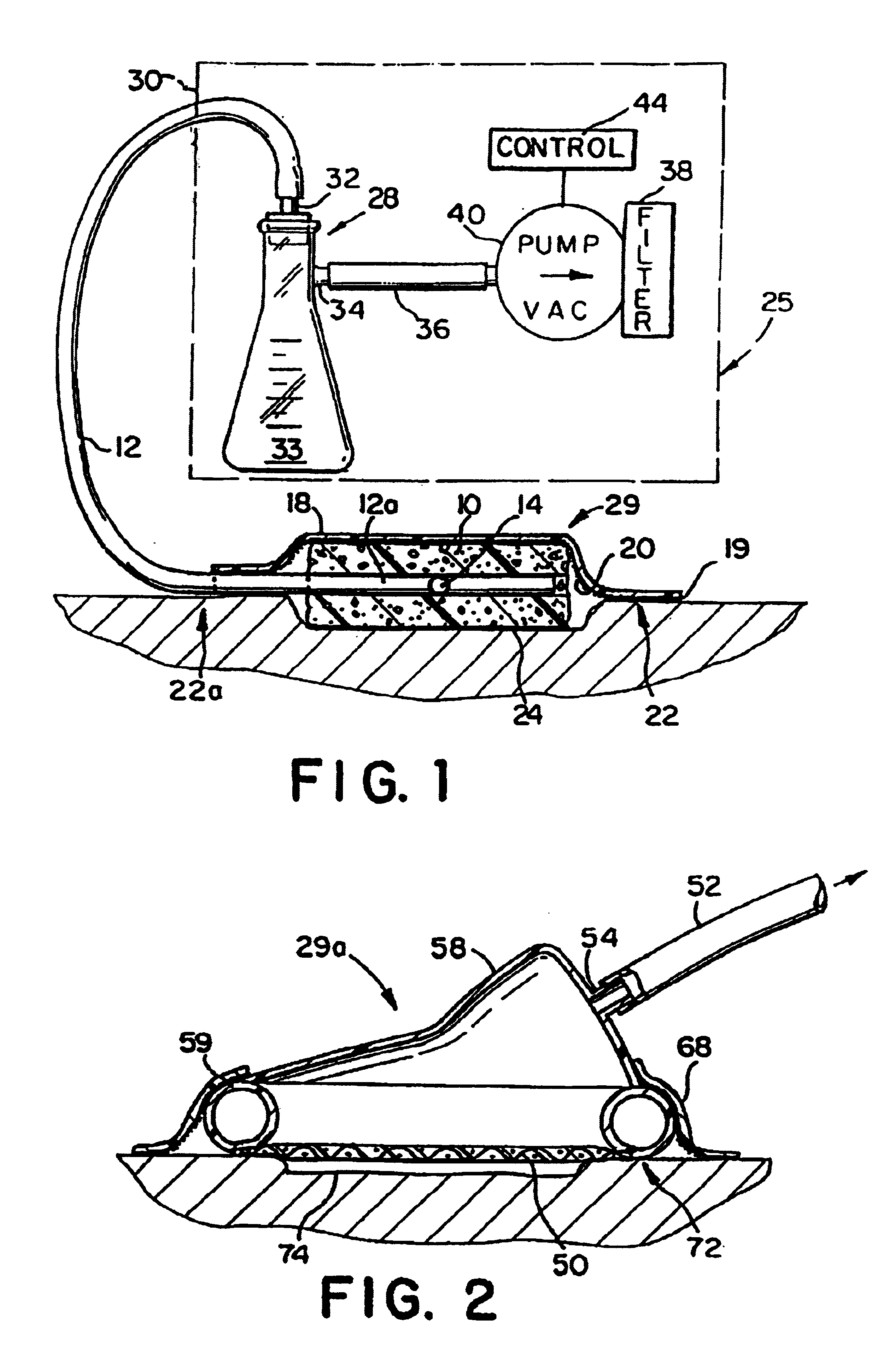
[0060]Five 15 kg pigs were obtained and wounds created as set forth in Example 1. Two 2.5 cm diameter defects were created on the dorsum of each pig using a sterile technique, with a 7.5 cm interval retained between the edges of the defects. Hemostasis was obtained by electrocautery. Prior to placement of the reduced pressure appliances, 108 organisms of Staphylococcus aureus in 1 ml saline solution were injected into each wound. The reduced pressure appliances of the type shown in FIGS. 2 and 11 were then attached as in Example 1, and a reduced pressure of 5 in. Hg below atmospheric pressure was applied to one of the wounds upo...

example 3

Treatment of Burns

[0064]Use of reduced pressure appliances upon burns has been found to retard the progression of partial thickness burns into full thickness burns. A partial thickness burn is a burn in which the depth of cell death due to thermal trauma does not extend below the level of the deepest epidermal structures (i.e., the base of hair follicles, sweat glands, sebaceous glands, etc.). A burn that is initially a partial thickness burn will often deepen and progress into a full thickness burn due to insufficient blood circulation to the epidermal cells beneath the partial burn.


PUM


Abstract

A method of treating tissue damage comprises applying a negative pressure to a wound sufficient in time and magnitude to promote tissue migration and thus facilitate closure of the wound. The method is applicable to wounds, burns, infected wounds, and live tissue attachments. A wound treatment apparatus is provided in which a fluid impermeable wound cover is sealed over a wound site. A screen in the form of an open-cell foam screen or a rigid porous screen is placed beneath the wound cover over the wound. A vacuum pump supplies suction within the wound cover over the treatment site.

Description

RELATED APPLICATIONS[0001]This application is a continuation of application Ser. No. 08 / 467,243, entitled “Wound Treatment Employing Reduced Pressure,” filed on Jun. 6, 1995, now ABN; which is a continuation of application Ser. No. 08 / 028,677, entitled “Wound Treatment Employing Reduced Pressure,” filed Mar. 9, 1993, now U.S. Pat. No. 5,636,643, issued Jun. 10, 1997, which is a continuation-in-part of application Ser. No. 07 / 792,001, entitled “Method of Treating Tissue Damage and Apparatus for Same,” Filed on Nov. 14, 1991, now U.S. Pat. No. 5,645,081, issued Jul. 8, 1997, each of such applications being incorporated herein by reference.FIELD OF THE INVENTION[0002]The present invention relates to an apparatus and method for treating a wound by applying reduced pressure to the wound.BACKGROUND OF THE INVENTION[0003]The treatment of open wounds that are too large to spontaneously close has long been a troublesome area of medical practice. Closure of an open wound requires inward migra...

Claims


Application Information

Patent Timeline
Patent Type & Authority Patents(United States)
IPC IPC(8): A61B19/00A61B17/00A61B17/08A61B17/30A61F13/00A61F13/02A61M1/00A61M1/08A61M16/06A61M27/00
CPCA61B19/00A61F13/02A61M1/0088A61M27/00A61M1/005A61B17/08A61B17/085A61B2017/00557A61B2017/306A61F2013/00919A61M1/0023A61M1/0037A61M1/0056A61M1/008A61M1/08A61M16/06A61M2205/3379A61F13/00068A61B90/00A61F13/0226A61F13/0246A61M2205/7518A61M2205/7545A61M1/75A61M1/782A61M1/79A61M1/84A61M1/91A61M1/982
Inventor ARGENTA, LOUIS C.MORYKWAS, MICHAEL J.
Owner WAKE FOREST UNIV HEALTH SCI INC
Why Eureka
Try Eureka