Rotavirus subunit vaccine

a subunit vaccine and rotavirus technology, applied in the field of rotavirus subunit vaccines, can solve the problems of high morbidity from rotavirus disease, high risk of reversion, and difficult generation of rotavirus vaccines, and achieve the effect of increasing the immunological protection of said individual against rotavirus infection

Inactive Publication Date: 2007-12-25
CHILDRENS HOSPITAL MEDICAL CENT CINCINNATI
View PDF20 Cites 20 Cited by
  • Summary
  • Abstract
  • Description
  • Claims
  • Application Information

AI Technical Summary

Benefits of technology

[0023]Another embodiment of the invention is a method of immunizing a subject in need of rotavirus immunity comprising the steps of: immunizing with a rotavirus vaccine; and sub

Problems solved by technology

While mortality has been controlled in developed countries by universal access to emergency medical care, morbidity from rotavirus disease remains high.
This ability has also made generation of a rotavirus vaccine extremely challenging.
However, as with all attenuated viruses, there is always a high risk of reversion.
Among some 2,200 infants enrolled in the double-blind, placebo controlled study, the vaccine was successful in reducing severe diarrheal illness in about 70% of subjects but was less effective at reducing the incidence of less serious diarrheal illness.
Recently, it was reported that there have been serious side effects such as bowel blockage associated

Method used


Image

  • Rotavirus subunit vaccine
  • Rotavirus subunit vaccine
  • Rotavirus subunit vaccine

Examples


example 1

Construction of Recombinant pMAL-c2 Plasmids

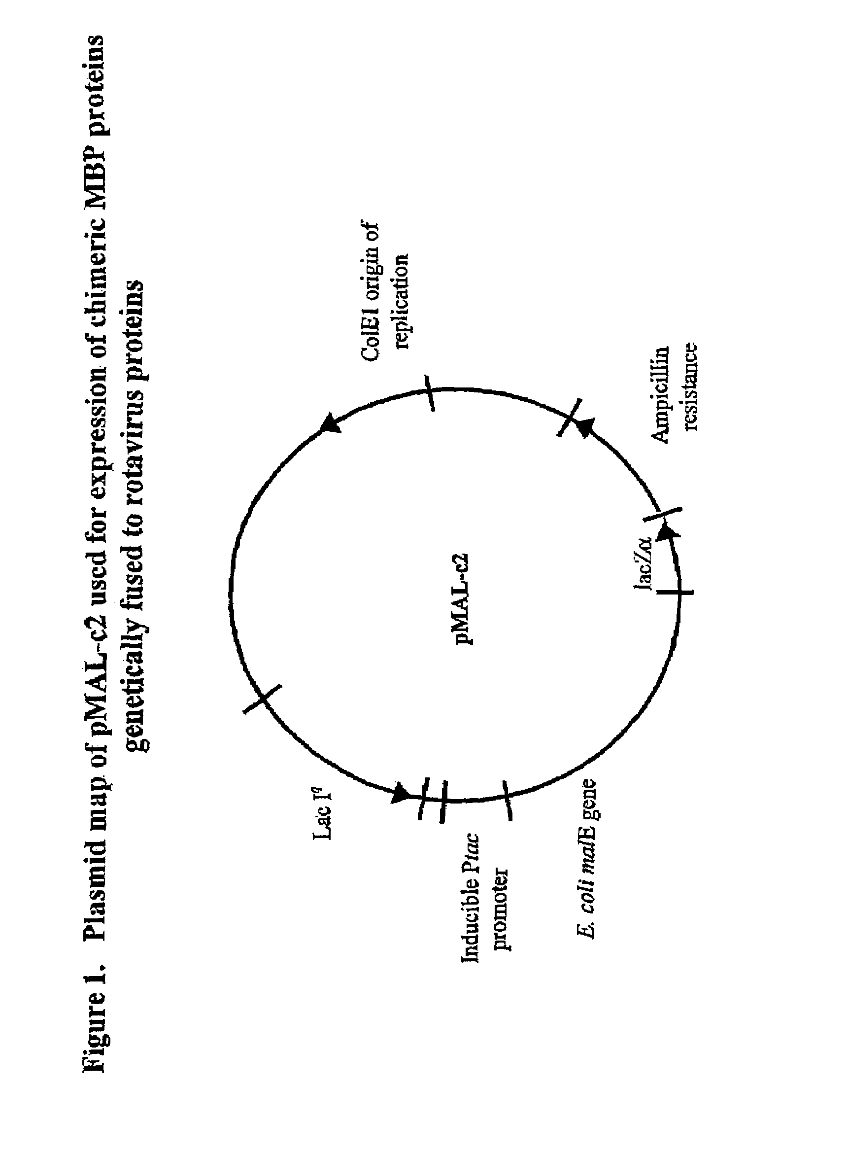
[0048]Recombinant plasmids pMAL-c2 / EDIM4, pMAL-c2 / EDIM6 and pMAL-c2 / LDIM7 were constructed using pMAL-c2 (New England Biolabs, Beverly Mass.) by insertion of cDNAs encoding full length VP4 or VP6, or a truncated form of VP7 (TrVP7) of rotavirus strain EDIM (FIG. 1). cDNAs were synthesized by polymerase chain reaction (PCR) using the plasmids pGEM-3Z / EDIM4, pGEM-3Z / EDIM6 and pGEM-3Z / EDIM7 as templates and gene specific primers determined by nucleotide sequencing of the gene inserts. The nucleotide sequences have been deposited into GenBaiik nucleotide sequence database and assigned with the Accession Numbers AF039219, U65988 and AF039220 for VP4, VP6 and VP7 gene respectively.

[0049]The murine EDIM strain of rotavirus used for the construction of the pGEM recombinant plasmids was originally isolated from the stool of an infected mouse and adapted to grow in cell culture by passage in MA-104 cells in the laboratory. A triply plaque-purified i...

example 2

Expression and Purification of Fusion Proteins

[0052]Recombinant bacteria were grown as an overnight culture (37° C., shaken at 215 rpm) in rich broth (tryptone, 10 gm; yeast extract, 5 NaCl, 5 gm; glucose, 2 gm and 100 mg of ampicillin per liter). On the following day, 10 ml of overnight cell culture were inoculated into 1 liter of rich broth containing glucose and ampicillin. The culture was grown until the optical density A600 reached 0.6. IPTG was then added to 0.3 mM to induce expression of fusion protein. Growth was continued for 3 hours.

[0053]Cells were harvested by centrifugation (4,000 g; 20 min at 4° C.), resuspended in PBS, and subjected to centrifugation. The pellet was frozen at −20° C., thawed slowly in cold water, and resuspended in a total of 50 ml of buffer L (5 mM NaH2PO4, 10 mM Na2HPO4, 30 mM NaCl, 10 mM 2-beta mercaptoethanol and 0.2% Tween 20, 1 mM PMSF, 25 mM benzamidine, and 200 mg / L of lysozyme). After digestion for 15 min at room temperature (rt), the suspens...

example 3

Biochemical Characterization of MBP::VP6 Fusion Protein

[0056]It has been shown that recombinant VP6 expressed by the bacculovirus expression system forms structures that resemble double-layered rotavirus particles when examined by electron microscopy. Purified MBP::VP6 fusion protein was analyzed by sucrose gradients to determine if these fusion proteins assembled into organized structures resembling virus particles that could be fractionated in a sucrose gradient. MBP::VP6 was subjected to centrifugation (SW 50, 35,000 g, 60 min) through a 4 ml sucrose gradient (20-50%) on a 1 ml cesium chloride cushion (60%). A total of 16, 300-μl fractions were collected. Distribution of MBP::VP6 in the sucrose gradient and cesium chloride cushion was analyzed by Western blot analysis and distribution of virus particles was analyzed by silver nitrate staining of the SDS-gel (FIG. 3). The results showed that MBP::VP6 remained in the top 4 fractions of the gradient, while double-layered virus parti...


PUM

PropertyMeasurementUnit
Volumeaaaaaaaaaa
Volumeaaaaaaaaaa
Volumeaaaaaaaaaa

Abstract

The present invention is directed to the generation and use of recombinant rotavirus fusion proteins as immunogens to produce a protective immune response from immunized individuals. In one embodiment, the present invention contemplates a recombinant rotavirus fusion protein vaccine composition comprising a rotavirus subunit protein or immunogenic fragment thereof, and an adjuvant in combination with the recombinant rotavirus subunit fusion protein. In one aspect of this embodiment, the recombinant rotavirus fusion protein comprises a rotavirus subunit protein and a fusion partner protein in genetic association with the rotavirus subunit protein, wherein the fusion partner protein does not interfere with expression and immunogenicity of the rotavirus subunit protein, the fusion partner protein prevents complex formation by the rotavirus subunit protein, and the fusion partner protein facilitates purification of the recombinant rotavirus fusion protein. In another aspect of this embodiment, the rotavirus subunit protein is selected from the group consisting of VP1, VP2, VP3, VP4, VP6, VP7, NSP1, NSP2, NSP3, NSP4 or NSP5. In yet another aspect of this embodiment, the rotavirus subunit protein is VP6.

Description

[0001]This application is a division of U.S. patent applicant Ser. No. 09 / 429,801, filed Oct. 29, 1999, now U.S. Pat. No. 6,589,529, which is a non-provisional application of provisional application Ser. No. 60 / 106,347, filed Oct. 30, 1998, now abandoned.BACKGROUND OF THE INVENTION[0002]Rotaviruses comprise a genus within the family Reoviridae, and are ubiquitous throughout the animal kingdom. Rotavirus infection is known to cause gastrointestinal disease and is considered the most common cause of gastroenteritis in infants. In fact, essentially every species of domestic animal tested has its own endogenous rotaviruses that cause diarrhea in newborns. Rotaviruses are also thought to be the most significant cause of gastroenteritis in young children and animals. Kapikian & Chanock, Rotaviruses, Virology, 2nd edition, Fields et al, eds., New York: Ravenpress, 1353-1404 (1990). Rotaviruses cause diarrheal disease primarily in the young, but infection and disease in older children and a...

Claims


Application Information

Patent Timeline
IPC IPC(8): A61K39/00A61K39/15C12Q1/70A61K35/76C12N15/09A61K38/00A61K39/39A61P1/00A61P1/04A61P31/14C07K14/14C07K19/00C12N1/15C12N1/19C12N1/21C12N5/10C12N15/46C12P21/02C12R1/19
CPCC07K14/005A61K39/00C12N2720/12322C07K2319/00A61P1/00A61P1/04A61P31/14
Inventor CHOI, ANTHONYWARD, RICHARD L.
Owner CHILDRENS HOSPITAL MEDICAL CENT CINCINNATI
Why Eureka
Try Eureka