Floppy, unstructured regions of proteins can play a dominant role in this problem; the
energetics and
kinetics of
crystallization are often less favorable than for fully structured proteins, and these regions are often more susceptible to degradation during purification than are structured regions, thus promoting sample heterogeneity.
Unfortunately, no robust technique exists to discern structured vs. unstructured regions of target proteins at the pace required for HT efforts, nor is there a general and robust method to effect the stabilization of unstructured regions within proteins that are to be subjected to crystallographic structure determination efforts.
Despite the utility of such exchange data, the methods used to obtain it have remained labor intensive and
time consuming, with substantial limitations in
throughput, comprehensiveness and resolution.
Despite the availability of many enhancements that facilitate such efforts, high
throughput production of stable protein constructs that suitably crystallize continues to be a serious
bottleneck.
While definition of successful constructs has long been a problem for conventional
crystallography, the inadequacies of current approaches are particularly acute and costly for structural
genomics efforts that presently show only a 5-20% success rate in target crystallization.
However the switch to higher eukaryotes, such as mouse and human, will entail even lower success rates, due in part to more complex and higher molecular weight proteins.
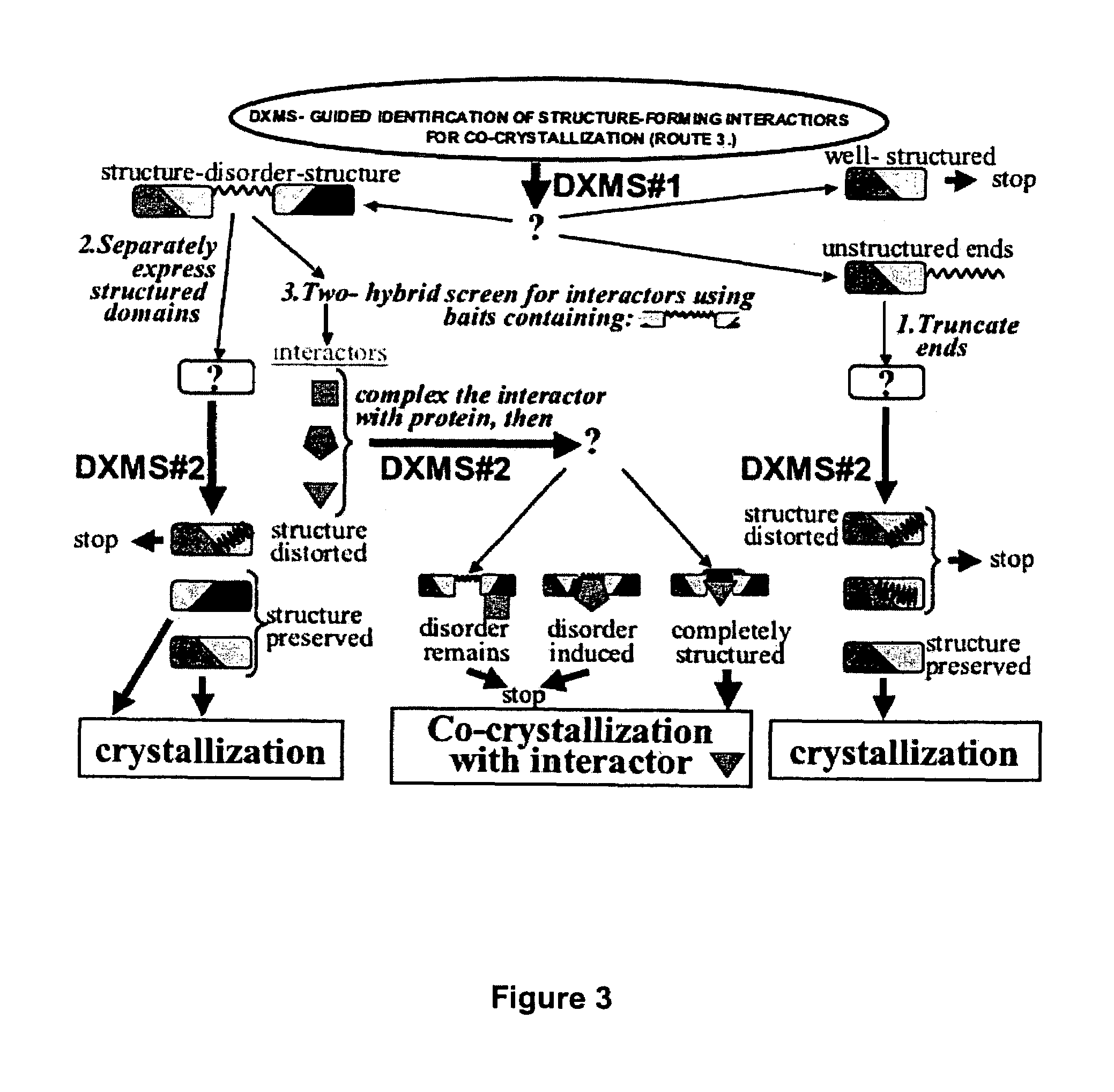
Unfortunately, while such unstructured regions may serve a function within the protein when it is interacting with binding partners in the normal cellular environment, such unstructured regions can inhibit or prevent crystallization of the entire protein when it is purified away from and therefore not bound to its structure-forming binding partners.
This unstructured protein subregion problem has been apparent for years, but its full extent is difficult to discern from the published literature.
Moreover, unstructured regions of proteins are particularly susceptible to inadvertent degradation by contaminating cellular
proteases in the course of purification and storage.
With NMR
spectroscopy, protein quantity, concentration, time needed, and size are limiting.
As such its use is
time consuming, frequently requiring that multiple proteolytic reactions be refined for optimal cleavage.
Interpretation of limited
proteolysis results is confounded by the possibility that
proteolysis may clip internal loops, leading to destabilization and subsequent further
proteolytic degradation of what was actually a structured region.
Unfortunately, the experimental definition of domain boundaries, even when they are anticipated, is often problematic, as it was for these proteins, and is usually addressed through
trial and error, by making many constructs and testing the outcome as far as expression,
solubility and crystallization.
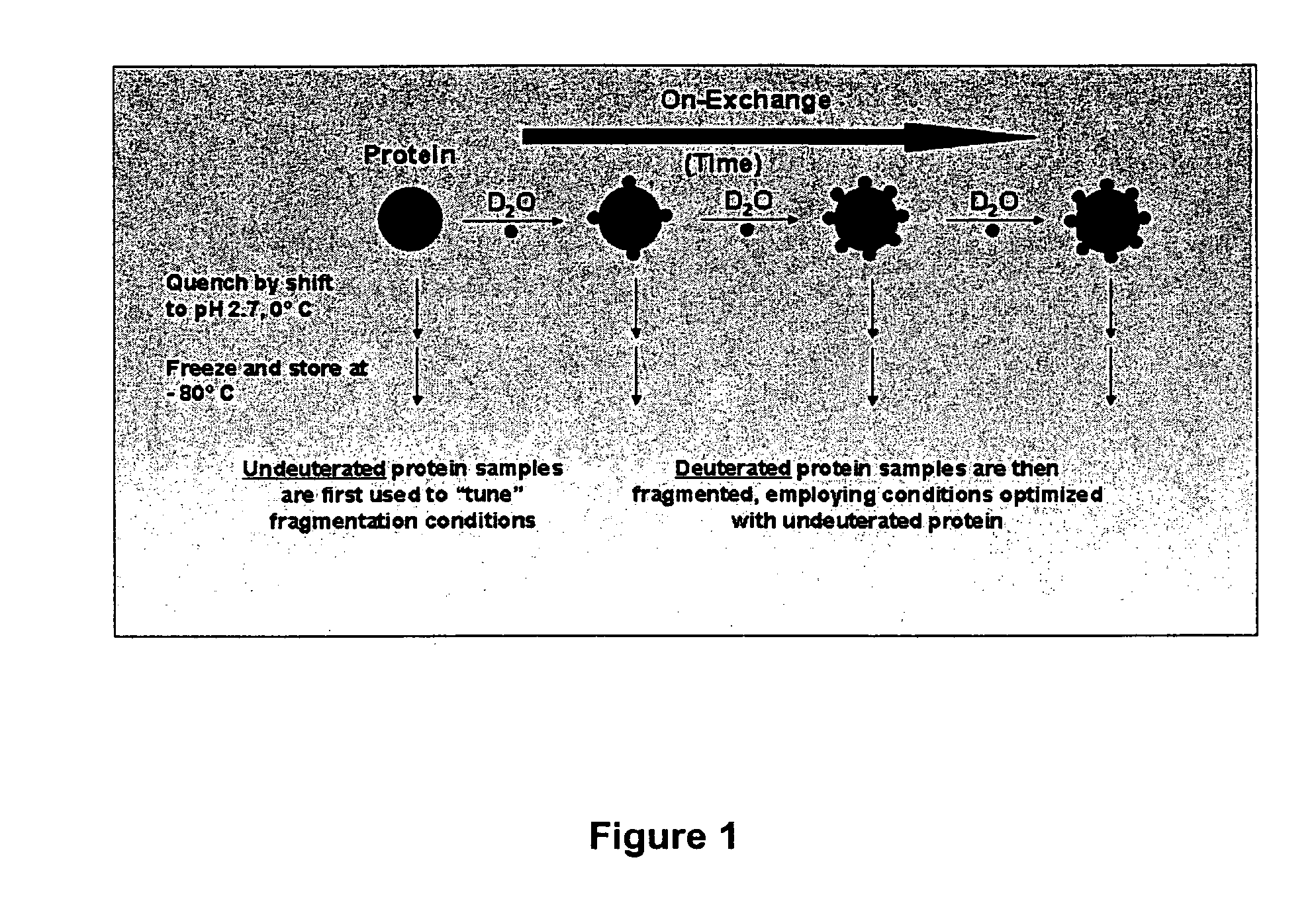
In structured regions of a protein, most
peptide amide hydrogens exchange much slower (up to 10^9 fold slower) than this maximal exchange rate, as they are not efficiently exposed to
solvent.
In general, the techniques that are the easiest to use and which give the quickest answers, result in an inexact and only approximate idea of the nature of the critical structural features.
Other techniques that are capable of finely characterizing polypeptide three-dimensional structure are considerably more difficult in practice.
While these techniques can ideally provide a precise characterization of relevant structural features, they have major limitations, including inordinate amounts of time required for study, inability to study large proteins, and, for X-
ray analysis, the need for protein and / or protein-binding partner crystals.
The process of generating protein crystals suitable for
structural analysis is commonly recognized as the most difficult and time-consuming step in the process of a crystallographic structure determination (see, e.g., Wiencek, Ann. Rev. Biomed. Eng.
Floppy, unstructured regions of proteins can play a dominant role in this problem; the
energetics and
kinetics of crystallization are often less favorable than for fully structured proteins, and additionally, these regions are often more susceptible to degradation during purification than are structured regions, thus promoting sample heterogeneity.
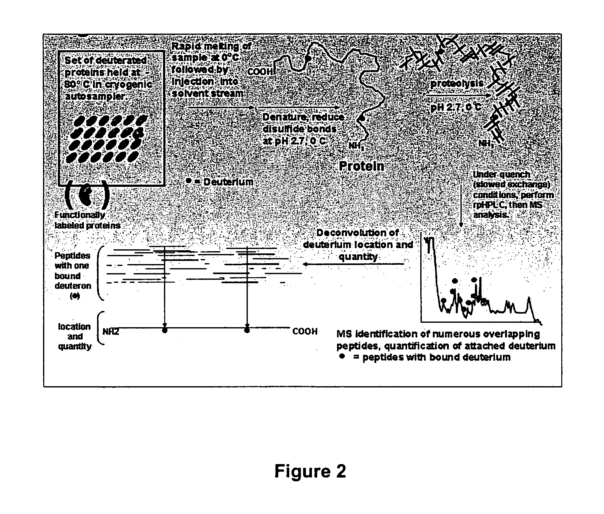
These studies do not allow a determination of the identity (location within the protein's primary
amino acid sequence) of the exchanging
amide hydrogens measured.
Again, determination of the identity of the particular
peptide amides experiencing changes in their environment is not possible with these techniques.
However, the techniques described by Rosa and Richards were of marginal utility, primarily due to their failure to optimize certain critical experimental steps.
However, as the fragmentation of
hemoglobin proceeds, each fragment's secondary and
tertiary structure is lost and the unfolded peptide amide hydrogens become freely accessible to H2O in the buffer.
Moreover, in Englander's work, there is no appreciation that a suitably adapted exchange technique might be used to identify the peptide amides which reside in the contacting surface of a protein
receptor and its binding partner.
Unfortunately, acid
proteases are very nonspecific in their sites of cleavage, leading to considerable HPLC separation difficulties.
Even then, the fragments were “difficult to separate cleanly”.
Over the succeeding years since this observation was made, no advances have been disclosed which address these critical limitations of the
medium resolution hydrogen exchange technique.
Efforts to improve the technology by employing other acid reactive
proteases other than
pepsin have not significantly improved the technique.
Furthermore, study of proteins by the NMR technique is not possible unless the protein is small (generally less than 30 kD), large amounts of the protein are available for the study, and computationally intensive
resonance assignment work is completed.
The resolution of the
deuterium-exchange
mass spectrometry work disclosed in these publications therefore remained at the 10-14
amino acid level, with the primary limitation of their art being the ability to generate only a small number of peptides with the
endopeptidase pepsin, as they employed it.
Despite the utility of such exchange data, the methods used to obtain it have remained labor intensive and
time consuming, with substantial limitations in throughput, comprehensiveness and resolution.
Despite the availability of many enhancements that facilitate such efforts, high-throughput production of stable protein constructs that suitably crystallize continues to be a serious
bottleneck.
While definition of successful constructs for protein production has long been a problem for conventional crystallography, the inadequacies of current approaches are particularly acute and costly for structural
genomics efforts that presently show only a 10-20% success rate in target crystallization.
However, a switch to higher eukaryotes, such as mouse and human, will entail even lower success rates, due in part to more complex and higher molecular weight proteins.

