Adsorption of nucleic acids to a solid phase

a nucleic acid and solid phase technology, applied in the field of nucleic acid adsorption to a solid phase, can solve the problems of interacting with the target component during lysis, difficult to isolate or measure, and many biological substances, especially nucleic acids, presenting special challenges in isolating them from their natural environmen

Active Publication Date: 2009-02-17
ROCHE DIAGNOSTICS OPERATIONS INC
View PDF23 Cites 27 Cited by
  • Summary
  • Abstract
  • Description
  • Claims
  • Application Information

AI Technical Summary

Benefits of technology

[0019]The inventors surprisingly found that performing a two-step lysis and binding procedure is advantageous, whereby in the first step lysis is effected by mixing the biological sample with an aequous lysis buffer containing a chaotropic agent and incubating the mixture; in the second step, the concentration of the chaotropic agent in the mixture is increased and the mixture is contacted with a solid phase capable of binding nucleic acids, whereby the nucleic acid in the liquid phase is adsorbed to the solid phase.
[0033]Guanidinium and thiocyanate ions are classified in the Hofmeister series as having enhanced chaotropic properties over, e.g. potassium or sodium cations, or iodide or chlorate anions, respectively. It is thus also preferred that in the mixture of step (b) the concentration of the chaotropic agent is between 2 M and 4 M.
[0041]When lysing a biological sample in order to set free the nucleic acids or when binding the nucleic acid to the solid phase it is further preferred to use a detergent in the procedures, that is to say an anionic, cationic, zwitterionic or non-ionic detergent. Such detergents are well known to the person skilled in the art. Generally, a “detergent” is a surface active agent, also known as a surfactant. A detergent is capable of lowering the surface tension of the medium in which it is dissolved, and / or the interfacial tension with other phases, and, accordingly, is positively adsorbed at the liquid / vapour and / or at other interfaces. Thus, detergents are amphipathic molecules with polar (water soluble) and nonpolar (hydrophobic) domains. They are capable of binding to hydrophobic molecules or molecular domains to confer water solubility. Depending on its ionic characteristics, a detergent can be categorized as an ionic detergent, a non-ionic detergent, and a zwitterionic detergent. Ionic detergents can be further classified into either cationic detergents such as SDS (sodium dodecyl sulfate), LiDS (lithium dodecyl sulfate), or cetyltrimethylammoniumbromide (CTAB), and anionic detergents such as deoxycholic acid, or sodium lauroyl sarcosine. Thus, these are usually highly protein denaturant. Non-ionic detergents such as are less protein denaturant. This is also true for zwitterionic detergents such as CHAPS or Sulphobetaine 14. Zwitterionic compounds, also known as zwitterions, inner salts or dipolar ions are neutral compounds having formal unit electrical charges of opposite sign.
[0042]Therefore, it is preferred that the lysis buffer of step (a) contains a detergent. It is more preferred that the lysis buffer of step (a) contains an anionic, cationic, zwitterionic or non-ionic detergent. It is even more preferred that the detergent in the lysis buffer of step (a) is selected from the group consisting of sodium dodecyl sulfate, lithium dodecyl sulfate, cetyltrimethylammoniumbromide, deoxycholic acid, sodium lauroyl sarcosine, TRITON X-100, TWEEN 20 (ICI Americas Inc.), octyl beta-D-glucoside, Nonidet P40, CHAPS or Sulphobetaine 14. However, other detergents are possible. Generally, when using the combination of a chaotropic agent, a detergent and a protease for lysing a biological sample, the skilled artisan selects a detergent and its concentration in the mixture of step (b) on the basis that proteolytic activity is preserved.
[0066]Particularly preferred is the TAQMAN (Roche Molecular Systems, Inc.) detection method disclosed in WO 92 / 02638 and the corresponding U.S. Pat. Nos. 5,210,015; 5,804,375; 5,487,972. This method exploits the exonuclease activity of a polymerase to generate a signal. In detail, the target nucleic acid component is detected by a process comprising contacting the sample with an oligonucleotide containing a sequence complementary to a region of the target nucleic acid component and a labelled oligonucleotide containing a sequence complementary to a second region of the same target nucleic acid component sequence strand, but not including the nucleic acid sequence defined by the first oligonucleotide, to create a mixture of duplexes during hybridization conditions, wherein the duplexes comprise the target nucleic acid annealed to the first oligonucleotide and to the labelled oligonucleotide such that the 3′-end of the first oligonucleotide is adjacent to the 5′-end of the labelled oligonucleotide. Then this mixture is treated with a template-dependent nucleic acid polymerase having a 5′ to 3′ nuclease activity under conditions sufficient to permit the 5′ to 3′ nuclease activity of the polymerase to cleave the annealed, labelled oligonucleotide and release labelled fragments. The signal generated by the hydrolysis of the labelled oligonucleotide is detected and / or measured. TAQMAN technology eliminates the need for a solid phase bound reaction complex to be formed and made detectable. In more general terms, a procedure for the purification of a target nucleic acid component followed by a detection step is disclosed wherein the amplification and / or detection reaction is a homogeneous solution-phase.

Problems solved by technology

Many biological substances, especially nucleic acids, present special challenges in terms of isolating them from their natural environment.
This makes them difficult to isolate or to measure, in particular in biospecific assays which allow the detection of specific nucleic acids, or the detection of specific properties of a nucleic acid.
A problem often encountered during the lysis is that other enzymes degrading the target component, e.g. desoxyribonucleases or ribonucleases degrading nucleic acids, come into contact with the target component during lysis.
Other components released during this process may be e.g. endotoxins belonging to the family of lipopolysaccharides which are toxic to cells and can cause problems for products intended to be used in human or animal therapy.
This is a limiting step, however, that prevents the procedure from being used to process large quantities of samples.
This procedure has a disadvantage, however, in that the precipitation is not selective for nucleic acids.
As a result, this procedure can not be used to remove significant quantities of any inhibitors of specific enzymatic reactions that may be present.

Method used


Image

  • Adsorption of nucleic acids to a solid phase
  • Adsorption of nucleic acids to a solid phase
  • Adsorption of nucleic acids to a solid phase

Examples


example 1

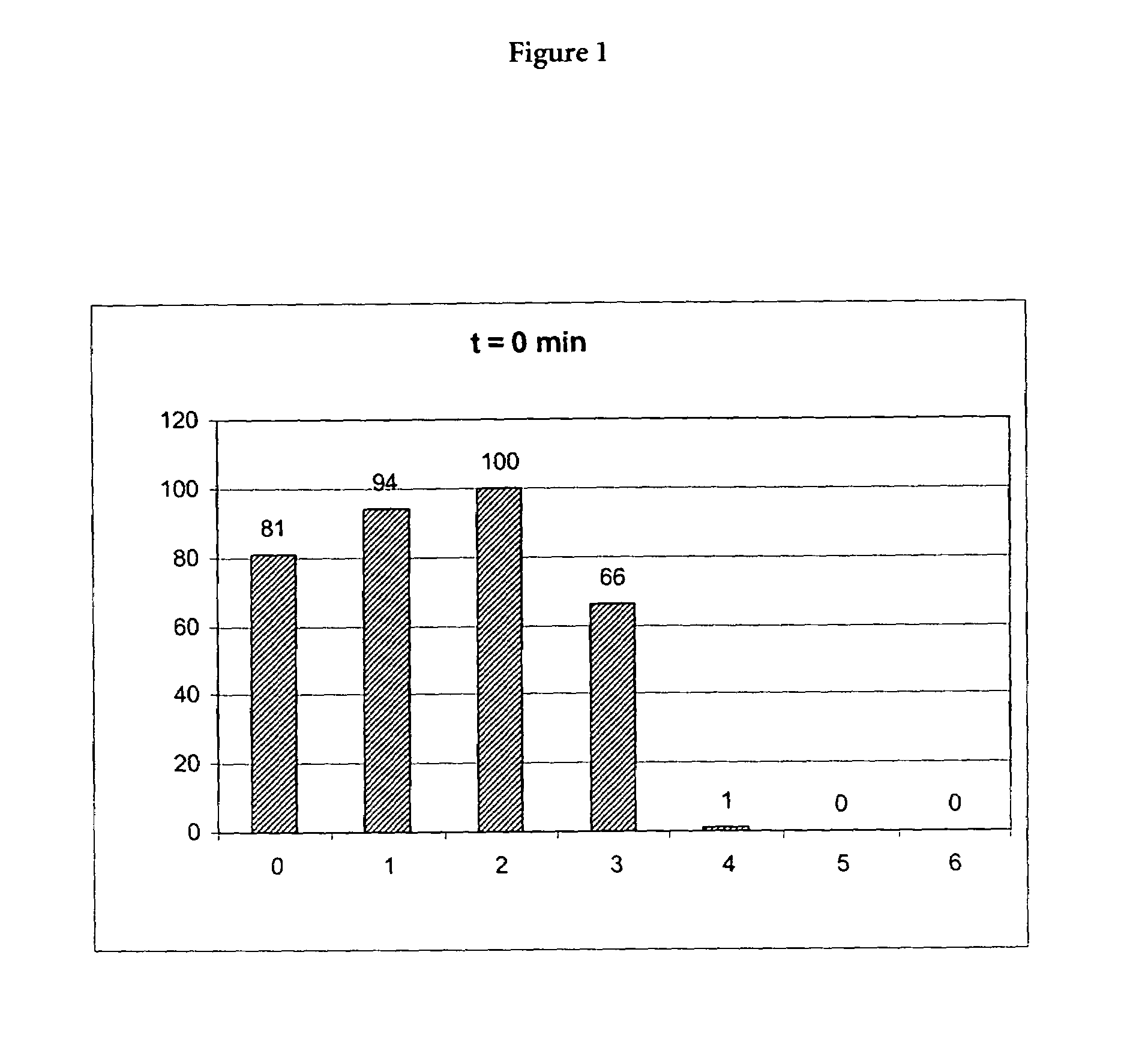
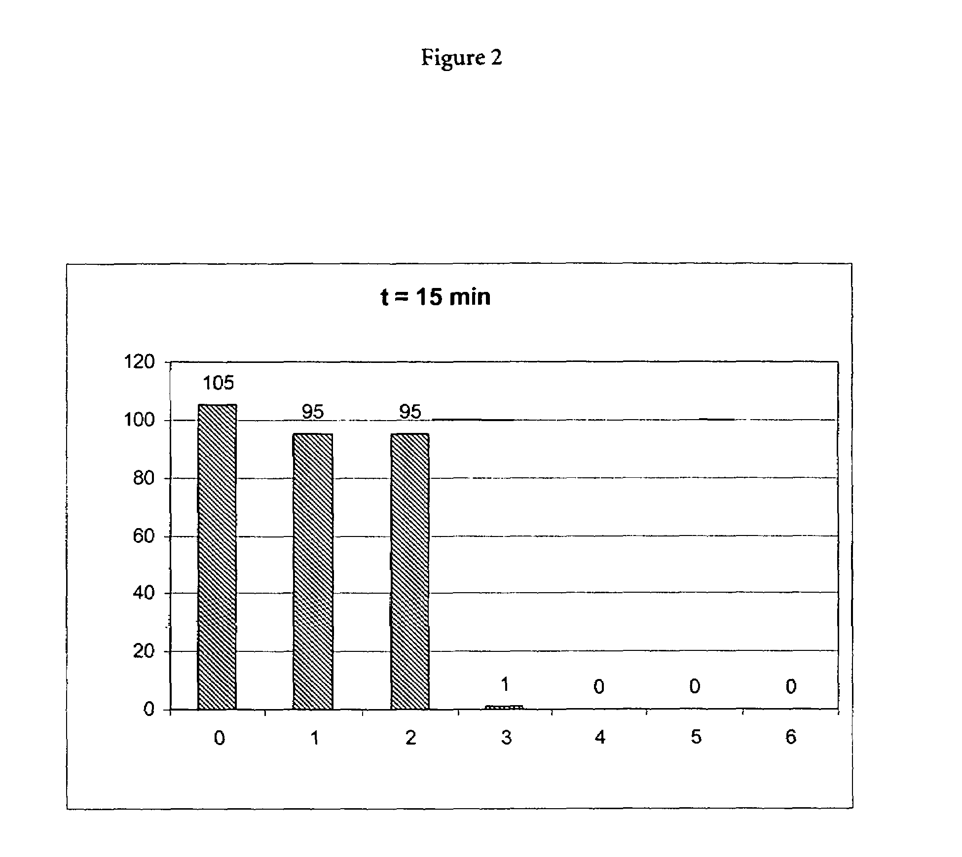
(A) Incubation of Proteinase K in the Presence of a Chaotropic Agent

[0073]10 μl of a 20 mg / ml stock solution of proteinase K (Roche Diagnostics GmbH, Mannheimm, catalogue no. 745723; 90 mg dissolved in 4.5 ml water) was mixed with a chaotropic buffer containing 50 mM Tris-HCl pH 6.0, 1% DTT, 20% TRITON X-100 and x M Guanidinum thiocyanate; x=0, 1, 2, 3, 4, 5, 6). Directly after mixing the proteinase K activity was measured in a first aliquot (10 μl) of the mixture using the assay described below (0 min activity value). 15 min after mixing the proteinase K activity was measured in a second aliquot (10 μl) of the mixture using the assay described below (15 min activity value).

(B) Assay to Determine Proteinase K Activity

[0074]10 μl of the proteinase K in chaotropic buffer (see above, (A)) were mixed with assay buffer, that is to say 980 μl 0.2 M Triethanolamin, 0.05% (weight by volume) PEG 6000, 0.1 M Calcium chloride, and 10 μl of a 200 mM substate solution. The substrate was Suc-Ala-...

example 2

(A) Reagents

[0078]a) proteinase K stock solution: recombinant proteinase K, PCR grade, 50 U / ml[0079]b) Lysis Buffer: 4 M guanidinium thiocyanate, 50 mM Tris-HCl, 20% TRITON X-100, pH 6.0[0080]c) Binding Buffer: 4 M guanidinium thiocyanate, 50 mM Tris-HCl, 20% TRITON X-100, pH 6.0[0081]d) Inhibitor Removal Buffer: 5 M guanidinium hydrochloride, 20 mM Tris-HCl, 38% ethanol, pH 6.6[0082]e) Wash Buffer: 20 mM NaCl, 2 mM Tris-HCl, 80% ethanol, pH 7.5[0083]f) Elution Buffer: 50 mM Tris-HCl, pH 8.2[0084]g) Ethanol (100%)

Additionally necessary: Commercially available spin columns containing a silica membrane, e.g. NucleoSpin Blood L, distributed by Machery & Nagel.

(B) Experiment 1: One-Step Procedure without Proteinase K Treatment[0085]1. pipette 1,000 μl EDTA blood into a 15 ml Falcon tube[0086]2. add 1,000 μl Lysis Buffer, vortex gently[0087]3. incubate for 15 min at room temperature on a roller mixer, agitate[0088]4. put a spin column in a new Falcon tube[0089]5. transfer the mixture of ...

example 3

(A) Reagents

[0153]a) Lysis buffer / binding Buffer, chaotropic (7M [lysis buffer] / 4M [binding buffer] guanidinium thiocyanate, 50 mM Tris-HCl, 20% TRITON X-100, pH 6.0)[0154]b) Inhibitor Removal Buffer (5M guanidinium HCl, 20 mM Tris-HCl, 38% ethanol, pH 6.6)[0155]c) Washing Buffer (20 mM NaCl, 2 mM Tris-HCl, 80% ethanol, pH 7.5)[0156]d) Elution Buffer (50 mM Tris, pH 8.1)[0157]e) Ethanol (absolute)

Additionally necessary: Glass fibre filter columns (with silica membrane, e.g. NucleoSpin Blood L, commercially available from Macherey-Nagel)

(B) Experiment 5, One-Step Procedure: Lysis Buffer: 7M, No Binding Buffer

[0158]1,000 μl EDTA blood was pipetted into a 15 ml Falcon tube, 1,000 μl Lysis buffer (7 M) was added and the tube was vortexed. The mixture was incubated for 15 min at 56° C. on a Thermo Mixer. A new Falcon tube with a glass fibre filter column was provided and the whole sample preparation (about 2,000 μl; chaotrope concentration 3.5 M) was transferred to the filter column. Aft...


PUM

PropertyMeasurementUnit
temperatureaaaaaaaaaa
ionic strengthaaaaaaaaaa

Abstract

The present invention is directed to a method for adsorbing, i.e. non-covalently binding, nucleic acids to a solid phase using a two-step procedure. Furthermore, the present invention pertains to a method for isolating nucleic acids from a biological sample. In the first step of the procedure, lysis is effected by mixing the biological sample with an aqueous lysis buffer containing a chaotropic agent and incubating the mixture; in the second step, the concentration of the chaotropic agent in the mixture is increased and the mixture is contacted with the solid phase, whereby the nucleic acids in the liquid phase is adsorbed to the solid phase.

Description

FIELD OF THE INVENTION[0001]The present invention is directed to a method for adsorbing, i.e. non-covalently binding, a nucleic acid to a solid phase. Furthermore, the present invention pertains to a method for isolating a nucleic acid from a biological sample.BACKGROUND[0002]Many biological substances, especially nucleic acids, present special challenges in terms of isolating them from their natural environment. On the one hand, they are often present in very small concentrations and, on the other hand, they are often found in the presence of many other solid and dissolved substances e.g. after lysis of cells. This makes them difficult to isolate or to measure, in particular in biospecific assays which allow the detection of specific nucleic acids, or the detection of specific properties of a nucleic acid. Such biospecific assays play a major role in the field of diagnostics and bioanalytics in research and development. Examples for biospecific assays are hybridisation assays, immu...

Claims


Application Information

Patent Timeline
Patent Type & Authority Patents(United States)
IPC IPC(8): C12Q1/68C07H19/00C07H21/00C12N15/09B01J20/00C07H1/08C07H21/02C07H21/04C12N1/08C12N15/10G01N33/15G01N33/53
CPCC07H21/02C07H21/04C12N15/1006C12Q1/6834C12Q2527/137C12Q2527/125C12Q2523/308F16D55/02F16D63/006F16D65/16F16D2125/28
Inventor ZIELENSKI, RALFGEISSLER, KLAUSWALTER, THOMAS
Owner ROCHE DIAGNOSTICS OPERATIONS INC
Why Eureka
Try Eureka