
Optical fiber scanner for performing multimodal optical imaging

a multi-modal, optical imaging technology, applied in the field of optical fiber scanners, can solve the problems of insufficient image resolution to provide the information required, unacceptably high false negative rate, and physicians' limited ability to detect and examine microscopic changes in early-stage neoplasia in vivo. , to achieve the effect of reducing back reflection

Inactive Publication Date: 2009-11-10
UNIV OF WASHINGTON
View PDF56 Cites 152 Cited by
  • Summary
  • Abstract
  • Description
  • Claims
  • Application Information

AI Technical Summary

Benefits of technology

[0025]The step of moving the focal point of the optical fiber scanner can be done in one of several different ways, including actuating an elastomeric polymer that changes length in response to an electric potential, changing a focus of a variable focus fluid lens in response to an electric potential, driving a motor to rotate a shaft that shifts the focal point longitudinally, applying either a hydraulic or pneumatic pressure to overcome a spring tension and thereby shifts the focus, or controlling a pressure applied to vary a separation between tissue at a site and the distal portion of the optical fiber scanner. Each of these various approaches can thus shift the focal point longitudinally to enable transverse scanning at each different depth. Another embodiment includes a beamsplitter that directs a portion of the light from the light source transversely relative to a longitudinal axis of the optical fiber scanner, and a deformable membrane mirror, which shifts the disposition of the focal point of the optical fiber scanner in response to an electric potential applied to the deformable membrane mirror, causing the disposition of the focal point to change in the direction in which the portion of the light is penetrating into the subject.
[0026]Preferably either an angled bevel is created on a distal end of an optical fiber through which light is conveyed in the optical fiber scanner, or an anti-reflection coating is applied to one or more possibly reflective surfaces, to substantially reduce back reflection.

Problems solved by technology

Unfortunately, biopsy is an invasive procedure and often results in unacceptably high rates of false negative diagnoses because of random sampling errors, particularly when the area of interest is small (as in cases of early cancers).
While x-rays, positron emission tomography (PET), magnetic resonance imaging (MRI), ultrasound, and surface tissue endoscopy have all played significant roles in the detection of macroscopic abnormalities (e.g., large tumors and strictures), physicians are still challenged by their limited ability to detect and examine microscopic changes in early-stage neoplasia in vivo, using current clinical imaging technologies, which often have insufficient image resolution to provide the information required.
Yet, such devices are typically too bulky and are thus often the limiting factor in imaging probe miniaturization.
Although it is possible to deliver an optical beam to internal organs using a single mode optical fiber, the integration of beam scanning, focusing, and collection using an endoscope only a few millimeters in diameter is a major engineering challenge that has not successfully been solved in the prior art.
A further challenge is the need for a focus-tracking mechanism that can maintain a high transverse resolution at varying depths, in particular when the focused spot sizes are small.
Although attempts have been made to integrate fiber-optic imaging bundles with scanning confocal microscopy for imaging internal organs, wherein the fiber-optic bundle relays tissue images from internal organs to a conventional scanning confocal microscope outside the human body, the resulting resolution is sub-optimal and generally unsatisfactory for most purposes.
The lower resolution is primarily due to cross-talk between fibers and limited fiber packing density.
In addition, MEMS scanners may also introduce wave front deformation to the imaging beam, since MEMS mirrors are thin and tend to warp during scanning.
Further size reduction is difficult, as a result of limitations imposed by the size of the electromagnetic actuator.
Yet, the scanning speed is severely limited to a few transverse scans per second.
Although GRIN lenses can be readily implemented in the scanning fiber endoscope, it is well known that GRIN lenses can cause chromatic aberrations.
However, low transverse resolution degrades image contrast.
In this conventional technique for OCT scanning, focus tracking means that the focus point is rapidly tracked at each different transverse location before moving to the next transverse location, which is very challenging to achieve.
Prior art advances in OCT and confocal imaging have not yet achieved this goal.
One major technical difficulty is the temporal broadening of femtosecond pulses through optical fibers due to the group velocity dispersion (GVD) and self-phase modulation (SPM), which results in a power-law decrease of multiphoton excitation efficiency.

Method used


Image

  • Optical fiber scanner for performing multimodal optical imaging
  • Optical fiber scanner for performing multimodal optical imaging
  • Optical fiber scanner for performing multimodal optical imaging

Examples


first embodiment

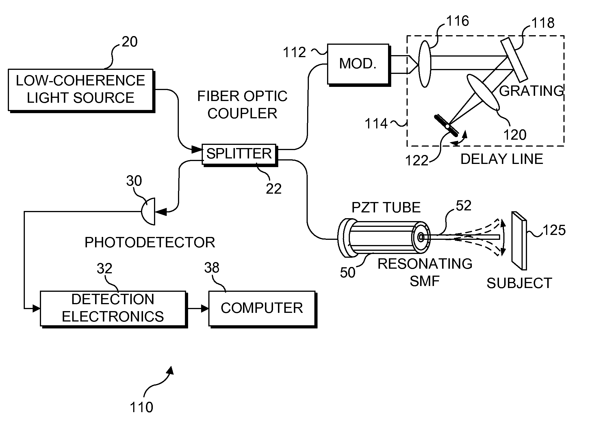
[0069]An important feature of the present invention is the relatively slow change in the position of the focal point in regard to depth in a subject tissue after each transverse scan has been completed, or continuously during the transverse scanning. Accordingly, it is important to provide an acceptable focus-tracking scheme for the scanning miniature endoscope and the other embodiments of the present invention. a depth focus-tracking system 90 shown in FIG. 9 includes a micromotor 92 disposed adjacent to scanning endoscope 70 to precisely longitudinally translate the scanning endoscope assembly, i.e., the PZT actuator and imaging optics, relative to a subject that is being imaged. The working principle of a micromotor is well known in the art. For this exemplary embodiment, micromotor 92 has a 1.9 mm diameter and rotatably drives a threaded hollow shaft 94. The threaded hollow shaft has an inner diameter of more than 250 μm so that optical fiber 86 can easily pass through the open ...

embodiment 130

[0080]An embodiment 130 for varying depth focus tracking is shown in FIG. 13. Light emitted from cantilevered optical fiber 52 is focused by a lens system that includes a single lens or multiple lenses. FIG. 13 exemplifies a lens system including a lens 136 and a lens 134. Lens 136 is fixed in position relative to the distal end of cantilevered optical fiber 52, while lens 134 moves with its sliding track longitudinally, to shift the position of the focal point in a subject tissue (not shown) in the directions indicated by a double headed arrow 132. To adjust the dept focus tracking, a motor 126 is energized, rotatably driving a threaded shaft 128. Threaded shaft 128 is threaded into a shuttle nut 131, and the shuttle nut is coupled to lens 134, so that when the shuttle nut moves longitudinally, lens 134 is also moved longitudinally. Alternatively, lens 134 can be fixedly mounted in the supporting tube, and lens 136 can be coupled to shuttle nut 131, so that lens 136 is moved longit...

embodiment 186

[0083]FIGS. 16C and 16D illustrate a schematic view for an embodiment 186 comprising a side viewing endoscope 187 according to the present invention, in which a deformable membrane mirror (DMM) 193 is used with a beamsplitter 189 and employed to vary a depth of the focal point of the endoscope in the tissue (not shown) of a subject that is to the side of the endoscope, thereby providing depth focus tracking in this tissue. Beamsplitter 189 changes the optical path of the light from being along the longitudinal axis of endoscope 187, by directing the light from the DMM laterally toward the side and through window 207. A lead 201 couples DMM 193 to ground, while a lead 199 connects to an electrostatic actuator 195, which is mounted behind the DMM. A differential voltage, ΔV, of up to about 100 VDC can thus be selectively applied between the DMM and the electrostatic actuator to deflect the DMM from its planar state. Two stops 197 are included peripherally around the electrostatic actu...


PUM

PropertyMeasurementUnit
Frequencyaaaaaaaaaa
Electric potential / voltageaaaaaaaaaa
Areaaaaaaaaaaa

Abstract

An optical fiber scanner is used for multiphoton excitation imaging, optical coherence tomography, or for confocal imaging in which transverse scans are carried out at a plurality of successively different depths within tissue. The optical fiber scanner is implemented as a scanning endoscope using a cantilevered optical fiber that is driven into resonance or near resonance by an actuator. The actuator is energized with drive signals that cause the optical fiber to scan in a desired pattern at successively different depths as the depth of the focal point is changed. Various techniques can be employed for depth focus tracking at a rate that is much slower than the transverse scanning carried out by the vibrating optical fiber. The optical fiber scanner can be used for confocal imaging, multiphoton fluorescence imaging, nonlinear harmonic generation imaging, or in an OCT system that includes a phase or frequency modulator and delay line.

Description

RELATED APPLICATION[0001]This application is a continuation-in-part of a U.S. patent application Ser. No. 09 / 850,594, filed on May 7, 2001 now U.S. Pat. No. 6,975,898, the benefit of the filing date of which is hereby claimed under 35 U.S.C. § 120.GOVERNMENT RIGHTS[0002]This invention has been funded at least in part through grants from the National Institutes of Health (NIH) under Federal Reporting note 1 R21 CA96633-01 and Federal Reporting note 1 R21 CA094303-01A1 and the U.S. government may have certain rights in this invention.FIELD OF THE INVENTION[0003]The present invention is generally directed to a method and optical fiber scanner that is used for either confocal imaging, optical coherence tomography (OCT), or multiphoton excitation imaging (including multiphoton fluorescence and harmonic generation imaging), and more specifically, to a method and optical fiber scanner in which a distal portion of the optical fiber scanner is moved in a desired pattern to scan transversely ...

Claims


Application Information

Patent Timeline
IPC IPC(8): A61B6/00B29C63/00B31B7/00B65D5/56G02B6/26G02B6/32G02B6/35G02B26/10
CPCA61B5/0062A61B5/0066A61B5/0068A61B5/6852G02B6/262G02B26/10G02B21/0028G02B21/0036G02B21/006G02B21/0076B31B2217/0084G02B6/3502G02B6/32A61B3/102B31B2105/001G02B26/103G02B7/08G02B23/2423
Inventor SEIBEL, ERICLI, XINGDELIU, XIUMEI
Owner UNIV OF WASHINGTON
Learn More