

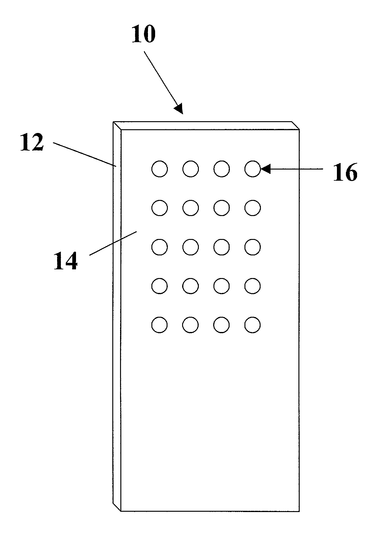
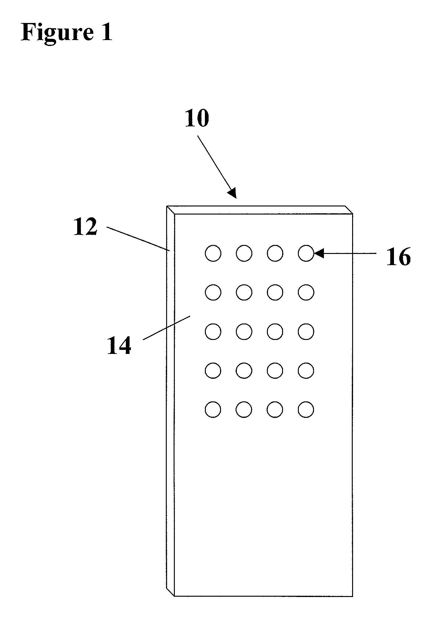
Arrays of biological membranes and methods and use thereof
a biological membrane and array technology, applied in the field of arrays of biological membranes, can solve the problems of cumbersome methods, difficult fabrication of protein arrays, and requiring the fabrication of patterned surfaces, and achieve the effect of enhancing the affinity of biological membrane microspots for substrates
- Summary
- Abstract
- Description
- Claims
- Application Information
AI Technical Summary
Benefits of technology
Problems solved by technology
Method used
Image

Examples
example 1
Materials
[0115]Membrane preparations of human β-adrenergic receptor subtype 1 (β1) and dopamine receptor subtype 1 (D1) were purchased from Biosignal Packard (Montréal, Canada). These receptor-associated membranes came suspended in a buffer solution containing 10 mM Tris-HCl, pH 7.4 and 10% glycerol. Human cloned neurotensin receptor subtype 1 (NT1R) and BODIPY-TMR-neurotensin (BT-NT) were purchased from Perkin Elmer Life Science (Boston, Mass.) and were received as membrane associated suspensions in a buffer solution containing 10 mM Tris-HCl (pH 7.4) and 10% sucrose. BODIPY-TMR-CGP12177 (BT-CGP) and BODIPY-FL-SCH23390 BF-SCH) were purchased from Molecular Probes (Eugene, Oreg.). CGP12177 and SCH23390 were purchased from Tocris Cookson, Inc (Ballwin, Mo.). Neurotensin was purchased from Sigma Chemical Co. (St. Louis, Mo.). Coming CMT-GAPS slides were used as received. The fluorescently labeled ligands and neurotensin were dissolved in DMSO and stored at −20° C. Before use, the liga...
example 2
Wheat Germ Agglutinin Surface Modification
[0127]Glass slides (Corning) were cleaned prior to use by soaking for 30 minutes in piranha etch (7:3 concentrated sulfuric acid: 30% hydrogen peroxide) followed by rinsing with distilled water. The glass slides were soaked in an ethanol solution containing 5% 3-isocyanatopropyltriethoxysilane for an hour, and then rinsed with ethanol and water, and finally dried with a flow of nitrogen. The silanized slides were used immediately for wheat germ agglutinin (WGA) coupling. The coupling was performed by soaking the slides in a solution containing 0.1 mg / ml WGA in 100 mM NaCl, 10 mM phosphate buffer, pH 7.5, followed by rinsing with water and drying. Human β-adrenergic receptor subtype 1 (β1) was purchased from Biosignal (Montreal, Canada). The receptor associated membranes were originally suspended in 10 mM Tris-HCl, 5 mM MgCl2 and 10% glycerol, pH7.4, and used directly for printing without further treatment. Eight separate grids of the β1 rece...
example 3
Family-Specific Arrays
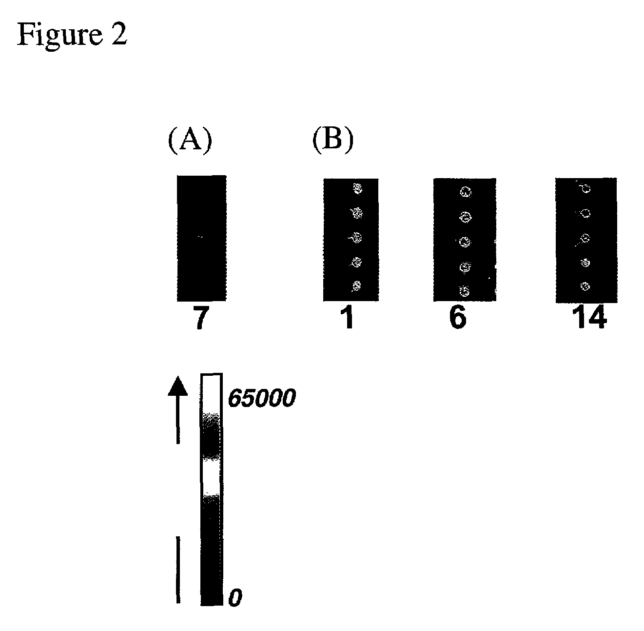
[0128]Three receptors belonging to the same family, adrenergic receptors, were arrayed within a single grid, and replicates of this grid were arrayed on a single slide. The receptors were β1, β2 and α2A, respectively. It has been reported that CGP12177 is a β1 / β2 antagonist, and a β3 partial agonist. Arrays were incubated with 5 nM BODIPY-TMR-CGP12177 in the absence and presence of varying concentrations of ICI118551. As illustrated in FIGS. 9A, 9B and 9C, only those spots corresponding to the β1 and β2 receptors light up. In the presence of 10 nM ICI118551 (a selective β2 agonist), the fluorescence intensity of the β2 receptors decreases, while the intensity of the β1 receptors remains almost at the same level. However, as the concentration of ICI118551 increases to 500 nM, the fluorescence intensity of β1 dramatically decreases, while the intensity of β2 is similar to the intensity observed in the presence of 10 nM ICI118551 These results illustrate that ICI1...
PUM
| Property | Measurement | Unit |
|---|---|---|
| contact angle | aaaaa | aaaaa |
| thick | aaaaa | aaaaa |
| thickness | aaaaa | aaaaa |
Abstract
Description
Claims
Application Information