[0020]The same advantages described above for internal multiphoton nonlinear spectroscopy through optical fibers penetrating a body cavity or tissue can be advantageously applied to probe other penetrable materials that are, or can be made, fluorescent, or those that generate nonlinear scattering signals or other nonlinear signals. Some examples of such other uses include probing materials such as food products, natural or manmade polymeric structures (e.g., collagen gels or scaffolding), or porous media, and rapid in vitro screening applications utilizing fluorescence. The methods of the present invention may also be coupled with high-throughput assays to identify potential drug candidates. The penetrable body of material may include non-liquid and liquid samples. The advantage in the latter example is that multiphoton illumination allows for efficient simultaneous excitation of different fluorophores using IR light (Xu et al., PNAS 93(20): 10763, (1996), which is hereby incorporated by reference in its entirety), eliminating the need for multiple excitation sources. Furthermore, the high NA (typically>0.6) of the microstructured fibers would allow for much more efficient collection of signal back through the fiber than possible with a conventional single mode fiber (NA<0.2) since collection scales with the fourth power of the NA (i.e., NA2).
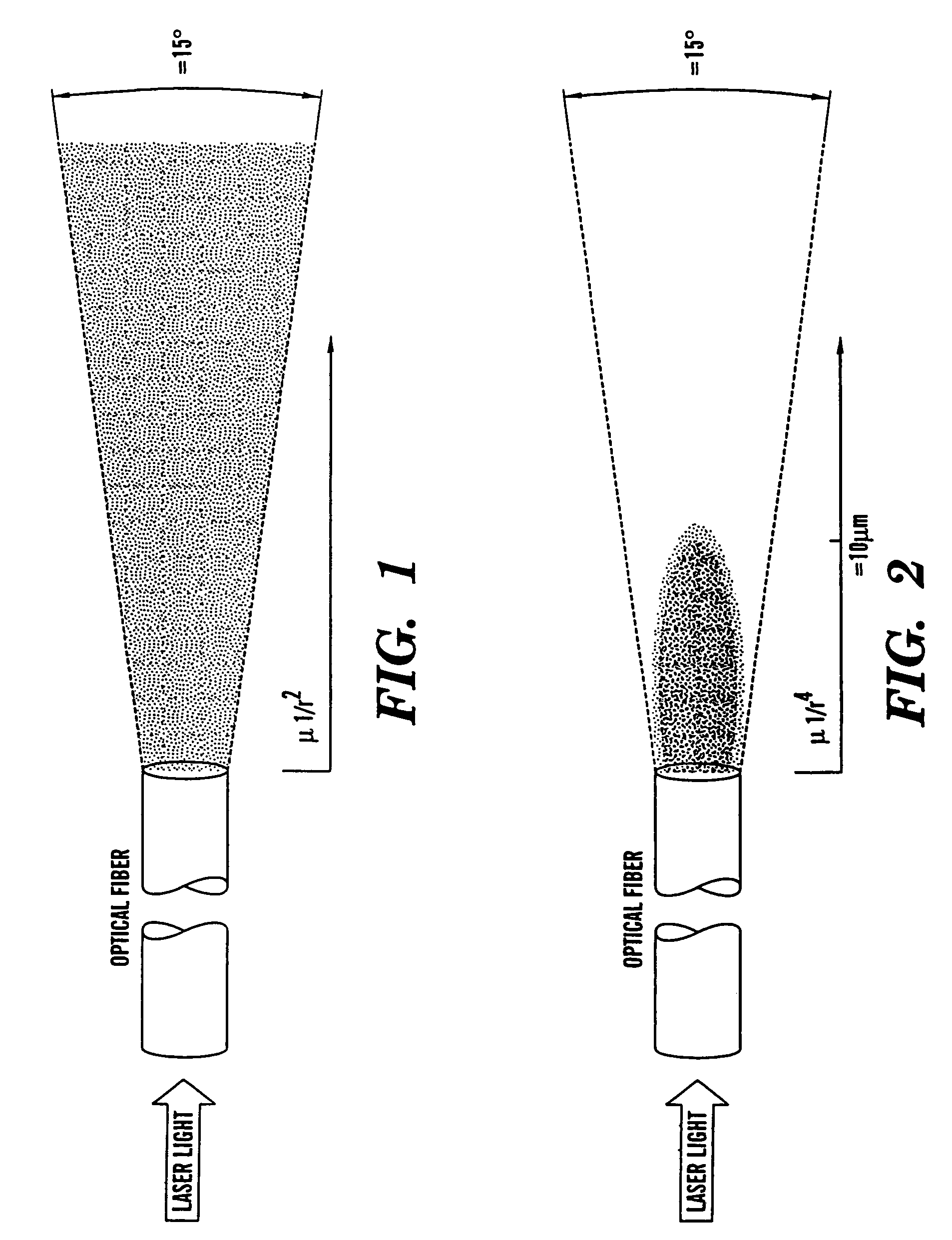
[0021]One important advantage of multiphoton excitation is that the illumination is not strongly absorbed by hemoglobin and myoglobin and, in fact, not strongly absorbed by any other common tissue components. Linear scattering of infrared light by tissue is also significantly less than scattering by ultraviolet wavelengths. The principal advantage of multiphoton excitation, however, for endoscopic fluorescence and nonlinear scattering spectroscopy is that the effective focal volume within which the sample is activated is well defined and highly localized (FIG. 2). The reason for this is that the rate of two-photon excitation of fluorescence or for generation of second harmonic signals is proportional to the square of the illumination intensity. For higher multiphoton processes, the power law exponent of the intensity is larger, cubic for three-photon excitation for example. In multiphoton laser scanning microscopy, this higher power law feature makes possible three-dimensional resolution without generating out-of-focus fluorescence that would have to be excluded by confocal spatial filtering.
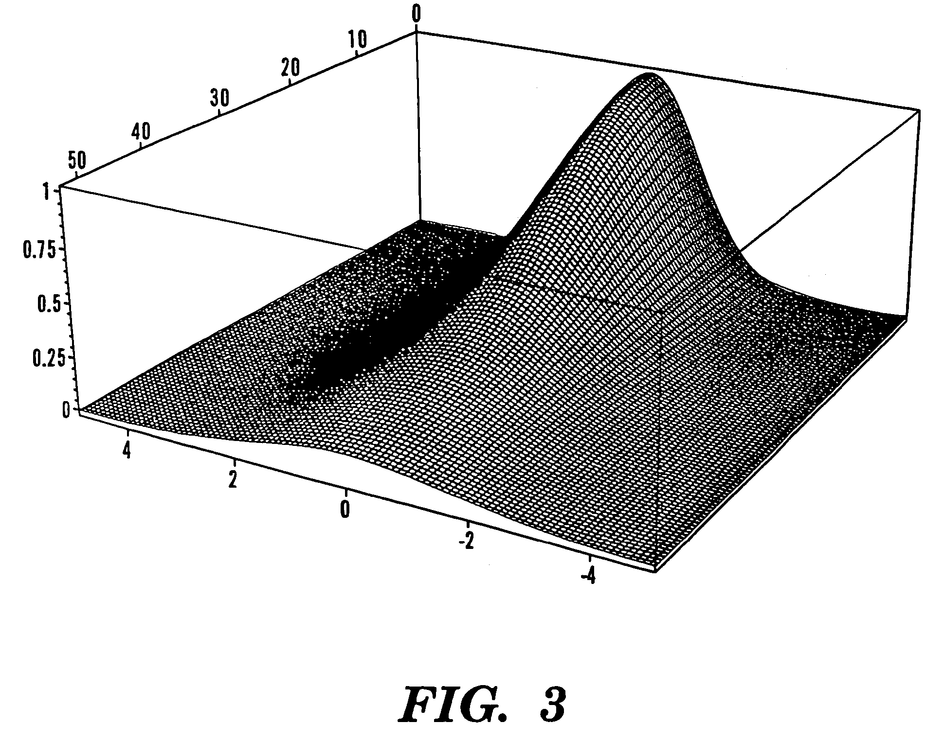
[0023]An analogous advantage applies to fluorescence multiphoton excited by laser light transmitted through an optical fiber as in application to endoscopic tissue fluorescence. Although the illumination intensity fans out just as for one-photon excitation with roughly equal total power at each value of radius from the end of the fiber or focal plane of any focusing lens, the excited fluorescence does not follow this fanned out illumination. In the case of two-photon excitation through a single mode optical fiber, the distribution of fluorescence is localized near the fiber tip in a shape resembling a candle flame beginning at the fiber tip. This effective focal volume is defined by the spatial distribution of the square of the illumination intensity for two-photon excitation. The square of the illumination intensity falls off roughly as the reciprocal fourth power ofthe distance from the fiber tip so that the fluorescence excitation is localized. Significant fluorescence is emitted only from this limited volume where the square of the excitation intensity is large, as illustrated in FIG. 3. Lenses, prisms, etc. can be used to shift the effective focal volume beyond the end of the optical fiber and / or to provide a side-looking orientation without losing the advantages of multiphoton excitation.
[0024]This effect provides a well-defined focal volume for the fluorescence excitation and allows useful spatial resolution which is sharp enough to resolve important anatomical structures. For example, in the colon, about 5 distinct layers should be distinguishable. At the surface of the endothelium, an array of crypts covers the area and is terminated in a cellular layer that closes the bottoms of the crypts, followed by several more layers including smooth muscle and connective tissue for a total of about 0.5 mm. These layers are readily resolved by multiphoton laser scanning microscopy exciting the intrinsic tissue fluorescence, which differs from layer to layer. Such layers and their perturbations by disease near the surface are thus distinguishable in the intact tissue by endoscopic spectroscopy of the tissue fluorescence with sufficient spatial resolution as provided by multiphoton excitation.
[0025]The multiphoton excitation of the present invention allows accurate spatial discrimination and permits quantification of fluorescence from small volumes whose locations are defined in three dimensions. This is especially important in cases where thicker layers of cells are to be studied. In this case, the fiber can penetrate the tissue to observe and resolve the multiphoton excited fluorescence of deeper layers, thus providing optical biopsy in situ. Furthermore, multiphoton excitation greatly reduces the background fluorescence and scattering artifacts.
[0026]The present invention incorporates a scheme that allows for simultaneous delivery of high-power sub-picosecond laser pulses and for collection of the generated fluorescence through the same large-core, high numerical aperture, microstructured optical fiber. Although these fibers are multimode, they are able to effectively propagate the pulses primarily in the single fundamental mode. Using only simple dispersion compensation at the input of the system, the large area of the fiber results in a small effective nonlinearity which allows for delivery of high peak-power laser pulses such that at the output of the fiber the pulse duration is approximately equal to that of the input pulse into the system. The high numerical aperture of the fiber permits efficient collection of fluorescence that is produced as a result of excitation at the output of the system by the sub-picosecond laser pulse. Such a system can effectively operate with input pulses over a large wavelength range spanning the visible and infrared regimes. The relatively large core-cladding index contrast also allows for the straightforward coupling of the input pulses from the laser source into the fundamental mode of the microstructured fiber. Such a system could prove to be extremely useful for applications such as in vivo multiphoton microscopy and endoscopy that require delivery of femtosecond pulses.