Stromal collagen in the diagnosis and characterization of breast cancer

a breast cancer and stromal collagen technology, applied in the field of stromal collagen in the diagnosis and characterization of breast cancer, can solve the problems of low two-photon absorption, radiation decay, fluorescence emission, etc., and achieve the effects of enhancing collagen signal, enhancing radial alignment pattern, and assessing the occurrence or potential of metastasis

Active Publication Date: 2010-09-28
WISCONSIN ALUMNI RES FOUND
View PDF11 Cites 16 Cited by
  • Summary
  • Abstract
  • Description
  • Claims
  • Application Information

AI Technical Summary

Benefits of technology

[0030]In an embodiment, a first, second, or third reference level can each independently relate to a reference tissue having a normal condition or a disease condition at a particular disease stage or historical time point. For example, a first, second, or third reference level can be a previously assessed level from the same or different test sample from the same patient or a different patient. When the reference level is derived from the same patient, there can be a particular diagnostic advantage in such level serving as an internal control, whether or not temporally synchronized with the test sample. In the specific example where a historical reference level itself reflects a disease state, the reference level can serve to evaluate the present test sample while also providing other information, e.g., regarding the level or rate of change. When the reference level reflects a disease state, there can be a diagnostic advantage in facilitating a comparison of the reference image with the test image to accurately assess the test sample. In a preferred embodiment, greater diagnostic information is achieved by accruing and evaluating multiple reference levels.
[0034]The methods of the present invention optionally include the step of quantitatively evaluating one or more tumor-associated collagen signatures identified in test images and test data to provide information relevant to the diagnosis of cancer. For example, some embodiments further comprise the step(s) of measuring the density, concentration, deposition, organization, and / or localization of collagen in a region having enhanced collagen signal, the concentration of collagen fibers having a taut configuration, the extent of tautness of collagen fibers, the degree of enhanced radial alignment, the proximity of taut and / or radially aligned collagen fibers to a tumor or precancerous tissue or any combination of these parameters so as to assess the state, composition, physical properties and invasiveness of cancerous or precancerous tissues in the test sample. Methods of the present invention optionally include the step of correlating the presence and extent of two or more tumor-associated collagen signatures identified in a test image or test data to provide enhanced diagnostic information.
[0036]In another aspect, the invention provides a method of diagnosing breast cancer with an improved accuracy, precision, and / or timing relative to a conventional breast cancer screening and / or diagnostic techniques. In an embodiment, the improved timing relates to an ability to detect cancer or a suspected cancer condition at an early stage of development.

Problems solved by technology

In two photon fluorescence excitation techniques, for example, absorption of two lower energy photons simultaneously excites an electronic transition in a fluorophor, thereby causing radiative decay resulting in fluorescence emission of a single higher energy photon.
As the probability of two photon absorption is relatively low (for example, as compared to single photon absorption), excitation in this technique is limited to a spatially confined focused region of the excitation beam having a sufficiently high intensity of photons.
Although the potential for such applications, including endoscopy and optical biopsy applications, is clear, these techniques have not yet matured to the point so as to provide a robust clinical tool.

Method used


Image

  • Stromal collagen in the diagnosis and characterization of breast cancer
  • Stromal collagen in the diagnosis and characterization of breast cancer
  • Stromal collagen in the diagnosis and characterization of breast cancer

Examples


example 1

Collagen Reorganization at the Tumor-Stromal Interface Facilitates Local Invasion

I. Abstract

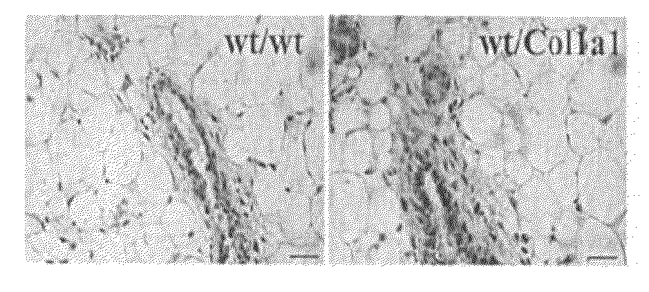
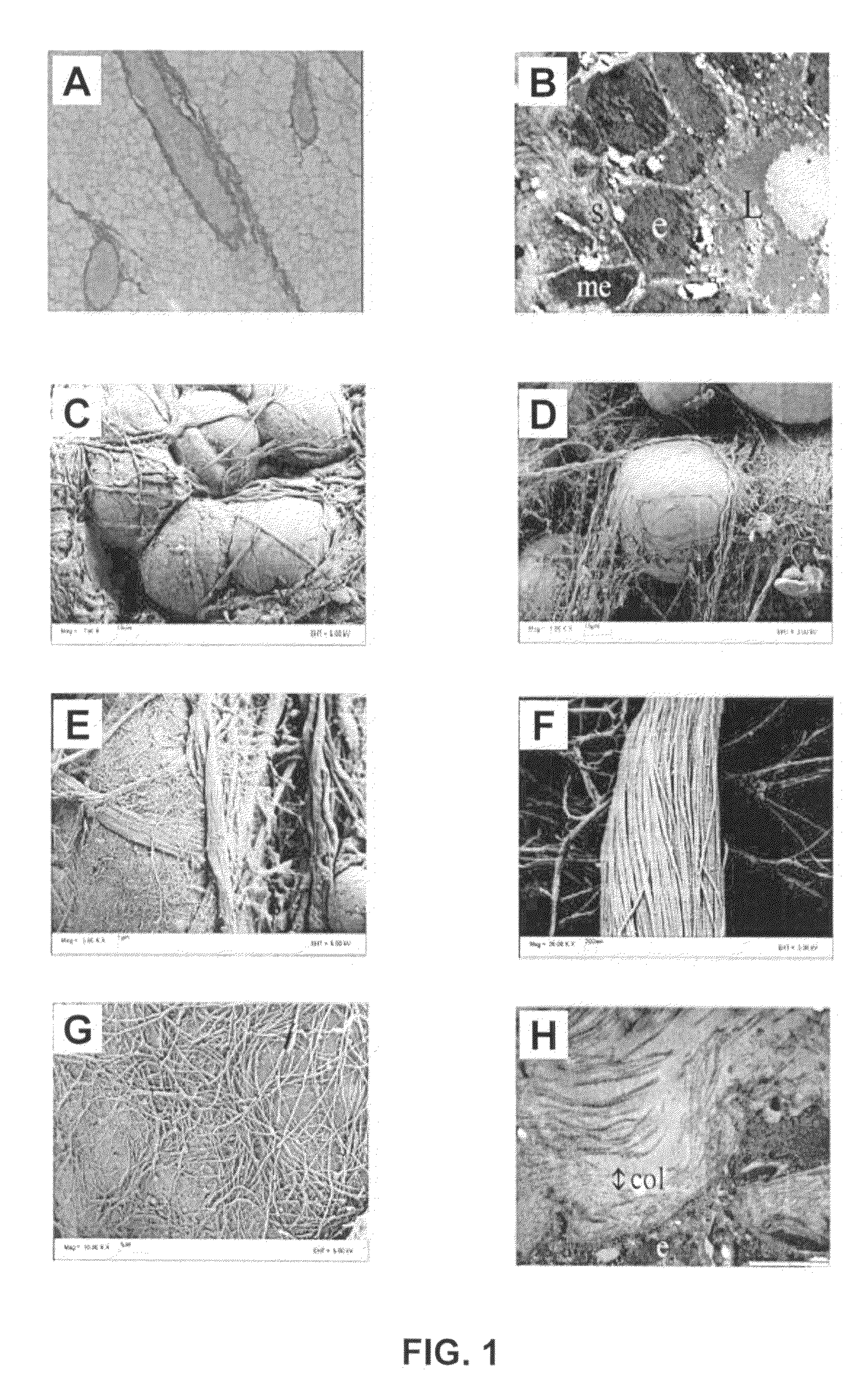
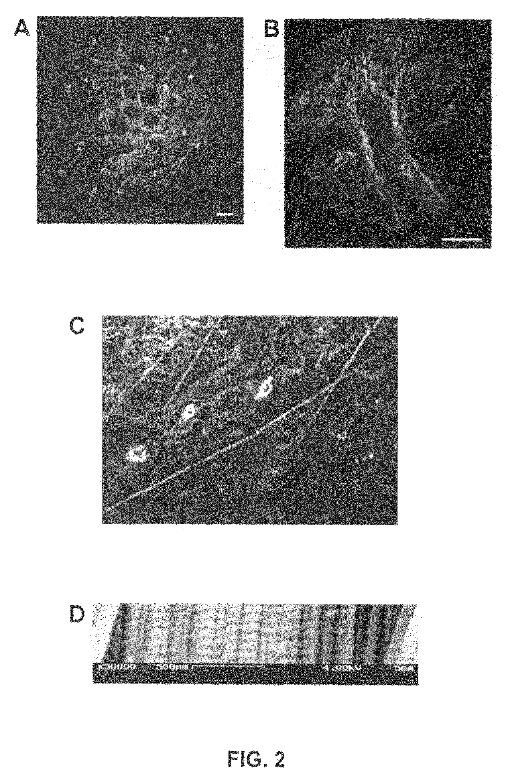
[0087]Stromal-epithelial interactions are of particular significance in breast tissue since misregulation of these interactions can promote tumorigenesis and invasion. Moreover, collagen-dense breast tissue increases the risk of breast carcinoma, although the relationship between collagen density and tumorigenesis is not well understood. As little is known about epithelial-stromal interactions in vivo, it is necessary to visualize the stroma surrounding normal epithelium and mammary tumors in intact tissues to better understand how matrix organization, density, and composition affect tumor formation and progression. In this example, we use both laser-scanning multiphoton and second harmonic microscopy to determine the organization of specific collagen structures around ducts and tumors in intact, unfixed and unsectioned mammary glands. Local alterations in collagen density are clearly seen, a...

example 2

Nonlinear Optical Imaging Systems for Detecting Cancer

[0106]It is a goal of the present invention to provide nonlinear optical imaging systems capable of generating information relevant to the diagnosis of cancer in biological materials, such as intact, unsectioned and unstained tissues. Useful imaging systems for some applications are noninvasive and nondestructive, and are capable of providing high resolution images of surface and subsurface components and features of tissue. Useful imaging systems are also capable of rapid in situ optical analysis, for example to provide real time imaging for facilitating therapeutic procedures, such as surgical removal of tumors and for quantitative analysis of the margins of a given tissue removal therapy.

[0107]In one embodiment, the present invention provides an optical system capable of generating, simultaneously or non-simultaneously (e.g. sequentially) both MP laser-scanning microscopy images and harmonic generation (i.e., first, second, th...

example 3

Nonlinear Optical Imaging of Cellular Processes in Breast Cancer

[0114]Nonlinear optical imaging techniques such as multiphoton and second harmonic generation microscopy used in conjunction with novel signal analysis techniques such as spectroscopic and fluorescence excited state life-time detection have begun to be used widely for biological studies. This is largely due to their promise to non-invasively monitor the intracellular processes of a cell together with the cell's interaction with its microenvironment. Compared to other optical methods these modalities provide superior depth penetration and viability and have the additional advantage in that they are compatible technologies that can be applied simultaneously. Therefore, application of these nonlinear optical approaches to the study of breast cancer holds particular promise as these techniques can be used to image exogenous fluorophores such as GFP as well as intrinsic signals such as second harmonic generation from collage...


PUM


Abstract

The present invention provides methods and systems for evaluating biological materials for the diagnosis of disease, such as gland abnormalities, and the cancerous and precancerous conditions. Nonlinear optical microscopy techniques, such as MP microscopy and harmonic generation microscopy, are used to generate high resolution, three dimensional images of a test tissue, such as a biopsy tissue sample and tissue in whole organisms, that are analyzed, optionally in combination, to detect, identify and characterize tumor-associated collagen signatures. The presence, abundance and extent of histological features and structural motifs comprising tumor-associated collagen signatures may be directly and accurately correlated with the onset and progression of cancer, such as breast cancer. The present methods are capable of providing an accurate and selective diagnosis of cancer, and provide diagnostic information complementary to conventional diagnostic methods.

Description

CROSS-REFERENCES TO RELATED APPLICATIONS[0001]This application claims priority under 35 U.S.C. 119(e) to U.S. provisional Patent Application 60 / 805,566, filed Jun. 22, 2006, and U.S. Provisional Patent Application 60 / 892,687, filed Mar. 2, 2007, which are both hereby incorporated by reference in their entireties.STATEMENT ON FEDERALLY SPONSORED RESEARCH OR DEVELOPMENT[0002]This invention was made with government support awarded by the following agencies: NIH grants CA076537 and EB000184 and Department of Defense Breast Cancer Research Program Grant Number BC031277. The United States government has certain rights in the invention.REFERENCE TO SEQUENCE LISTING, A TABLE, OR A COMPUTER PROGRAM LISTING COMPACT DISK APPENDIX[0003]Not ApplicableBACKGROUND OF THE INVENTION[0004]Biomedical imaging plays a central role in a large number of diagnostic and therapeutic procedures including visualizing external and internal anatomical and physiological structures, features, and systems; evaluatin...

Claims


Application Information

Patent Timeline
Patent Type & Authority Patents(United States)
IPC IPC(8): A61B6/00G01J3/00
CPCA61B5/0091A61B5/4312A61B6/502G06T7/0012A61B5/726G06T2207/10056G06T2207/30068G06T2207/30096G06T2207/30101A61B5/0068
Inventor KEELY, PATRICIA JOPROVENZANO, PAOLO P.WHITE, JOHN GRAHAMELICEIRI, KEVIN WILLIAM
Owner WISCONSIN ALUMNI RES FOUND
Why Eureka
Try Eureka