

Embryo modification and implantation
a technology which is applied in the field of embryo modification and implantation, can solve the problems of significant financial and emotional cost of each individual ivf treatment, ivf derived embryos might be compromised, and the practice of oocyte in vitro maturation (ivm) is as yet clinically unaccepted
- Summary
- Abstract
- Description
- Claims
- Application Information
AI Technical Summary
Benefits of technology
Problems solved by technology
Method used
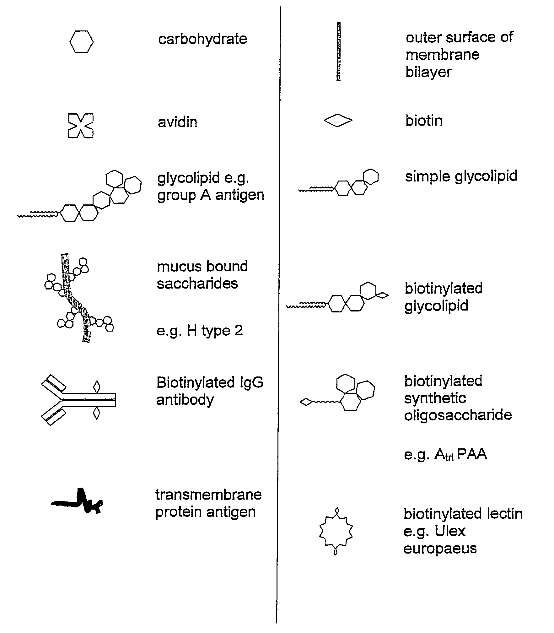
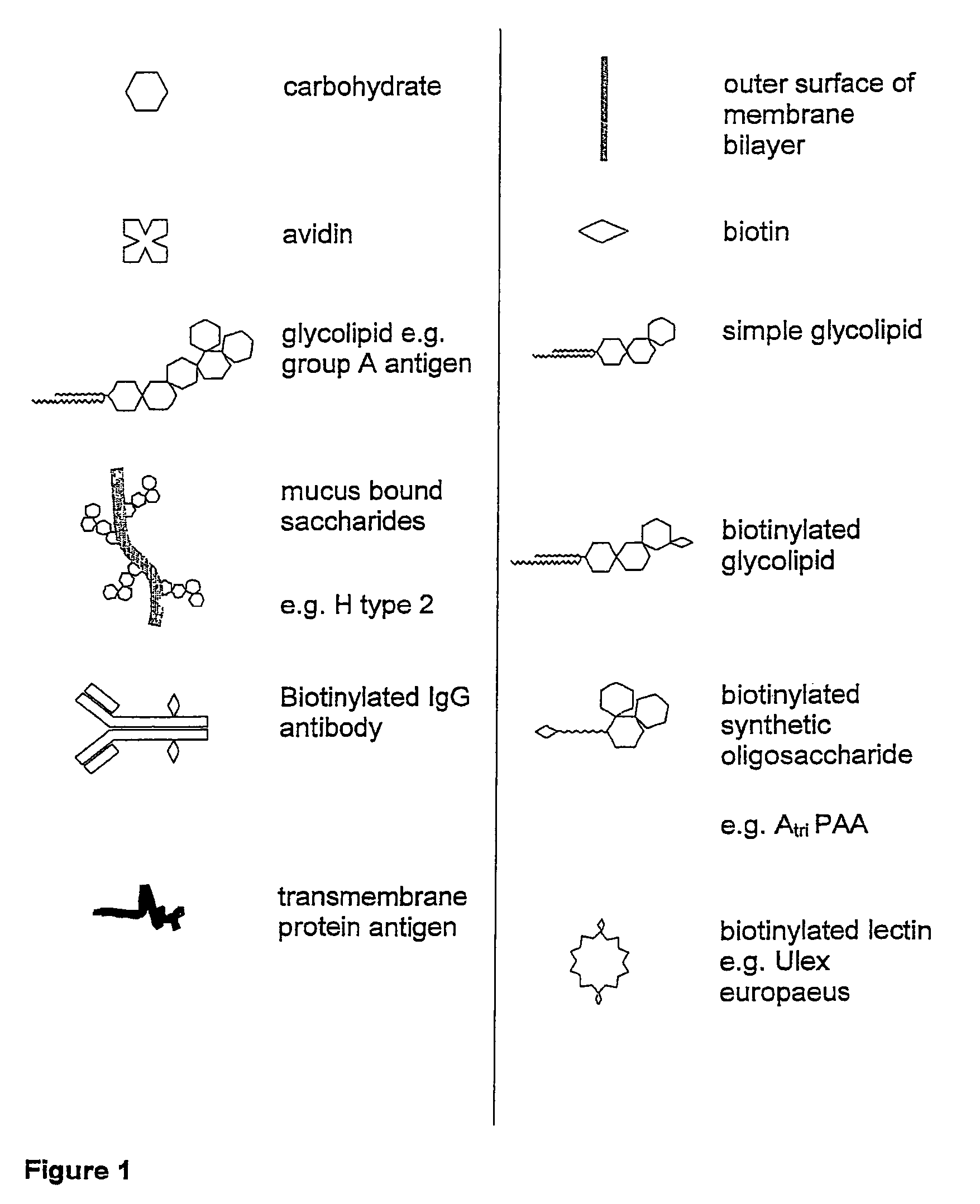
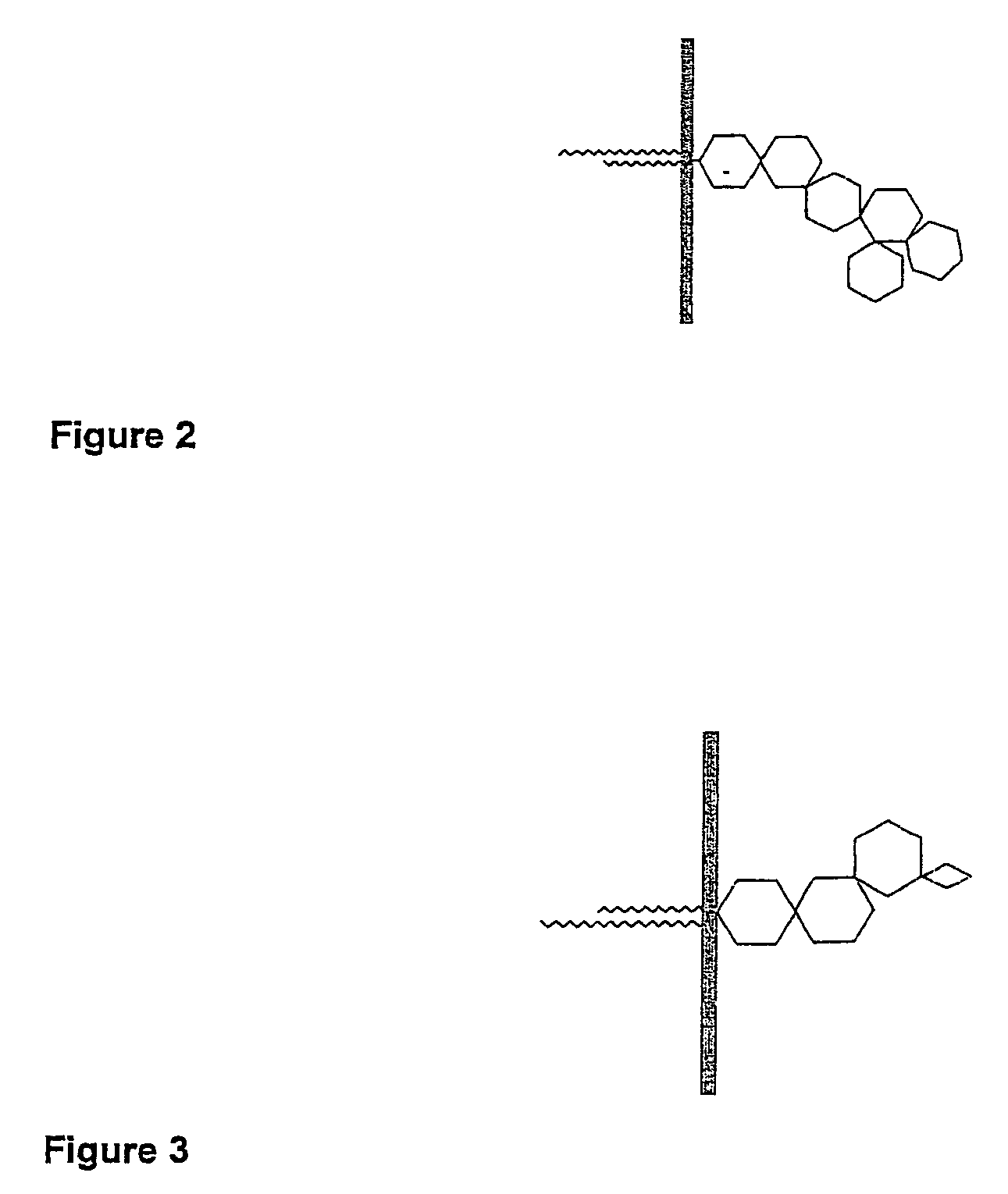
Image

Examples
example 1
[0116]Biotinylated gangliosides (BioG) were prepared using a modified procedure described by Wilchek and Bayer (1987). The extraction and purification of porcine gangliosides is carried out using established techniques (Karlsson 1987, Ladisch et al. 1987, Ledeen et al. 1982).[0117]1. Dried gangliosides purified from porcine brains, were reconstituted in PBS with the aid of sonification.[0118]2. The ganglioside sialic residues were oxidized by the addition of sodium m-periodate.[0119]3. The solution was subjected to 24 hr dialysis to remove the resulting peroxide.[0120]4. The oxidised ganglioside was incubated with biotin amidocaproyl hydrazide (Sigma B-3770) for 1 hr.[0121]5. The solution was subjected to further overnight dialysis in water to remove excess biotin amidocaproyl hydrazide.[0122]6. The resulting solution was dried via rota evaporation and reconstituted in 50% methanol water. Further evaporation was performed under nitrogen gas in a reduced pressure desiccator overnight...
example 2
[0123]Biotinylated immunoglobulin G was prepared using a method described by O'Shannessy 1990). Using similar procedures to those outlined in Example 1, the IgG was oxidised with a periodate solution and incubated with biotin amidocaproyl hydrazide.
example 3
[0124]Optimum BioG insertion concentrations and conditions were established by labelling the inserted BioG RBCs with avidin-FITC. The results are outlined in Table 1.
[0125]
TABLE 1Fluorescent signal of human RBCs inserted with arange of BioG and labelled with avidin-FITC concentrations.Avidin-FITCBioG mg / mlmg / ml1052.500.90+++++++++++−0.45+++++++++++−0.33+++++++++++−0.16+++++++++++−0−−−−
[0126]The optimum insertion concentration of BioG was 5 mg / ml. The minimum concentration of avidin required for adequate detection of BioG at 5 mg / ml concentration, was 0.16 mg / ml. The optimum minimum insertion time was determined to be 1 hour as seen in Table 2.
[0127]
TABLE 2Fluorescent signal of human RBCs incubated with BioG fora variety of times, then labelled with avidin-FITC.Hours of incubation0.250.50.7511.524626+++++++++++++++++++++++++++++++++
[0128]The amount of fluorescent signal score for the 2 hr incubation tube reduced from 4+ to 3+ after avidin labelling 5 days post insertion, suggesting m...
PUM
| Property | Measurement | Unit |
|---|---|---|
| molecular mass | aaaaa | aaaaa |
| concentration | aaaaa | aaaaa |
| concentration | aaaaa | aaaaa |
Abstract
Description
Claims
Application Information