Method and apparatus for performing transgastric procedures

a transgastric and ophthalmology technology, applied in the field of minimally invasive approaches, can solve the problems of inaccurate in many instances, no known non-invasive method that can directly measure the pressure, and no simple non-invasive way of directly measuring the left atrial pressur

Inactive Publication Date: 2011-12-06
MICROACCESS
View PDF10 Cites 4 Cited by
  • Summary
  • Abstract
  • Description
  • Claims
  • Application Information

AI Technical Summary

Benefits of technology

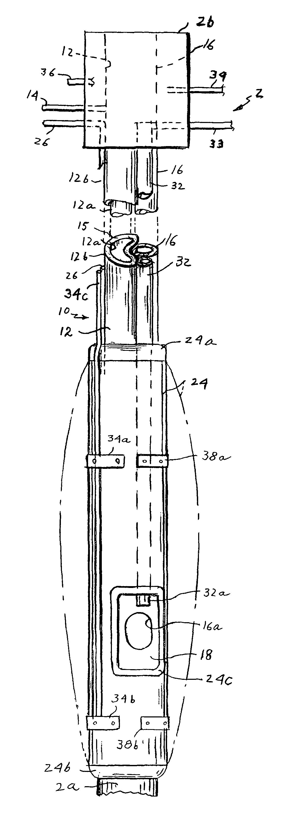
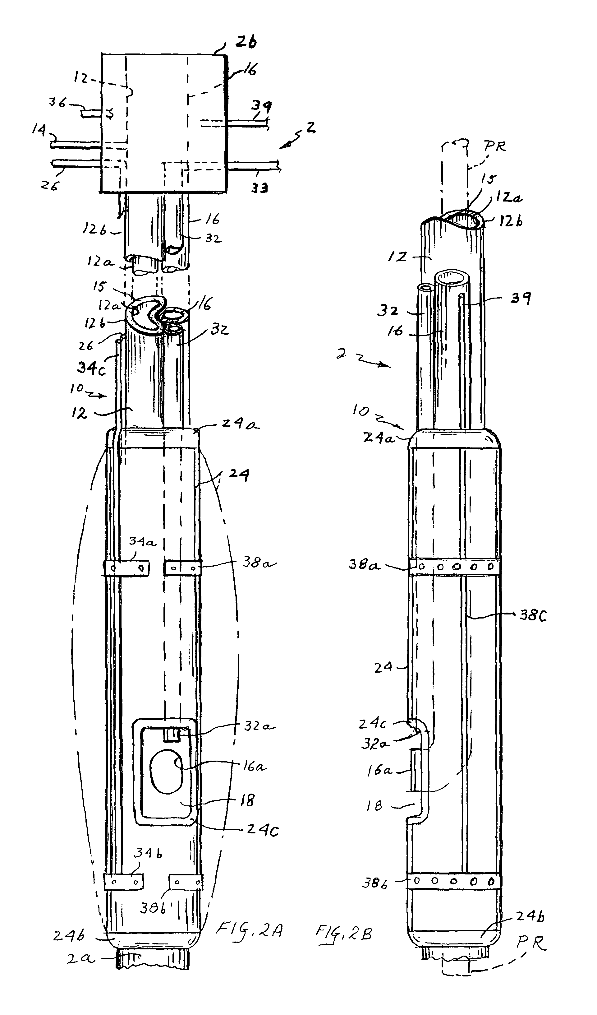
[0027]A second lumen of the AMU is the transesophageal access lumen (TAL) which is a longitudinal lumen that extends from the proximal end of the AMU and travels along its length to make a 90° angle at its lower end and exits perpendicular to the main axis of the AMU in the center of the AEF. The TAL carries elongated flexible devices including an access side unit (ASU) that penetrates through the esophageal wall and creates an access to the desired target area outside the esophagus. A third tubular lumen inside the AMU is the suction lumen (SL) used to create a negative pressure in the space of the AEF between the outer surface of the AMU and the esophageal mucosa. The SL also allows for suctioning of any secretions that may escape into the AEF.
[0031]The apparatus also includes a transesophageal intracardiac access (TIA) which is similar in structure to the ASU but smaller in diameter to fit inside the larger lumen of the ASU. It can create an opening of 6 mm or more in a cardiac chamber. The TIA allows the passage of elongated flexible devices to the interior of the heart to perform various procedures. In a preferred embodiment, the TIA uses an umbrella at its distal end to seal the penetration through the cardiac chamber wall and to make a purse string suture that is used at the end of the procedure to secure the penetration site. In another embodiment, the sealing means at the distal end of the TIA can be an inflatable balloon, double balloons, or an expandable flange. The short length and wide diameter of the TIA allow for the precise positionability and control of the intracardiac devices.
[0033]The TIA may also include a valve for hemostasis in the inner lumen to prevent blood back flow inside the lumen of the TIA after opening a penetration to the interior of the heart. The valve may be located at the proximal end of the TIA or more preferably at the distal end thereof. The TIA can then be flushed across the hemostatic valve with normal saline to create a blood-saline interface to avoid air bubbles in the lumen of the TIA. If the TIA is positioned in a low pressure chamber like the right or left atrium, the back-pressure of the blood my be low enough to obviate the need for a valve mechanism in the blood-saline interface. After the placement of the TIA inside a cardiac chamber, preferably the left atrium, a wide, short and almost straight access is created from outside the body across the esophagus into the left atrium that allows various devices to be described to perform diagnostic and therapeutic procedures.

Problems solved by technology

There is no known non-invasive method that can directly measure the pressure in any chamber of the heart.
Unfortunately, there is no simple non-invasive way of directly measuring the left atrial pressure.
Even with invasive measurement as in pulmonary artery catheterization, the measured value reflects an indirect estimation of the left atrial pressure, and thus can be inaccurate in many instances.
Both techniques have their inherent side effects and complications.
The invasiveness of the current techniques limits the early implementation of a shunt closure especially in children, which is a curative intervention if done before irreversible vascular changes.
Other techniques use the catheter transvascular approach with limited success due to lack of control and torque at the end of a long flexible, narrow catheter used in the procedure.
Chronic anticoagulation in itself carries serious risk of internal bleeding added to the toxicity of chronic anti-arrhythmic medications used to stabilize atrial fibrillation.
The invasiveness of the open chest approach has limited the number of the Maze-like procedures used to radically prevent the fibrillation impulses from being conducted to the ventricles.
Also, the catheter-based approach is inaccurate, tedious, time consuming (up to 12 hours) and not definitive in creating enough linear ablations to prevent impulse conduction.
The thoracoscopic approach is easier than the catheter-based transvascular approach but the side access to the posterior heart limits the linearity of ablation especially around the entrance of the pulmonary veins, which results in incomplete Maze, and recurrence of disease.
The three known accesses to the heart namely, the open chest, the catheter-based transvascular, and the thoracoscopic approaches suffer from serious limitations and complications which, in turn, limit the therapeutic options for most of patients.
First, the risk of stopping the circulation with the possibility of causing marked decrease in tissue perfusion or ischemic damage that may involve vital tissues like the brain, heart or kidneys.
Second, the risk of embolization of dislodged tissues in the aorta due to aortic manipulation including clamping and catheterization.
Third, opening the chest wall by cutting through all layers including the bony sternum with great force applied for rib retraction produces significant pain after the surgery with post surgical morbidity and, if severe, mortality.
The post surgical wound care and pain may require rehabilitation in complicated cases with longer hospital stays and increased expenses.
Fourth, concomitant morbid states or age extremes may adversely increase all of the above-mentioned risks.
Again, these procedures have fundamental disadvantages.
For the transvascular approach to the treatment of heat defects, the disadvantages are inherent in the fact that the procedural tools have to go through and stay in a blood vessel or a cardiac chamber.
Thus, the tools can only be long, narrow, flexible catheters.
This affects the controllability and the force generation at the tip of the catheter which, in turn, decreases the accuracy and precision of the procedure.
To reach the left heart, a septostomy opening is made in the interatrial septum that decreases the control over the catheter and makes the manipulation of the catheter tip more difficult as the catheter has to pass through the narrow right atrium and the small septostomy opening.
Another main limitation is the caliber of the lumen of peripheral vessels through which the catheter has to travel.
This is even a more limiting factor in young children whose smaller vessels raise the risk of vessel injury that may be acute, such as intimal dissection, or chronic such as major vascular obliteration and fibrosis.
In the elder population, the occurrence of peripheral fat embolization is another risk for manipulations of a catheter in the aortic lumen for procedures like coronary angiography.
This may result in temporary acute or chronic ischemia to the lower or upper extremities.
The small size of the vessels limits the size of catheters used in ablation techniques.
That limitation gives rise to a lack of control and positionability due to the flexibility, increased distance and decreased force at the tip of such catheters.
The transvascular approach has a limited use in surgical procedures like septa defect repair by using an introductory device due to the above-mentioned limitations.
In addition to the above-mentioned limitations, the use of patches to close a septal defect using the transvascular approach with lack of distal force at the delivery tip may result in inadequate fixation of the patch to the defect plus the inability of patch repositioning after its application to the defect site.
The detachment of the patch from the defect site may lead to serious patch embolization and failure of repair.
The disadvantages of such an approach can be considered.
The inaccessibility of the posterior aspect of the heart to the rigid scopes passed from either side of the heart, which is mandatory to create an atriostomy opening in the posterior aspect of the atrium, is a problem.
However, due to the narrow intercostals spaces in the anterior rib aspect, it is difficult to make such a stitch which makes the posterior aspect of the heart a difficult area to access.
Another limitation is the need to have a second monitoring system to assess catheter position and convey accurate measurements as the procedure is not performed under direct visual examination.
Such monitoring devices make the approach more complicated, may lack accurate precision, and may need to be invasive, e.g. removal of the fourth rib for visualization, or add a potential risk of ionizing radiation exposure with CT scanning, or be cumbersome and slow with MRI scanning, adding to the complexity of the technique.
Second, the limited windows between the ribs as a limiting border from above and below the access, the surrounding intercostals muscles and underlying vital structures including the lungs, pleura, nerves, and great vessels can limit the access specially in young children with narrow intercostals spaces, and patients with deformities.
The need to access multiple intercostal openings to use a plurality of thoracoscopes counteracts the main objective of the technique to be minimally invasive and may turn out to be more invasive and cause significant tissue damage as it may also require removal of the fourth rib.
The need to deflate the lung to widen the surgical field adds to the invasiveness and increases the complication potential of the procedure.
The inability to explore the posterior mediastinum and related structures on the posterior aspect of the heart as the catheter is advanced from an anterior position makes procedures involving the posterior aspect of the heart less accessible as in ablation around the entrance of the four pulmonary veins in the left atrium.
Indeed, the thoracoscopic approach has been criticized lately by a number of studies that show numerous post-surgical complications.
None of the currently available techniques directly accesses the posterior aspect of the heart and coronary circulation or the posterior mediastinum.

Method used


Image

  • Method and apparatus for performing transgastric procedures
  • Method and apparatus for performing transgastric procedures
  • Method and apparatus for performing transgastric procedures

Examples


Embodiment Construction

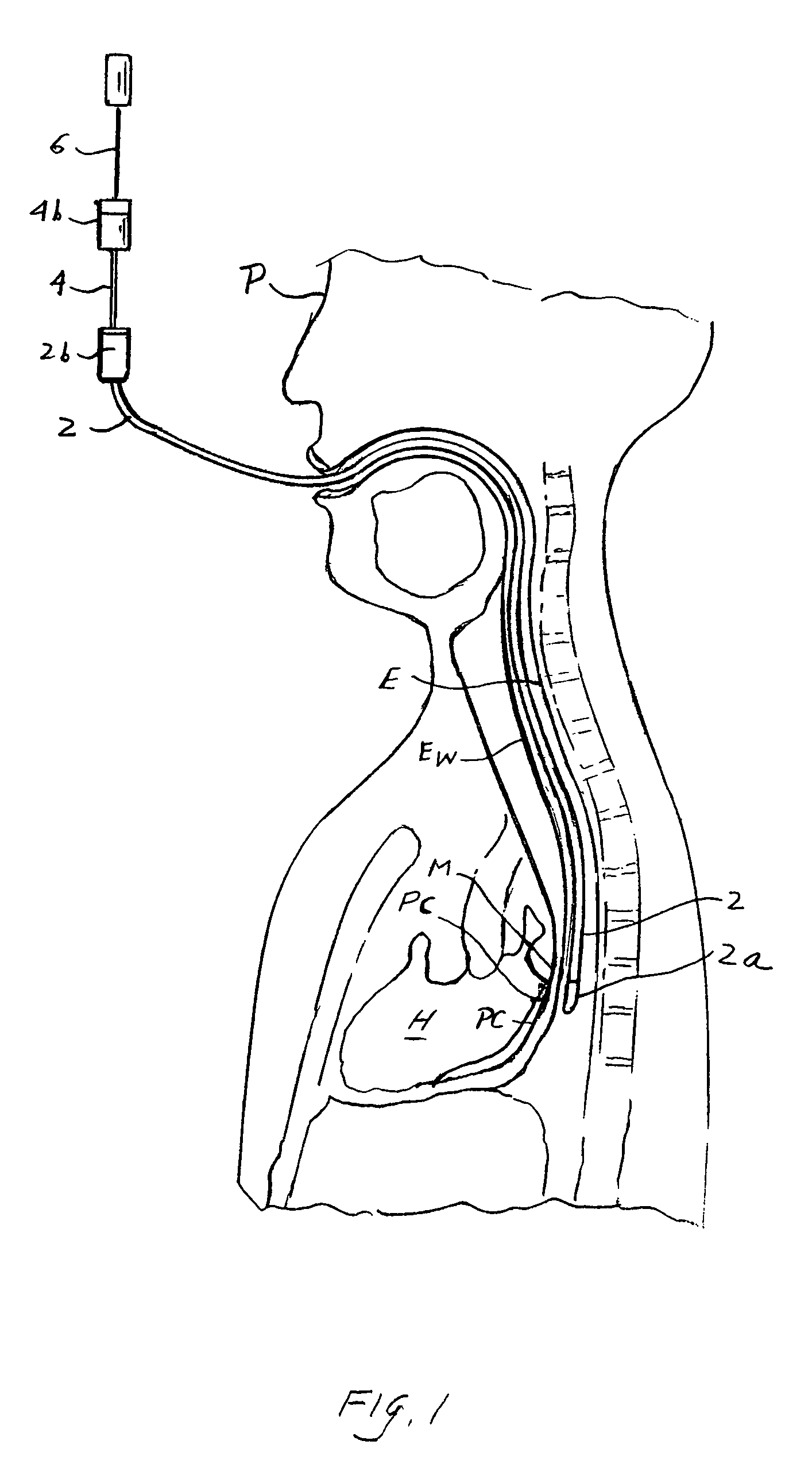
[0057]Referring to FIG. 1 of the drawings, my apparatus comprises an access main unit (AMU) 2 which may be inserted via the month into the esophagus E of a patient P so that the distal or working end 2a of AMU 2 is located behind the patient's heart H. The apparatus also includes an access side until (ASU) 4 which is received in AMU 2 and whose distal end protrudes from AMU end 2a and is designed to penetrate the esophageal wall Ew to access extraesophageal regions in the patient's body such as the posterior mediastinum M. Another component of the apparatus is a transesophageal intercardiac access device (TIA) 6 which is received in ASU 4 and whose distal end may protrude from the ASU so that it may penetrate the pericardium PC and access heart H.

1) The Access Main Unit (AMU) 2

[0058]Referring to FIGS. 1, 2A and 2B of the drawings, AMU 2 may be inserted into the lumen of the esophagus E so as to isolate a segment of the esophagus behind the heart H.AMU 2 comprises an elongated flexib...


PUM


Abstract

Apparatus for performing a transgastric procedure includes an elongated tubular main access device having the first lumen with open proximal end and a side opening distal end, and a second lumen with open ends. A balloon having a wall on the outside of the device surrounds the distal end and a fluid conduit extends along the device for inflating the balloon so that when the device is inserted into a patient's esophagus and the balloon is inflated, the portion of the esophagus opposite the distal end is isolated from the remainder of the esophagus. The apparatus may also include a side access unit having an elongated flexible coaxial inner and outer tubes having proximal and distal ends and being movable relatively in the axial direction. First seals are mounted to the distal end of the outer tube, and second seals are mounted to the distal end of the inner tube, an actuator adjacent to the proxable ends of the tubes move the tubes relatively so as to vary the axial spacing the first and second seals. Various methods for using the apparatus are also disclosed.

Description

CROSS-REFERENCE TO RELATED APPLICATIONS[0001]This is a continuation of Ser. No. 10 / 700,113, filed Nov. 3, 2003, now abandoned, which is a continuation of Ser. No. 09 / 718,642, filed Nov. 22, 2002 2000, now U.S. Pat. No. 6,689,062, which has the benefit of provisional application No. 60 / 167,147, filed Nov. 23, 1999.BACKGROUND OF THE INVENTION[0002]1. Field of the Invention[0003]The present invention relates generally to a minimally invasive approach to perform transesophageal cardiovascular and mediastinal procedures. More specifically, the invention relates to implementing the concept of transesophageal access to the heart, great vessels and related structures. The invention describes devices and methods to create a transesophageal access to the heart and surrounding structures to perform a body of surgical procedures within a beating heart and on other surrounding structures.[0004]2. Background of the Invention[0005]The access to the human heart has always been a source of active re...

Claims


Application Information

Patent Timeline
Patent Type & Authority Patents(United States)
IPC IPC(8): A61B8/00A61B8/12A61B17/00A61B17/34
CPCA61B8/12A61B18/1492A61B17/00234A61B17/3209A61B18/20A61B17/3421A61B2017/00243A61B2017/00247A61B2017/00269A61B2017/3419A61B2017/3445A61B2017/3484A61B2017/3488A61B2017/3492A61B2018/00392A61B5/285A61B5/287A61B1/32A61B5/6853A61B2018/00351A61B2018/00488A61B2018/00577
Inventor MESALLUM, SAMEH
Owner MICROACCESS
Why Eureka
Try Eureka