
Method for the treatment of valvular disease

a valvular disease and compound technology, applied in the field of medical methods and compounds, can solve the problems of calcific injury, aortic valve stenosis, difficulty in determining whether the valve is bicuspid or tricuspid, etc., to slow down or stop reverse valvular stenosis, and slow down the progression of valvular stenosis

Inactive Publication Date: 2012-04-24
MONTREAL HEART INST
View PDF65 Cites 7 Cited by
  • Summary
  • Abstract
  • Description
  • Claims
  • Application Information

AI Technical Summary

Benefits of technology

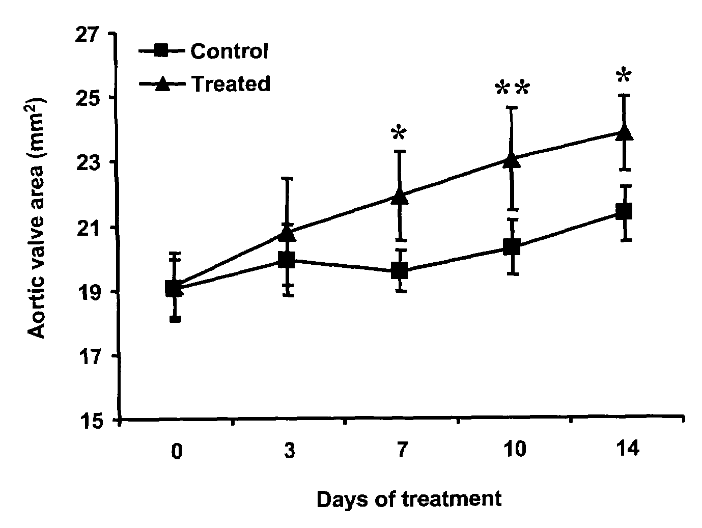
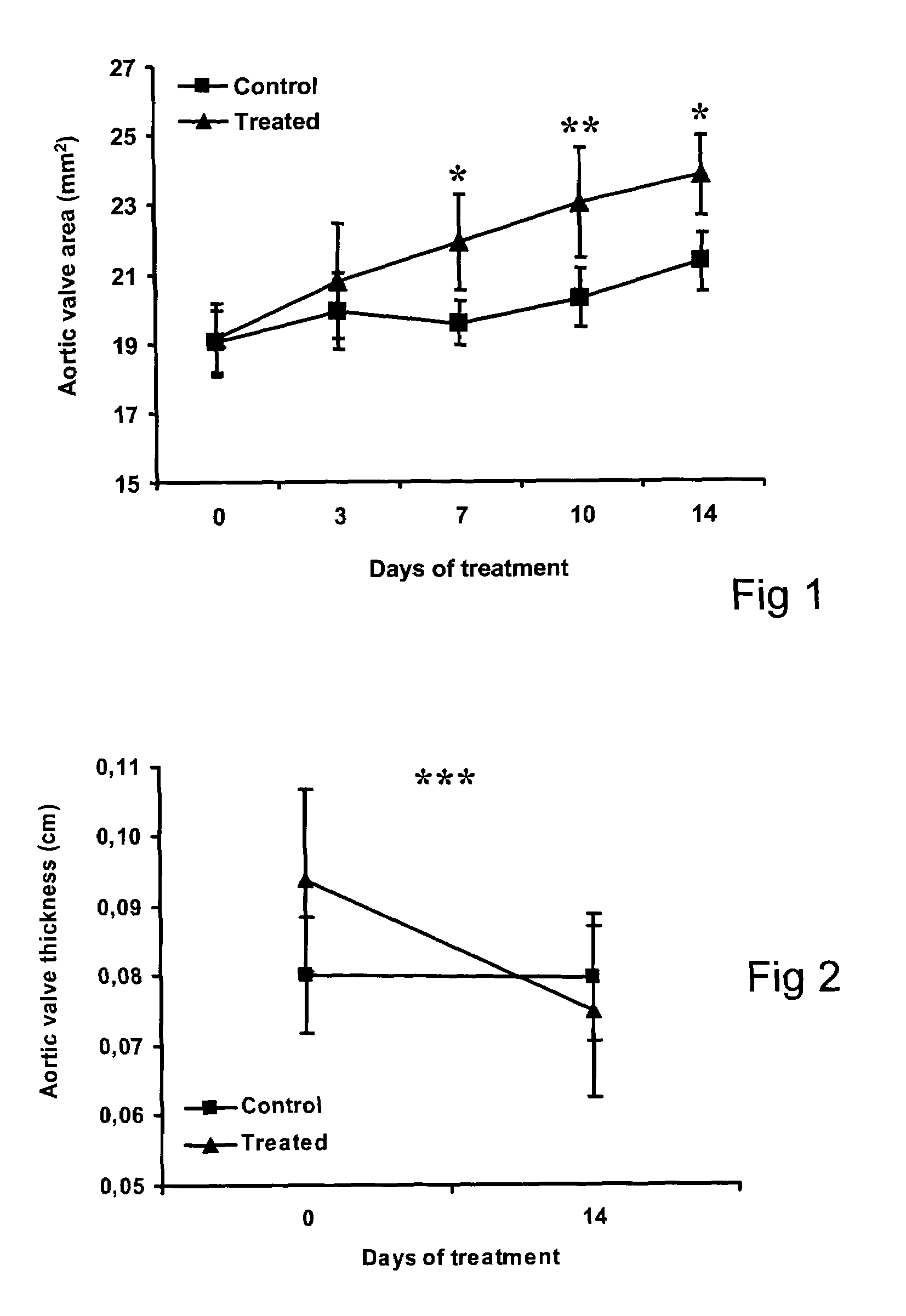
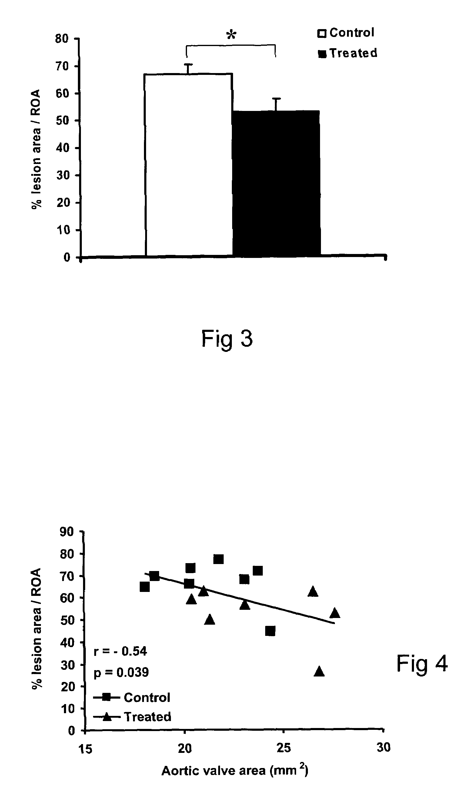
The method effectively reduces aortic valve stenosis progression and reverses existing stenosis, as demonstrated by significant improvements in aortic valve area and thickness in experimental models, with potential for regressing calcifications and improving patient outcomes without the need for invasive surgery.

Problems solved by technology

For any of several reasons, it can happen that the aortic valve is somehow damaged and may become stenosed.
Indeed, mechanical sheer stress typically leads to calcific injury.
More than 50% of adults with aortic stenosis are found to have a bicuspid valve, but fibrosis and calcification may make it difficult to determine whether the valve is bicuspid or tricuspid.
The less compliant, thickened left ventricle becomes more dependent on the atrial contribution to diastolic filling, such that left ventricular performance can deteriorate when atrial contraction is lost, for example during atrial fibrillation or atrial-ventricular dissociation.
The abnormal relaxation and increased stiffness of the thickened left ventricle during diastole also result in diastolic dysfunction and elevations of left ventricular and left atrial diastolic pressures.
Thus, significant aortic stenosis creates conditions in which high myocardial oxygen demands are inadequately supported by reduced oxygen supply, which leads to subendocardial ischemia.
Further elevation of the left ventricular end diastolic pressure (secondary to diastolic dysfunction with or without systolic dysfunction) results in pulmonary venous hypertension.
The increased myocardial oxygen demands in aortic stenosis with the underperfused subendocardial myocardium can produce angina pectoris, arrhythmias, and even sudden death.
Hence, aortic stenosis is associated not only with high mortality but also with substantial morbidity.
These nodules decrease the flexibility of the leaflets, thereby limiting their mobility and capacity to fully open.
The operative mortality for this procedure, particularly in the elderly, is relatively large.
Mechanical heart valves are durable and, hence, more likely to result in long-lasting function but require careful chronic anticoagulation because of thrombo-embolic risk.
Chronic anticoagulation therapy, however, carries with it a risk of haemorrhage similar in incidence to that of the residual risk for thrombotic events.
Such valves, however, carry a significantly higher risk of calcification than mechanical valves.
Since treatment of a functionally compromised bio-prosthetic heart valve frequently requires replacement with a new valve (and hence a second open-heart surgery), limitations on the useful life expectancy of a bio-prosthetic heart valve are both a serious medical problem for the patient and a financial drain on the medical system.
Furthermore, all prosthetic heart valves are somewhat stenotic.
Prosthetic dysfunction secondary to thrombosis or calcification can lead to increased obstruction or the development of regurgitation.
Regurgitation can also result from a perivalvular leak, that is a leak in the area of the sewing ring of the valve.
Turbulence associated with valve dysfunction can cause haemolysis and anemia.
Even normally functioning prosthetic valves can cause haemolysis in some patients.
Furthermore, some patients have aortic dimensions that are not large enough to easily accommodate conventional replacement valves.
Hence, there is a significant number of patients for whom valve replacement is impossible, impractical, or undesirable.
Nevertheless, this technique still requires the patient to be healthy enough to survive and recuperate from thoracic surgery, and involves all the costs and risks attendant with such surgery.
Although this procedure eliminates many of the risks and disadvantages attendant with the preceding two techniques, re-stenosis is very common within one year, limiting the usefulness of the technique to temporarily mitigating symptoms for those patients who are poor surgical candidates or refuse surgery.

Method used


Image

  • Method for the treatment of valvular disease
  • Method for the treatment of valvular disease
  • Method for the treatment of valvular disease

Examples


example

[0253]A complex animal model of aortic valve stenosis has been developed in rabbits. The model resulted in aortic valve stenosis characterized by a calcification similar to what is observed in a clinical setting in humans.

[0254]Methods

[0255]Animals and Experiments

[0256]An animal model adapted from that described by Drolet et al. (12) was used. Fifteen male New-Zealand White rabbits (2.7-3.0 kg, aged 12-13 weeks) were fed with a 0.5% cholesterol-enriched diet (Harlan, Indianapolis, Ind.) plus vitamin D2 (50000 UI per day; Sigma, Markham, Canada) in the drinking water until significant AVS, as defined by a decrease ≧10% of aortic valve area (AVA) or of the transvalvular velocities ratio (V1 / V2), could be detected by echocardiography (12.9±2.4 weeks).

[0257]The animals then returned to a standard diet (without vitamin D2) to mimic cholesterol lowering and were randomly assigned to receive either saline (control group, n=8) or the ApoA-1 mimetic peptide (treated group, n=7). Rabbits were...


PUM


Abstract

A method for treating valvular stenosis. The method involves the administration of a therapeutically effective amount of a reverse lipid (in particular cholesterol) transport agonist to a mammal. Most preferred is an Apolipoprotein A-1 mimetic peptide / phospholipid complex, the peptide of which is defined by the amino acid sequence of SEQ ID NO 1.

Description

[0001]This application is a U.S. national stage of International Application No. PCT / CA2007 / 000895 filed May 23, 2007, which claims the benefit of U.S. Provisional Patent Application Ser. No. 60 / 809,850 filed Jun. 1, 2006, the disclosure of which is hereby incorporated by reference in its entirety.FIELD OF THE INVENTION[0002]The present invention relates to the general field of medical methods and compounds and is particularly concerned with a method and compound for the treatment of valvular disease.BACKGROUND OF THE INVENTION[0003]The function of the heart is to supply the energy required for the circulation of blood in the cardiovascular system. Blood flow through all organs is passive and occurs only because arterial pressure is kept higher than venous pressure by the pumping action of the heart. The right heart pump provides the energy necessary to move blood through the pulmonary vessels and the left heart pump provides the energy that causes flow through the systemic organs.[...

Claims


Application Information

Patent Timeline
Patent Type & Authority Patents(United States)
IPC IPC(8): A61P9/10A61P3/06A61K38/16C07K14/775
CPCA61K31/00A61K31/683A61K31/688A61K38/1709A61K2300/00A61P3/06A61P9/00A61P9/10
Inventor TARDIF, JEAN-CLAUDE
Owner MONTREAL HEART INST
Learn More