Methods of evaluating a test agent in a diseased cell model

a cell model and test agent technology, applied in the field of disease models, can solve the problems of inability to translate animal models into human immunology, inability to accurately predict human outcomes, and inability to use animal models at all, so as to improve the level of interferon gamma (ifn)-inducible protein 10, and protect against glanders

Active Publication Date: 2012-10-30
SANOFI PASTEUR VAX DESIGN
View PDF58 Cites 3 Cited by
  • Summary
  • Abstract
  • Description
  • Claims
  • Application Information

AI Technical Summary

Benefits of technology

[0051]A report indicated that pretreatment of Balb/c mice with an oligodeoxynucleotide (ODN) containing CpG motifs (CpG ODN 7909) protected them against aerosol challenge with B. mallei. This protection was found to be associated with enhanced levels of interferon gamma (IFNγ)-inducible protein 10 (IP-10), IL-12, IFNγ, and IL-6. Thus, treatment with CpG ODN 7909 provided an effective pre-exposure therapy to protect against glanders. CpG ODN 7909 is an agonist of TLR9. TLRs 7 and 9 are thought to share the MyD88-dependent pathway that activates interleukin-1 receptor-associated kinases (IRAKs) and TRAF6 located downstream (Kawai & Akira (2005) Arthritis Res. Ther. 7, 12-19). These, in turn, activate NF-κB and mitogen-activated protein (MAP) kinases, leading to activation of inflammatory cytokine genes. Thus, TLR agonists activating TLR7/9 or other TLRs may act to protect against lethal infection with glanders.
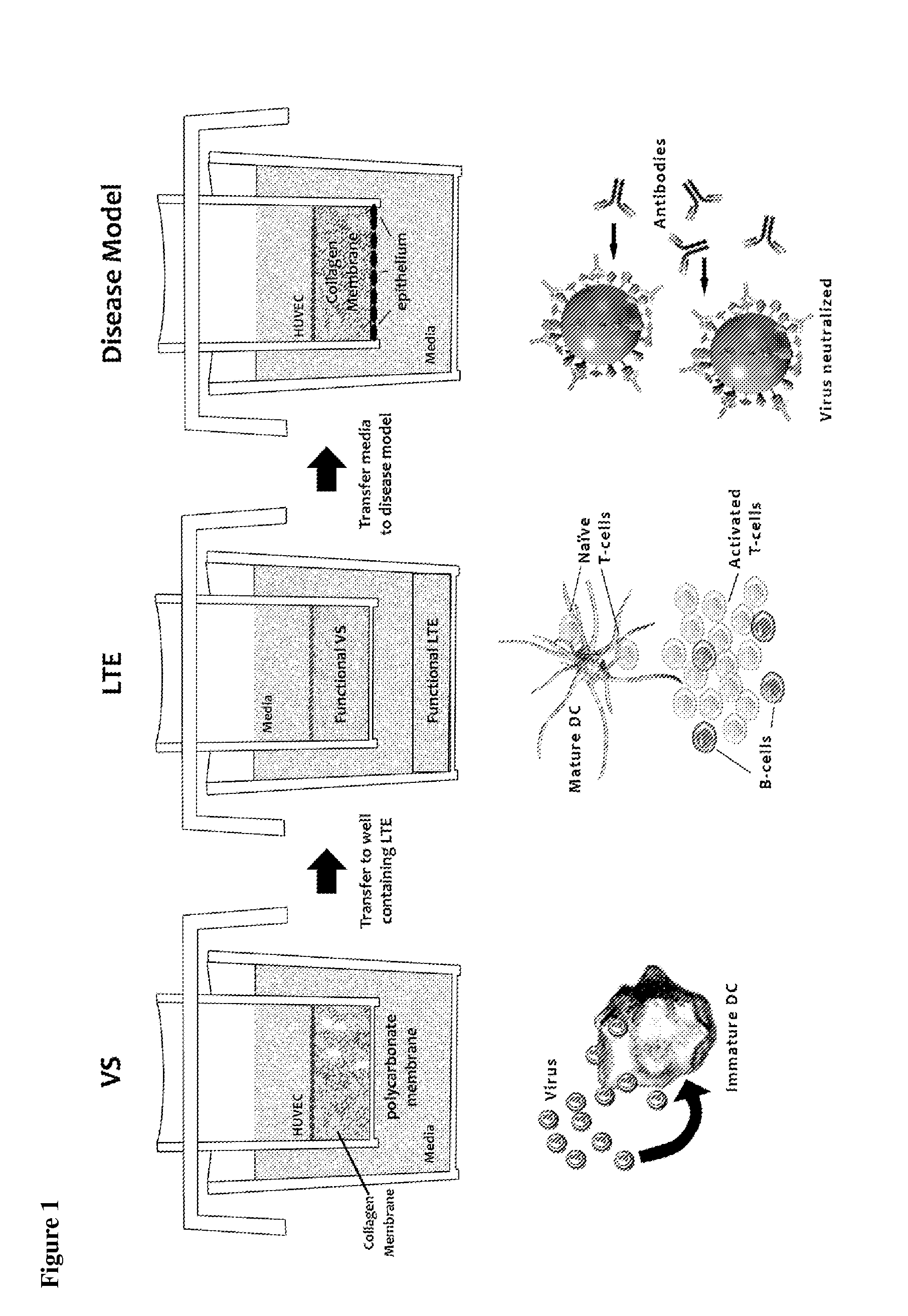
[0052]In summary, in the diseases described above, the pathogenesis of HIV, Yersinia, Burkholderia, filoviruses, and tularemia all involve antigen-presenting cells being infected with a pathogen, which they then transport to regional lymph nodes where pathogen replication occurs. As an example, the in vitro infectious disease model can be comprised of two modules: an infection module, where the macrophages/DCs are infected by the pathogen, which can be represented in model systems of the present invention by the VS, and the disease module, which represents the process...

Problems solved by technology

However, animal models of disease are often only approximations of the human disease state, and in some cases animal models are not available at all (e.g., pathogens that infect only humans).
Thus, animal models of disease may not accurately predict outcomes in human studies or...

Method used


Image

  • Methods of evaluating a test agent in a diseased cell model
  • Methods of evaluating a test agent in a diseased cell model
  • Methods of evaluating a test agent in a diseased cell model

Examples


example 1

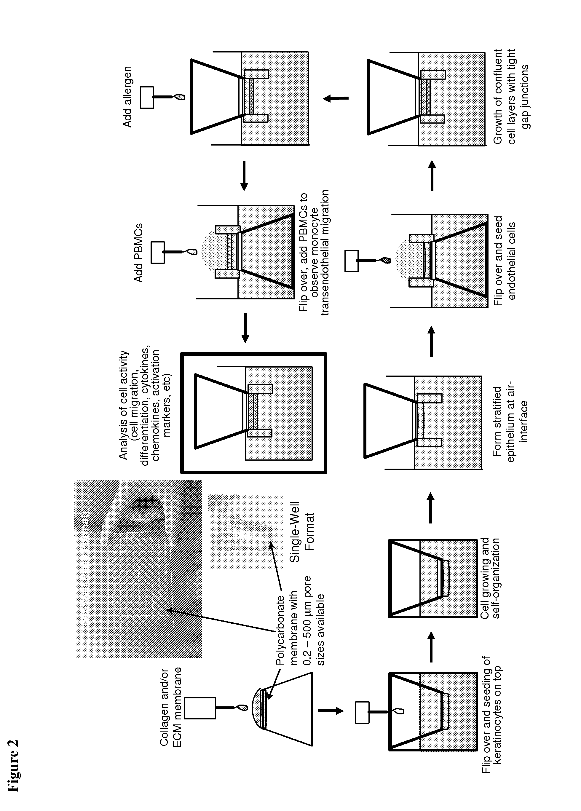
[0064]Generic tissue construct for a 3D in vitro disease model. FIG. 2 illustrates a 3D heterogeneous tissue construct, comprising the addition of cells on the top and bottom of the construct, to create endothelial and epithelial layers. This model is an improvement on our established 3D endothelial cell-only construct, which has been used for transendothelial migration and for monocyte to dendritic cell and macrophage differentiation (the vaccination site, VS).

[0065]The 3D model of this example can be used to study immunophysiological reactions when subjected to various diseases and vaccine formulations. This is a generic construct because most tissues involve a 3D extracellular matrix with associated endothelial and epithelial layers. The disease, whether viral, bacterial, or tumoral, is introduced into the generic tissue construct. The various immunocytes and biomolecules from the AIS (e.g., antibodies, T cells, cytokines, chemokines) can then be delivered to the disease model to...

example 2

[0066]Tumor modeling in the AIS using melanoma cells. Many in vitro model systems have been used for examining the effects of anti-cancer therapeutics and tumor growth in adult and childhood cancers, using both primary cells and various cell lines (see, e.g., Houghton et al. (2002) Clin Cancer Res 8, 3646-57). Such models have proven useful for assessing tumor metabolic states, inhibition of proliferation, and decreases in overall biomass (see, e.g., Monks et al., (1991) J Natl Cancer Inst 83, 757-66; Scherf et al., (2000) Nat Genet 24, 236-44).

[0067]Animal models of human cancers have not been good predictors of human therapeutic outcome because of species differences (see, e.g., Houghton et al.,(2002) Clin Cancer Res 8, 3646-57; Bridgeman et al.,(2002) Cancer Res 60, 6573-6; Batova et al.,(1999), Cancer Res 59, 1492-7).

[0068]As with any tumor model, the primary end goal is to increase patient survival and overall well being and to decrease tumor burden. The most predictive model w...

example 3

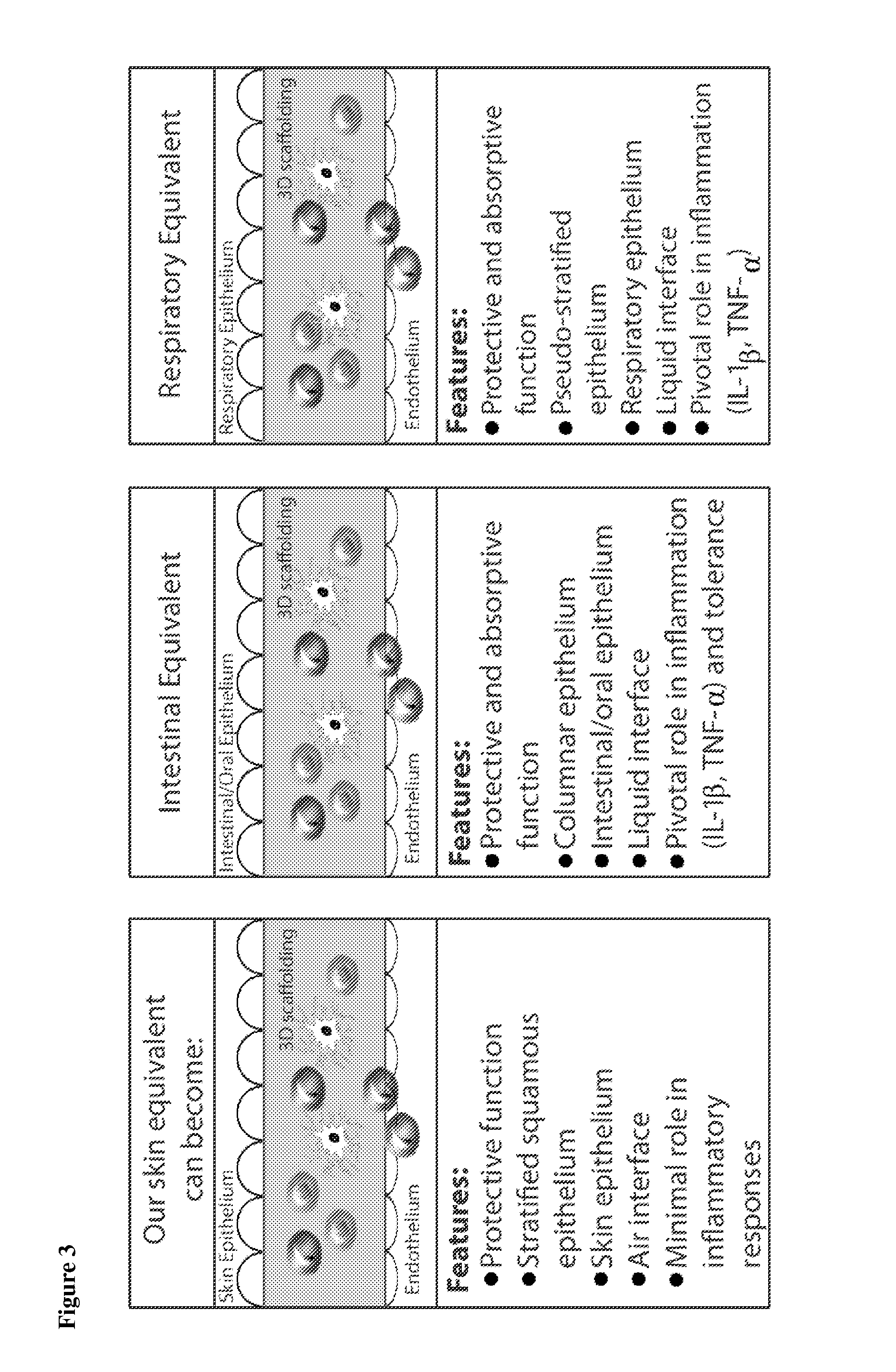
[0071]Heterogeneous tissue constructs with the addition of cells on the top and bottom of the tissue construct to form endothelial and epithelial layers. A schematic representation of the development of the generic disease model and how it can be tested with a particular disease is shown in FIG. 3. As an example, we used a polycarbonate membrane support structure to prepare a 3D ECM matrix, comprising either collagen, synthetic or natural materials (e.g., hydrogels, PLA, PLGA, gelatin, hyaluronic acid), or combinations thereof. We have established an ECM that is capable of supporting two cell layers. We first grow a layer of epithelial cells (e.g., human keratinocytes) on one side of the matrix. An advantage of this model is that other epithelial cells can be used, such as respiratory epithelial cells, skin epithelial cells, or intestinal / oral epithelial cells (as schematically illustrated in FIG. 3). The basement membrane zone between the epithelium and the matrix is important to t...


PUM


Abstract

The present invention relates to methods of constructing an integrated artificial immune system that comprises appropriate in vitro cellular and tissue constructs or their equivalents to mimic the tissues of the immune system in mammals. The artificial immune system can be used to test the efficacy of vaccine candidates and other materials in vitro and thus, is useful to accelerate vaccine development and testing drug and chemical interactions with the immune system, coupled with disease models to provide a more complete representation of an immune response.

Description

CROSS-REFERENCE TO RELATED APPLICATIONS[0001]This application is a divisional of U.S. application Ser. No. 12 / 833,640, filed Jul. 9, 2010, now issued as U.S. Pat. No. 8,062,889, which is a divisional of U.S. application Ser. No. 12 / 047,107, filed Mar. 12, 2008, now issued as U.S. Pat. No. 7,771,999, which was a continuation-in-part of U.S. application Ser. No. 11 / 453,003, filed Jun. 15, 2006, now issued as U.S. Pat. No. 7,709,256, which was a continuation-in-part of U.S. application Ser. No. 11 / 116,234, filed Apr. 28, 2005, now issued as U.S. Pat. No. 7,855,074, which claims the benefit of priority of both U.S. Provisional Application Ser. No. 60 / 565,846, filed Apr. 28, 2004, and U.S. Provisional Application Ser. No. 60 / 643,175, filed Jan. 13, 2005. Each of these applications is hereby incorporated by reference in its entirety.STATEMENT REGARDING FEDERALLY. SPONSORED RESEARCH[0002]This invention was made with government support under contract number NBCHC060058, awarded by the Defen...

Claims


Application Information

Patent Timeline
IPC IPC(8): C12N5/00C12N5/02C12N5/071
CPCG01N33/5082C12N5/0698C12N2502/11C12N2502/1192C12N2502/1323C12N2502/28C12N2502/30C12N2503/04C12N2531/00C12N2533/50C12N2533/54C12N2535/10C12N2501/21C12N2502/091C12N2502/094
Inventor WARREN, WILLIAM L.FAHLENKAMP, HEATHERHIGBEE, RUSSELL G.MISHKIN, ERIC M.SANCHEZ-SCHMITZ, GUZMANRIVARD, MICHAEL D.PAWAR, SANTOSH
Owner SANOFI PASTEUR VAX DESIGN
Why Eureka
Try Eureka