Biocompatible substrate for facilitating interconnections between stem cells and target tissues and methods for implanting same

a biocompatible substrate and target tissue technology, applied in the field of substrates, can solve the problems of reducing the ability of individuals to perceive visual images, affecting the function of the macula, and loss of vision in the center of the visual field, so as to reduce the risk of damage to the substrate during implantation and reduce tissue trauma

Active Publication Date: 2014-08-19
RGT UNIV OF CALIFORNIA +3
View PDF136 Cites 15 Cited by
  • Summary
  • Abstract
  • Description
  • Claims
  • Application Information

AI Technical Summary

Benefits of technology

[0013]In several embodiments, the substrate apical surface is substantially homogeneous and suitable for the growth of cells thereon. In some embodiments, the thickness of the substantially homogeneous apical surface ranges from about 0.1 to about 4 microns. In one embodiment, the thickness of the substantially homogeneous apical surface is between about 0.1 and about 0.5 microns. In some embodiments, the substantially homogeneous apical surface is roughened or otherwise treated to allow a population of cells to have a non-planar surface to grow on. The substantially homogeneous apical surface need not therefore be completely uniform in all embodiments. Rather, several embodiments comprise apical surfaces that are non-uniform, yet still substantially homogeneous. In several embodiments, the thickness of the substantially homogeneous apical surface for the growth of cells prohibits passage of proteins larger than about 60 kDa through the substrate. The thickness can be altered in different embodiments depending on the cell type and the size of proteins that are to be restricted (or allowed). The tailored dimension allows for the passage of nutrients through the substrate (or the removal of metabolic byproducts through the substrate).
[0016]In several embodiments the outer edges and corners of the substrate are rounded, in order to minimize risk of damage to the substrate during implantation (e.g., by catching or snagging on tissue during implantation).
[0017]In some embodiments, the substantially homogeneous apical surface further comprises a raised lip surrounding the surface, wherein the raised lip has a height ranging from about 10 to about 1000 microns and a width ranging from about 10 to about 1000 microns. In several embodiments the lip serves not only to protect the cells growing on the substrate, but also to reduce trauma to the tissue during implantation.

Problems solved by technology

Numerous pathologies can compromise or entirely eliminate an individual's ability to perceive visual images, including trauma to the eye, infection, degeneration, vascular irregularities, and inflammatory problems.
The function of the macula may be adversely affected by age related macular degeneration (wet or dry), diabetic macular edema, idiopathic choroidal neovascularization, high myopia macular degeneration, or advanced retinitis pigmentosa, among other pathologies.
Age related macular degeneration typically causes a loss of vision in the center of the visual field.
Resulting hardened lipids (lipofuscin) impede the reciprocal exchange of nutrients and waste products between the retina and choroid, and lead to PR death.
The walls of these newly formed vessels are mechanically weak, and extremely susceptible to rupture.
Hemorrhage usually results in loss of vision extremely quickly compared with dry AMD.
In conjunction with the loss of functional cells in the eye, the newly formed blood vessels are fragile and often leak blood and interstitial fluid, which can further damage the macula.
Many tissues are structurally or functionally dynamic in that the tissues flex during normal function, are subject to fluid flow or other shear stresses, or have numerous specialized cell types in close juxtaposition, thereby limiting the selection of target sites for cell delivery.

Method used


Image

  • Biocompatible substrate for facilitating interconnections between stem cells and target tissues and methods for implanting same
  • Biocompatible substrate for facilitating interconnections between stem cells and target tissues and methods for implanting same
  • Biocompatible substrate for facilitating interconnections between stem cells and target tissues and methods for implanting same

Examples


example 1

Determination of Pore Diameter that Retains RPE Cells

[0177]In the eye, a healthy Bruch's membrane functions as a molecular sieve that regulates the exchange of nutrients and metabolic wastes between the retina and the choroid. Based on the sub-retinal location of certain ocular-directed substrates disclosed herein, the porosity of the substrate would ideally simulate these functions of a healthy Bruch's membrane.

[0178]To investigate the pore diameter range that would allow for function, cell migration assays were performed. Circular parylene discs were manufactured with varying pore diameters (1, 2, 3, and 5 um) and placed on top of Transwell polyester (PET) cell culture inserts with 8 μm pores. Inserts were placed into 24-well culture plates. RPE cells were seeded on the parylene discs with serum free medium inside the PET cell culture inserts and medium containing 20 μg / ml recombinant human PDGF (as a chemoattractant) outside the inserts. After overnight culture, the PET insert me...

example 2

Biodegradable Polymer Substrates

[0179]As discussed above, parylene substrates are used in several embodiments. Biodegradable polycaprolactone (PCL) material compounded with polyethylene glycol (PEG) is used to test the formation of pores in substrates. During polymerization, the PCL-PEG material is spin-coated onto glass coverslips to make thin films of a desired thickness, as discussed above. Cyclic amino acids (Arg-Gly-Asp; cRGD) covalently bound to a PEG-PCL copolymer is annealed onto the surface of the films. Upon treatment with aqueous buffer or media, the water soluble PEG is washed away and creates pores in the film. The ratio of the PCL-PEG affects the degradation rate of the substrate. These pores increase the surface area of the polymer exposed to water and therefore increase the degradation rate. Through this approach both the porosity of the films as well as the degradation rate may be manipulated based on the amount of PEG added to the polymer blend.

example 3

In Vitro Analyses of hESC-RPE

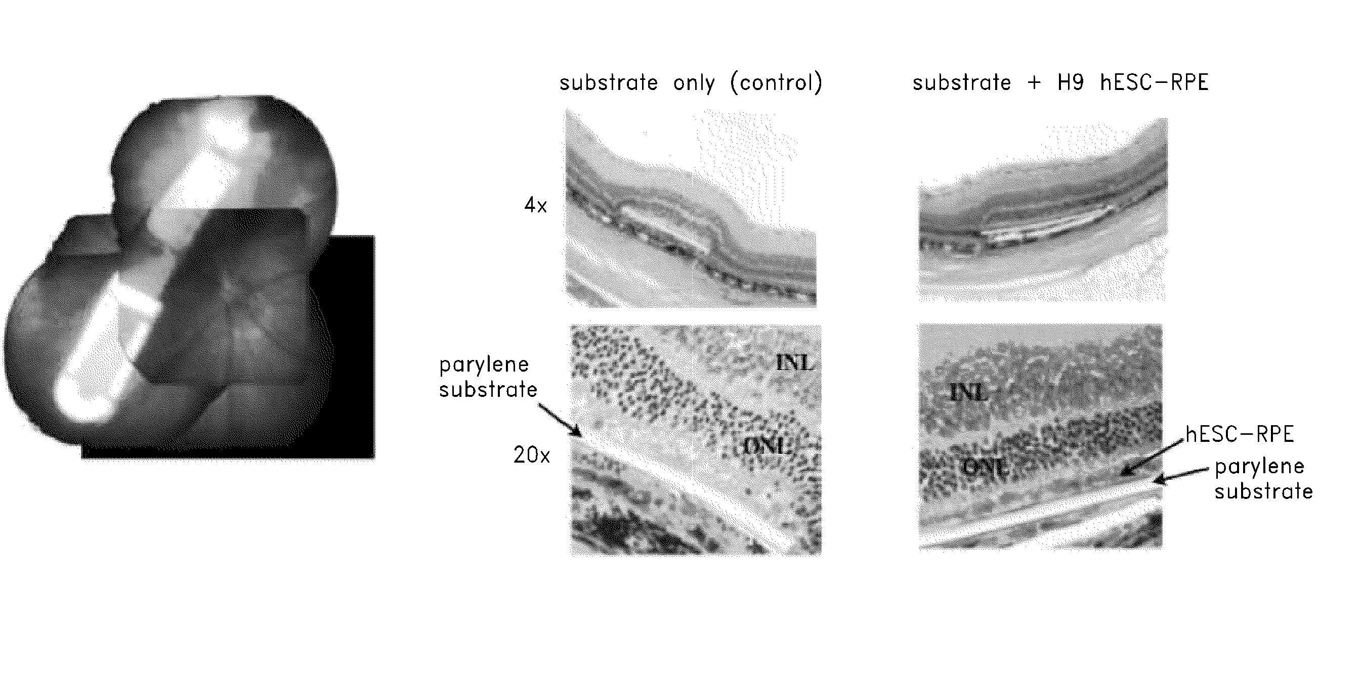
[0180]The present example demonstrates that hESC colonies grown to superconfluency, in the absence of FGF2, on mouse or human feeder layers or on matrigel, routinely produce discrete pigmented foci, which grow in size. These pigmented foci can be excised and expanded to produce highly differentiated monolayers that are similar to cultured fetal RPE or adult human cultured RPE. See FIG. 11. Cells can be produced in quantities required for clinical use at 99% purity, as determined by microscopic analysis of pigmented cells as well as quantitative PCR for nanog and Oct-4, markers of possible contaminating undifferentiated hESC. Monolayer cultures are phenotypically stable after prolonged culture (11 months) without passage. Furthermore phenotype and normal karyotype can be maintained for up to at least 4 passages. Based on cell numbers in culture, some embodiments employ about 1.5×10⁵ cells on a 5 mm diameter substrate are used for each treated eye.

[0181]Mu...


PUM

PropertyMeasurementUnit
molecular weightaaaaaaaaaa
thicknessaaaaaaaaaa
heightaaaaaaaaaa

Abstract

Disclosed herein are substrates for cell delivery to target tissues requiring treatment for various diseases that induce cell death, damage or loss of function. The substrates are configured to provide seeded cells, including stem cells, with a structural support that allows interconnection with and transmission of biological signals between the cells and the target tissue.

Description

RELATED CASES[0001]This application claims the benefit of U.S. Provisional Application Ser. Nos. 61 / 363,630, filed on Jul. 12, 2010 and 61 / 481,004, filed on Apr. 29, 2011, the contents of each of which is expressly incorporated in its entirety by reference herein.BACKGROUND[0002]1. Field of the Invention[0003]The present application relates generally to substrates that facilitate the administration of stem cells to target tissues in the context of stem cell therapy as well as to tools for manipulating and implanting such substrates into a target tissue.[0004]2. Description of the Related Art[0005]The scope of human disease that involves loss of or damage to cells is vast and includes, but is not limited to, ocular disease, neurodegenerative disease, endocrine diseases, cancers, and cardiovascular disease. Cellular therapy involves the use of cells, and in some cases stem cells to treat diseased or damaged tissues. It is rapidly coming to the forefront of technologies that are poised...

Claims


Application Information

Patent Timeline
Patent Type & Authority Patents(United States)
IPC IPC(8): A61K35/30B32B3/00A61F2/02A61K9/00A61K35/545A61K47/34A61L27/38
CPCA61L2430/16A61L27/3813A61L27/3834A61K47/34A61K35/545A61K35/30A61K9/0051A61L27/18A61L27/56A61L2430/00Y10T428/24479C12N5/0621C12N2533/30C12N2535/00A61P27/00A61L27/34A61L27/3808C08L65/04C08L67/04C08L83/08C08L89/00
Inventor HUMAYUN, MARKAHUJA, ASHISHTAI, YU-CHONGHINTON, DAVIDGRUBBS, ROBERTCLEGG, DENNISJOHNSON, LINCOLN V.HIKITA, SHERRY T.
Owner RGT UNIV OF CALIFORNIA
Why Eureka
Try Eureka