Controlled release microparticles
a microparticle and controlled release technology, applied in the field of drug delivery, can solve the problems of small implant size, inability to deliver large, inconvenient intraocular injection, etc., and achieve the effects of reducing burst, increasing duration, and increasing stability
- Summary
- Abstract
- Description
- Claims
- Application Information
AI Technical Summary
Benefits of technology
Problems solved by technology
Method used
Image
Examples
example 1
Preparation of Microparticles (Oil-in-Water)
[0136]Formulations were prepared via an oil-in-water solvent extraction / evaporation method. Macugen®; pegaptanib sodium ((OSI) Eyetech, Inc., NY, N.Y.) and PLGA were dissolved in methylene chloride and an emulsion was formed according to the process disclosed in PCT publication No. WO 2005 / 003180, which is incorporated herein by reference in its entirety. Following solvent extraction from the emulsion particles, the hardened microparticles were sieved through a 45 μm screen. Microparticles≦45 μm were collected by centrifugation and dried by lyophilization.
example 2
Preparation of Pegaptanib Microparticles (Water-in-Oil-in-Water)
[0137]A batch size of 200 milligrams dry microspheres containing pegaptanib was prepared according to the following procedure:
Step 1. Preparation of Primary Aqueous Phase
[0138]a. 30 mg Pegaptanib[0139]b. 300 μL water[0140]c. The mixture was vortexed to dissolve components
Step 2. Preparation of Organic Phase[0141]a. 200 mg PLGA (i.e. 50:50 lactide:glycolide, IV=0.37 dL / g)[0142]b. 7 ml methylene chloride (CH2Cl2)[0143]c. The mixture was vortexed to dissolve components
Step 3. Preparation of Secondary Aqueous Phase / Quench Solution[0144]b. 10.2 g polyvinyl alcohol[0145]c. 104 g sucrose[0146]d. 1.25 mL 1M Tris, pH 8.0[0147]e. 1 mL 0.5M EDTA, pH 8.0[0148]f. All components were dissolved in ˜800 mL water. The pH was adjusted to 7.4 and the final volume to was brought to 1 L.
Step 4.
[0149]The organic solution was homogenized at 20000 RPM for a total of 2 minutes using a Virtis homogenizer. While homogenizing, the primary aqueous ...
example 3
Morphology Analysis of Microparticles
[0156]Microparticle morphology was observed by Scanning Electron Microscopy (SEM) analysis. Microparticles were sputter coated with gold using an Anatech LTD Hummer 6.2 system. Scanning electron microscopic images were taken using a JEOL JSM-5600 scanning electron microscope and accompanying software at an accelerating voltage of 5-10 keV. For selected samples, SEM analysis of the internal microsphere structure was made after embedding microparticles in L.R. White Resin and then splitting the preparation after the resin hardened.
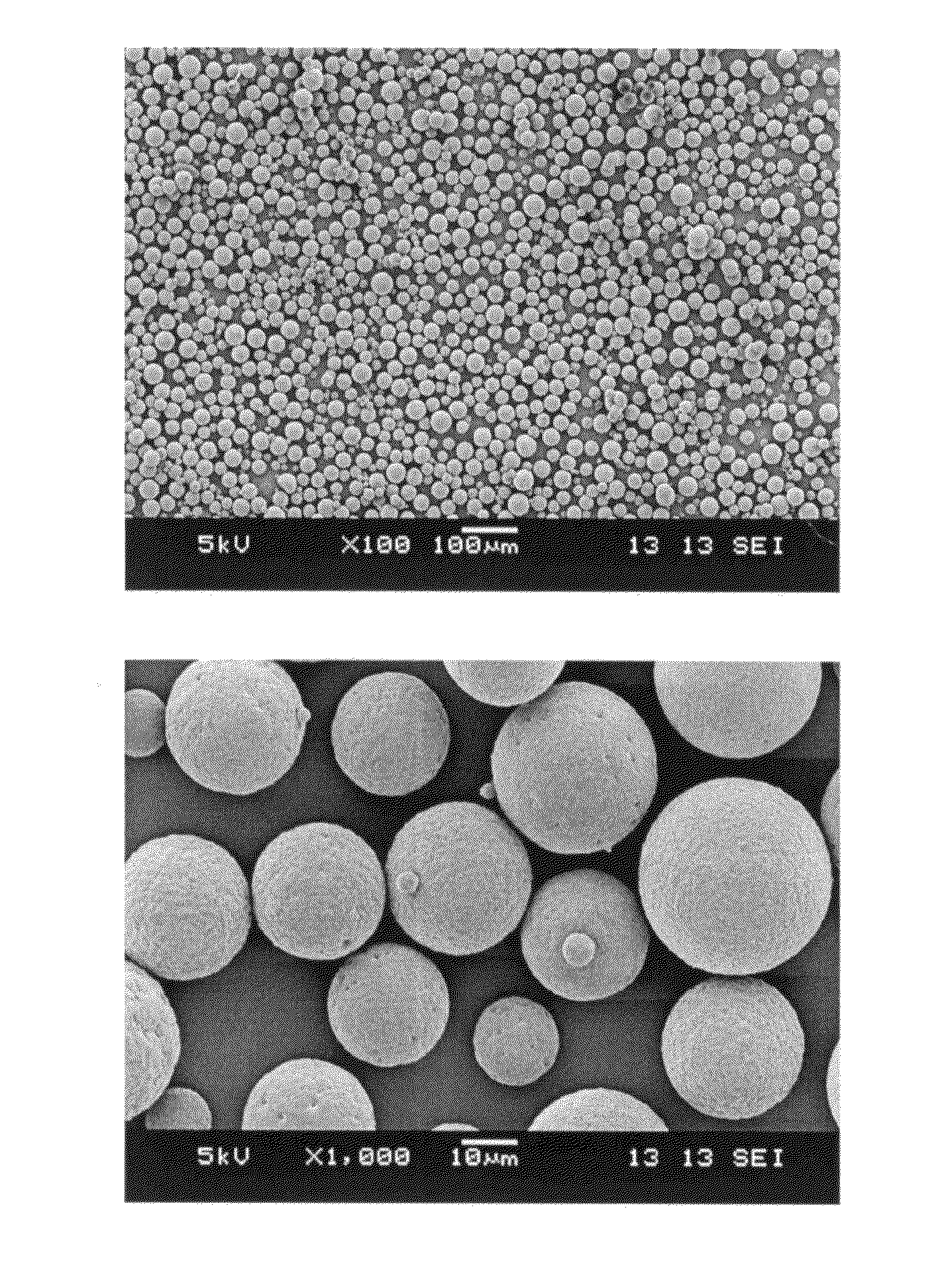
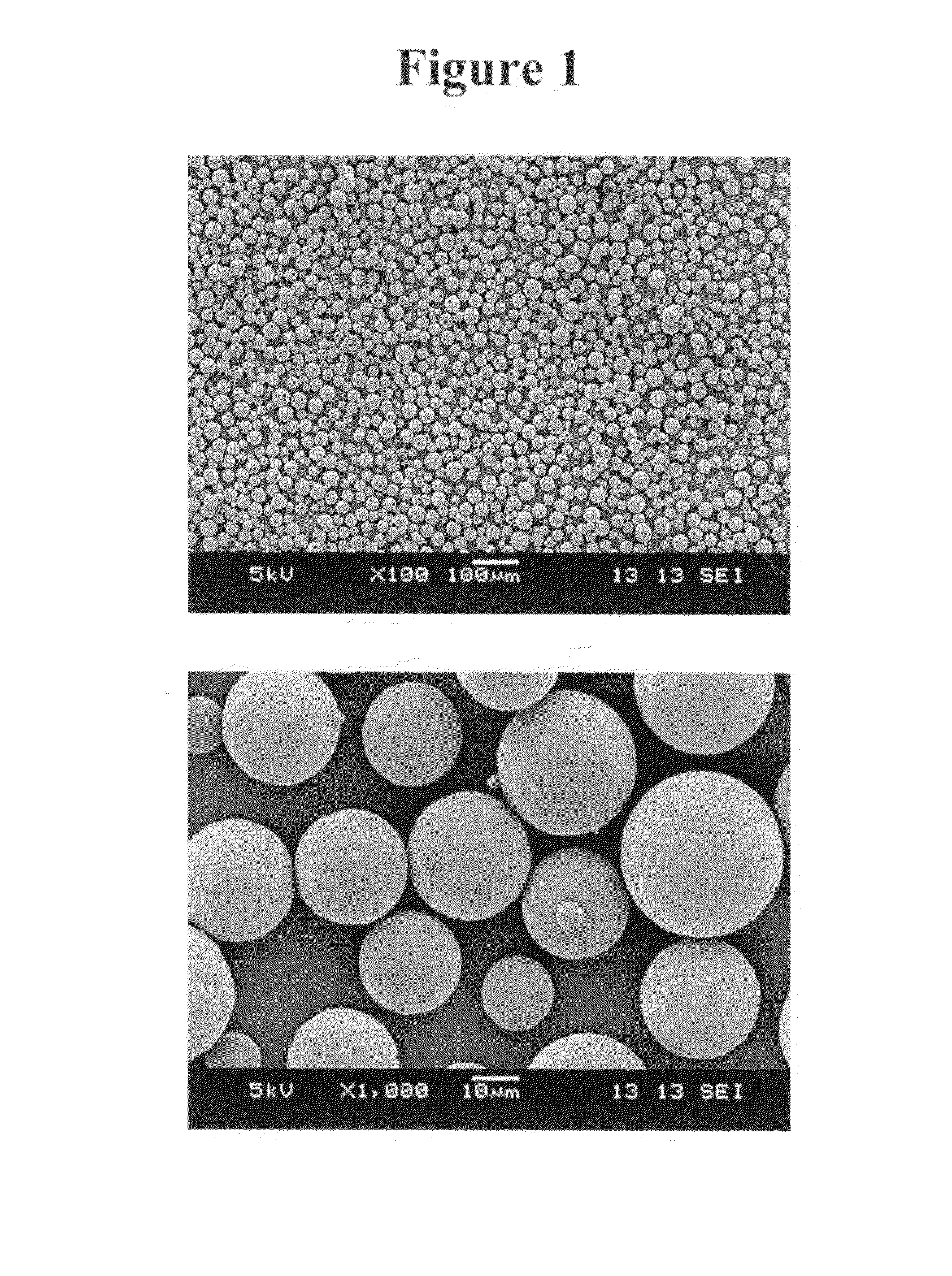
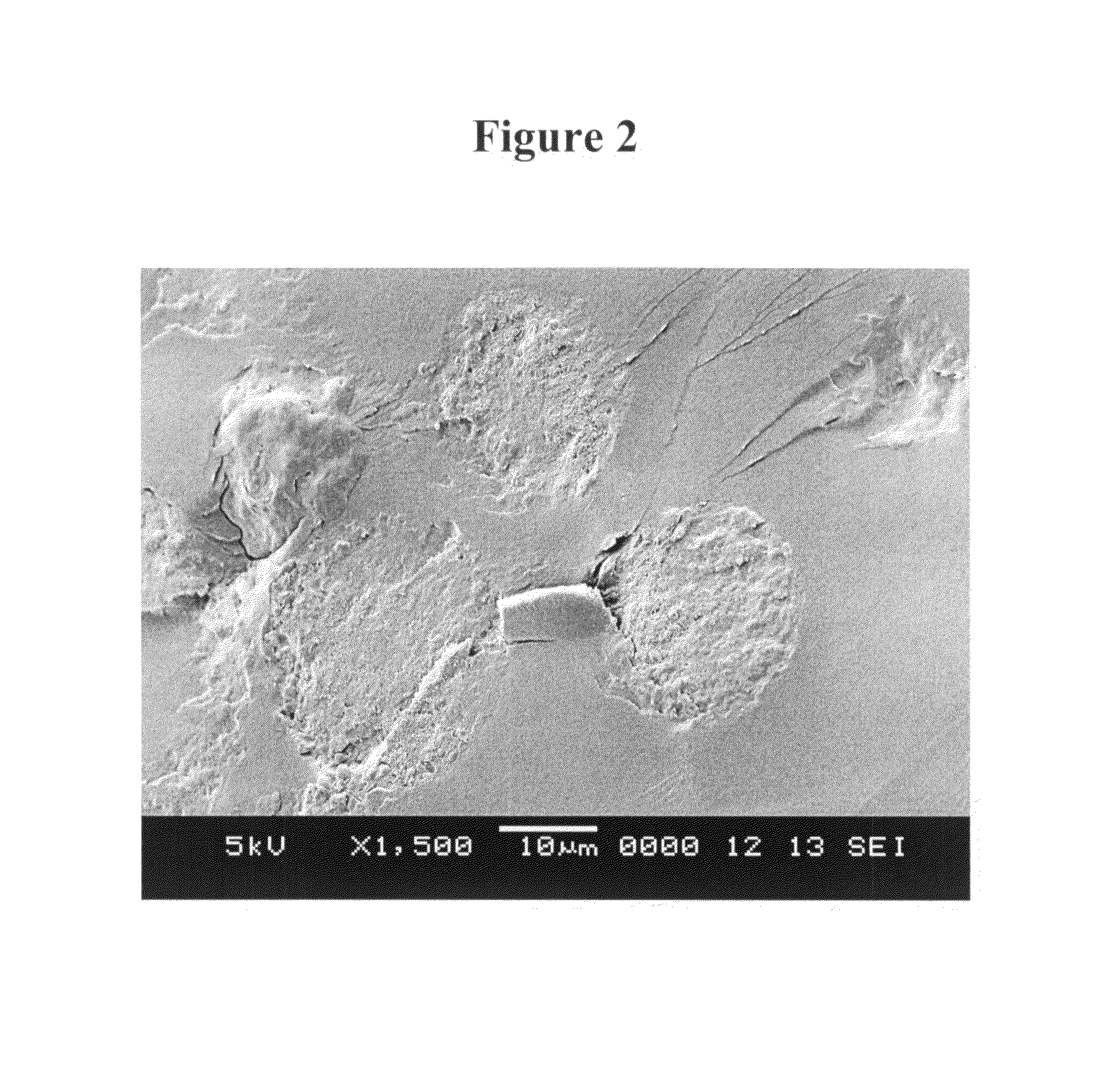
[0157]Scanning electron micrograph images of microparticles formed by the process as set forth in Example 1 are shown in FIGS. 1 and 2. The images of FIG. 1 show that the microparticles have a smooth external morphology. The image of FIG. 2 shows that the microparticles have a monolithic internal morphology.
[0158]A scanning electron micrograph image of microparticles formed by the water-in-oil-in water process as set fort...
PUM
| Property | Measurement | Unit |
|---|---|---|
| diameter | aaaaa | aaaaa |
| mean diameter | aaaaa | aaaaa |
| mean diameter | aaaaa | aaaaa |
Abstract
Description
Claims
Application Information
 - R&D
- Intellectual Property
- Life Sciences
- Materials
- Tech Scout
- Unparalleled Data Quality
- Higher Quality Content
- 60% Fewer Hallucinations
Browse by: Latest US Patents, China's latest patents, Technical Efficacy Thesaurus, Application Domain, Technology Topic, Popular Technical Reports.
© 2025 PatSnap. All rights reserved.Legal|Privacy policy|Modern Slavery Act Transparency Statement|Sitemap|About US| Contact US: help@patsnap.com


