
Exon grouping analysis

a technology of exons and groups, applied in the field of exon grouping analysis, can solve the problems of high cost, difficult identification of complex gene mutations contributing to diabetes disorders, and high cost of southern blotting, and achieves rapid examination of mutations, high throughput, and simple results.

Inactive Publication Date: 2014-12-09
DANA FARBER CANCER INST INC
View PDF3 Cites 0 Cited by
  • Summary
  • Abstract
  • Description
  • Claims
  • Application Information

AI Technical Summary

Benefits of technology

The Exon Grouping Analysis (EGAN) technology allows for simultaneous examination of mutations in multiple regions of a nucleic acid by amplifying them using multiplex PCR and analyzing them by conformation dependent electrophoresis. This method can detect mutations in up to 300,000 sites in a person's genome. The optional step of further analyzing any mutations or polymorphisms detected by DNA sequence provides a detailed understanding of the potential for disease or other health-related issues. Overall, EGAN provides a simple, high-throughput, and fast way to examine mutations in many target regions of a nucleic acid.

Problems solved by technology

Although substantial progress has been made in identifying the genetic basis of many human diseases, current methodologies used to develop this information are limited by prohibitive costs and the extensive amount of work required to obtain genotype information from large sample populations.
These limitations make identification of complex gene mutations contributing to disorders such as diabetes extremely difficult.
Southern blotting is both expensive and time-consuming when performed on large numbers of samples, such as those required to identify a complex genotype associated with a particular phenotype.
Other methods for detecting mutations are also prohibitively expensive and impractical.
While such an analysis is possible with current technology, the cost associated with the analysis of 6 billion bases is at this time prohibitive for routine diagnosis outside of the research environment.
While this technique is possible for common (Single Nucleotide Polymorphisms or SNP's) and known mutations, it is not economically feasible for unknown DNA mutations that may occur in a gene.
It is also not practical for the detection of insertions and deletions in DNA.
Each of the methods described above, and variations thereof, are limited in their applicability to complex mutational analysis.
Specifically, tumor samples are contaminated with normal cells (lymphocytes, stroma, etc.) which results in heterogeneous, impure DNA populations and makes it quite difficult to find mutations with the current gold standard of DNA sequencing.
Moreover, the current techniques suffer from large costs and lack of sensitivity in non-pure DNA in the case of DNA sequencing and low sensitivity and throughput in the case of the structural analysis based methods.
Additionally all these procedures suffer from low throughput which limits the utility and scope of analysis.
These phenomena present unique difficulties in designing clinical screening methods that can accommodate large numbers of sample DNAs.

Method used


Image

  • Exon grouping analysis
  • Exon grouping analysis
  • Exon grouping analysis

Examples


example 1

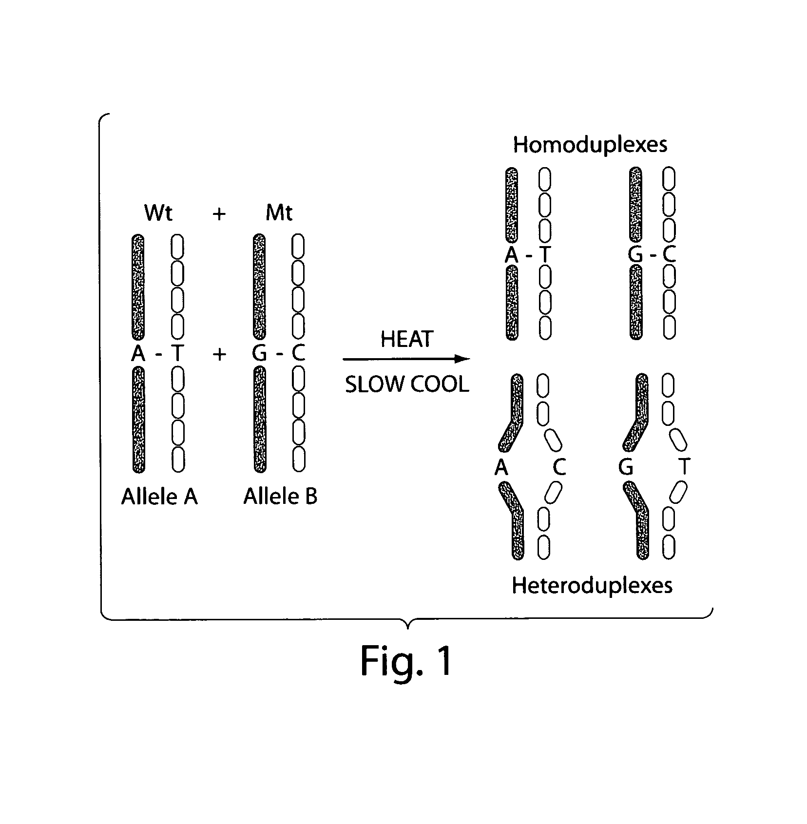
Amplification of Target DNA and the Formation of Homoduplexes and Heteroduplexes

A) Preparation of Sample DNA from Blood

[0098]DNA may be collected from blood. White blood cells collected from the peripheral blood were lysed with two washes of a 10:1 (v / v) mixture of 14 mM NH4Cl and 1 mM NaHCO3. The nuclei were resuspended in nuclei-lysis buffer (10 mM Tris, pH 8.0, 0.4M NaCl, 2 mM EDTA, 0.5% SDS, 500 ug / ml proteinase K) and incubated overnight at 37° C. Samples were then extracted with a one-fourth volume of saturated NaCl and the DNA was precipitated in ethanol. The DNA was then washed with 70% ethanol, dried, and dissolved in TE buffer (10 mM Tris-HCl, pH 7.5, 1 mM EDTA.). Ninety-five DNA samples from breast cancer patients and controls were obtained at DFCI under IRB approved protocols.

[0099]Primers for 88 PCR fragments (34 BRCA1 and 54 BRCA2) were chosen to span the coding region of BRCA1 and BRCA2. The PCR fragments ranged from 232 to 558 base pairs in BRCA1 and 194 to 567 base ...

example 2

Analysis of Amplified DNA by Electrophoresis and DNA Sequencing

Multiplexed PCR Lanes

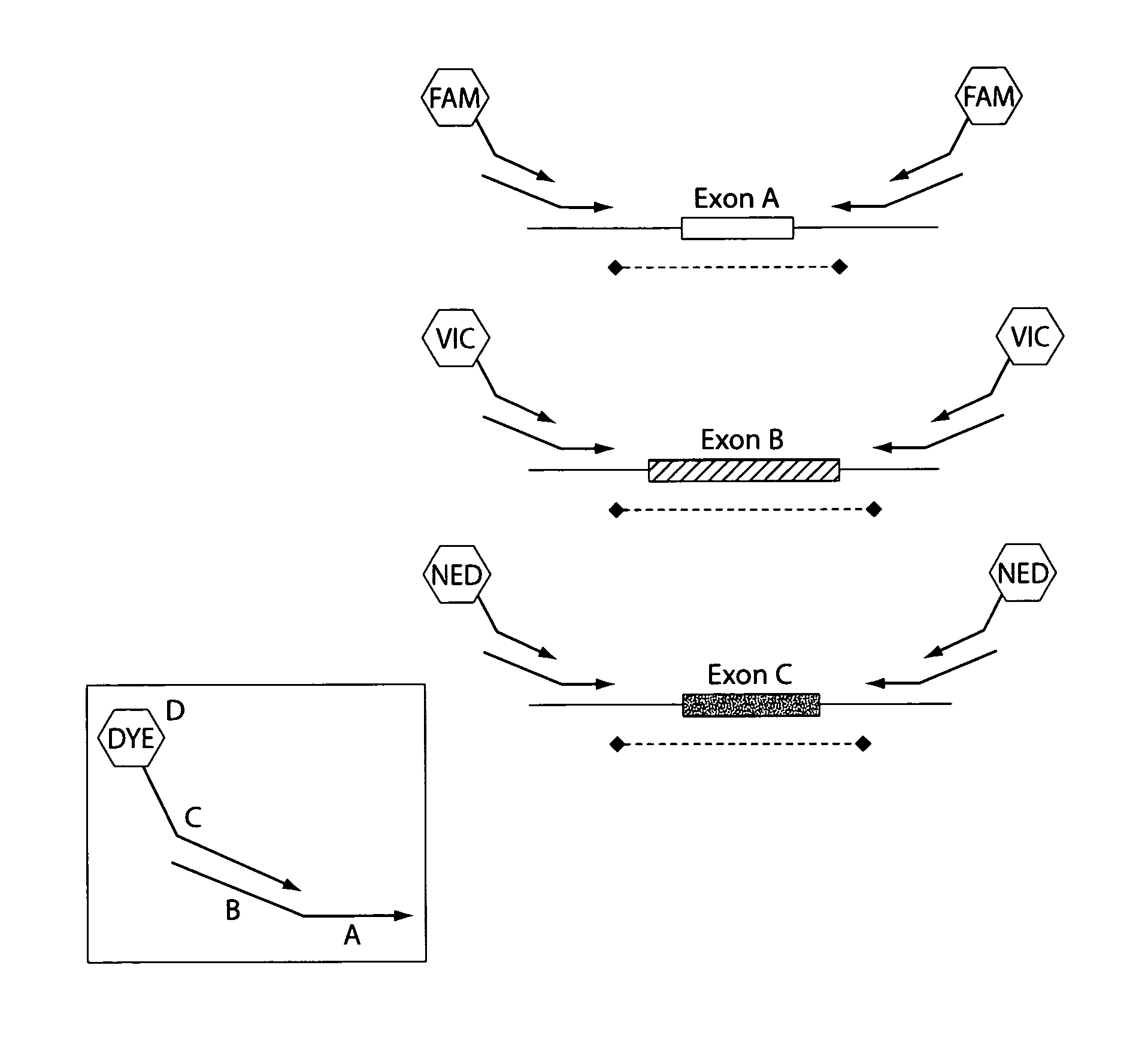
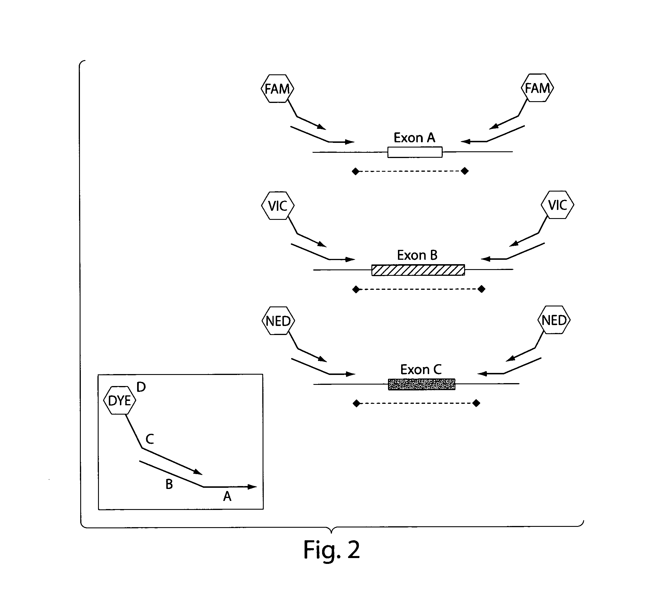
[0104]The 5 multiplexed PCR lanes consist of 6 FAM-labeled primers, 6 VIC-labeled primers and 6 NED-labeled primers. Following the heteroduplex formation, primers labeled in the same color from each patient are mixed together in 25 μL of PCR buffer (0.05M Tris, 0.05M KCL, pH 8.0). These multiplexed PCR fragments are then combined in a 3 FAM: 4 VIC: 6 NED ratio. The approximate final dilutions of PCR product are as follows: 1:100 FAM; 1:70 VIC; 1:45 NED. Finally the FAM, VIC, NED multiplexed mixture is combined in a 1:1 ratio with our ROX labeled size standard.

Scanning of Multiplexed PCR Products

[0105]The 0.2-mm thick gel was prepared with 1.13×MDE Gel Solution (Cambrex), 22.5% Formamide (Fisher Scientific), 15% Ethylene Glycol (Fisher Scientific), 0.05% ammonium persulfate (Fisher Scientific) and 0.005% N,N,N′,N′tetramethylethylenediamine (Fisher Scientific) in 0.6×TTE (44.4 mM Tris / 14.25 mM Taurine / ...

example 3

Validation of Exon Grouping Analysis

[0112]Exon Grouping Analysis (EGAN) on a library of 216 distinct test amplicons were tested and validated. For testing and evaluation, amplicons of different characteristics were tested. These characteristics includes size, GC richness (which can be expressed as % GC, % pyrimidines or % purines), and the nature of mutation. The nature of mutation may be the change of any one base of a nucleic acid sequence to any other base. A total of 12 types of mutations, such as A to G, A to C, A to T, G to A, G to T, G to C, C to G, C to T, C to A, or T to A, T to G, and T to C, can occur. Of course, point mutations that do not alter sequence, such as A to A, G to G, C to C and T to T are not detected. These parameters are plotted in FIG. 8A. In FIG. 8A, the amplicons range in size from 200 to 700 base pairs in the context of 40%, 50% and 60% GC richness. Each possible combination of GC richness and basepair length has a point mutation introduced where a base...


PUM

PropertyMeasurementUnit
pHaaaaaaaaaa
pHaaaaaaaaaa
pHaaaaaaaaaa

Abstract

This invention pertains to the identification of specific disease-causing DNA sequences in a subject. The methods of the present invention can be used to identify genetic alterations, to determine the molecular basis for genetic diseases, and to provide carrier and prenatal diagnosis for genetic counseling.

Description

RELATED APPLICATIONS[0001]This application claims the benefit of U.S. Ser. No. 60 / 919,810, filed Mar. 23, 2007 the contents of which is incorporated herein by reference in its entirety.INCORPORATION-BY-REFERENCE OF SEQUENCE LISTING[0002]The contents of the text file named “DFCI—042—001US_SubSeqList.txt”, which was created on Jul. 30, 2014 and is 8 KB in size, are hereby incorporated by reference in their entirety.BACKGROUND OF THE INVENTION[0003]Genomic DNA varies significantly from individual to individual, except in identical siblings. Many human diseases arise from genomic variations and mutations. The genetic diversity amongst humans and other life forms explains the heritable variations observed in disease susceptibility. Diseases arising from such genetic mutations include Huntington's disease, cystic fibrosis and Duchenne muscular dystrophy. Each of these diseases is associated with a single gene mutation. Diseases such as multiple sclerosis, diabetes, Parkinson's disease, Al...

Claims


Application Information

Patent Timeline
Patent Type & Authority Patents(United States)
IPC IPC(8): C12P19/34C12Q1/68
CPCC12Q1/6827
Inventor MIRON, ALEXANDERIGLEHART, J. DIRK
Owner DANA FARBER CANCER INST INC
Learn More