Preparation method of sample used for electrophoresis imaging analysis of single-cell gelatin and reagent kit

A single-cell gel and image analysis technology, which is applied to the sample preparation of single-cell gel electrophoresis image analysis, and the field of kits for implementing this method, can solve problems affecting popularization and application, threats to user health, difficult disposal of experimental waste, etc. problem, to achieve the effect of improving sensitivity and image quality, safe and reliable use, and high success rate

Inactive Publication Date: 2009-12-23
JINAN UNIVERSITY
View PDF4 Cites 0 Cited by
  • Summary
  • Abstract
  • Description
  • Claims
  • Application Information

AI Technical Summary

Problems solved by technology

For a long time, the sample slides of single-cell gel electrophoresis image analysis are generally stained with ethidium bromide (Ethidium Bromide, EB) as a nucleic acid dye, but the strong carcinogenicity of EB not only threatens the health of users, but also Experimental waste is difficult to deal with, polluting the environment
In addition, the existing sample slides for single-cell gel electrophoresis image analysis need to be covered with three layers of gel, the preparation process is more complicated, and other preparation steps are also relatively cumbersome, making it often difficult to prepare suitable samples, which also affects this method. promotion and application

Method used


Image

  • Preparation method of sample used for electrophoresis imaging analysis of single-cell gelatin and reagent kit
  • Preparation method of sample used for electrophoresis imaging analysis of single-cell gelatin and reagent kit
  • Preparation method of sample used for electrophoresis imaging analysis of single-cell gelatin and reagent kit

Examples


Embodiment 1

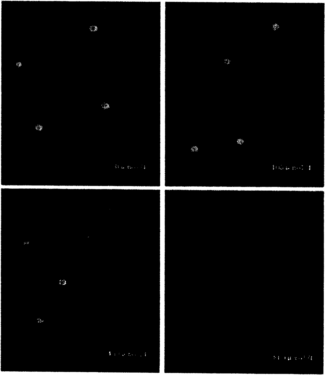
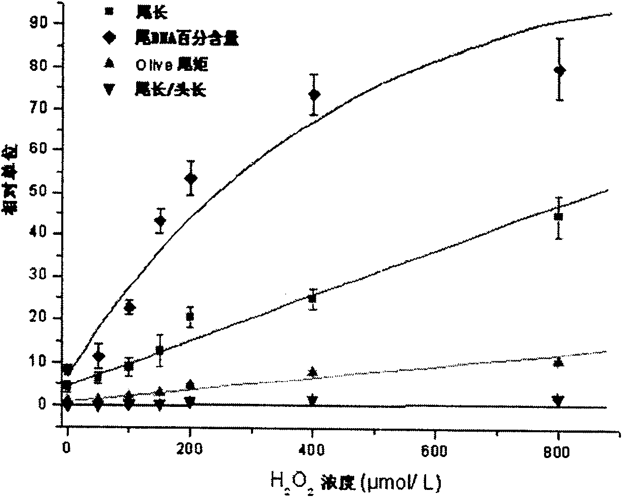
[0025] (1) Preparation of lymphocyte samples. 2ml of venous blood was drawn from young healthy male volunteers with no smoking history and no recent medication history, and anticoagulated with heparin. Then draw the diluted blood and add it to the lymphocyte separation medium (1:1), and centrifuge at 2000r / m for 20min. Aspirate the lymphocyte layer, wash twice with PBS, 1500r / m, 10min. The precipitate was diluted into a suspension with RPMI-1640 and counted (1×10⁷ / mL). Then with H 2 o 2 Exposure: Divided into 6 dosage groups, H 2 o 2 The final concentrations were 50 μM, 100 μM, 150 μM, 200 μM, 400 μM, and 800 μM, respectively. A blank control group (group 0) was set up and placed in a 37°C water bath for 1 hour to prepare lymphocyte samples.

[0026](2) Laying glue. Preheat a frosted glass slide at 40°C, and then evenly coat 1% eutectic point agarose gel prepared with calcium- and magnesium-free phosphate buffer solution (PBS) on the frosted glass slide, and store at lo...

Embodiment 2

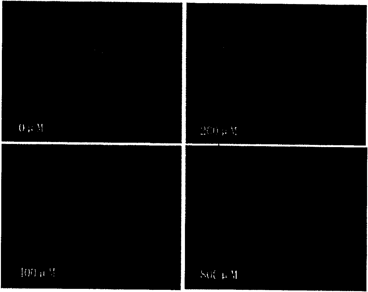
[0035] (1) Prepare bone marrow cell samples. Bone marrow cells were obtained from femurs of Kunming mice (Guangzhou Experimental Animal Center) aged 5 to 6 weeks and weighing 22 to 25 g, washed twice with PBS, and then centrifuged at 1500 r / m for 10 minutes. The precipitate after centrifugation was diluted into a suspension with RPMI-1640 and counted (1×10⁷ / mL); 2 o 2 Infected and divided into 6 dose groups, H 2 o 2 The final concentrations were 50 μM, 100 μM, 150 μM, 200 μM, 400 μM, and 800 μM, respectively. A blank control group (group 0) was set up and placed in a water bath at 37° C. for 1 hour to prepare bone marrow cell samples.

[0036] (2) Laying glue. Preheat a frosted glass slide at 41°C, and then evenly coat 1% eutectic point agarose gel prepared with calcium- and magnesium-free phosphate buffer solution (PBS) on the frosted glass slide, and store at low temperature. Cured under the hood to obtain a glass slide covered with the first layer of gel. Preheat 20 μ...


PUM


Abstract

The invention discloses a sample preparation method for single-cell gel electrophoresis image analysis, including the steps of gel laying, cell lysis, unspinning and electrophoresis, neutralization and washing, and fluorescent staining. The gel has two layers; the fluorescent staining step uses an asymmetric cyanine dye, that is, a SGD dye solution, and the SGD dye solution is a solution in which SYBR Gold dye is diluted with dimethyl sulfoxide at a volume ratio of 1:10000. The present invention also provides a kit specially used for implementing the above sample preparation method, including 7 prepared reagents and 6 processed frosted glass slides. The present invention uses SGD dye solution for fluorescent dyeing, which is non-toxic and harmless, and the experimental waste produced will not pollute the environment, and has higher fluorescence sensitivity; the paved gel has only two layers of glue, which is different from traditional sample glass slides. Compared with the three-layer gel process, the sample preparation process is simple, easy to operate, and has a high success rate.

Description

technical field [0001] The invention relates to cell detection technology, in particular to a sample preparation method for single-cell gel electrophoresis image analysis and a kit for implementing the method. Background technique [0002] Single cell gel electrophoresis image analysis (single cell gel electrophoresis, SCGE) technology, also known as comet assay (comet assay), is a simple, sensitive and rapid method for the research and quantitative detection of DNA damage in single cells developed in the late 1980s. new technology. Under the induction of related factors, if the DNA of the cell is damaged, it will cause changes in its high-level structure, causing its superhelical structure to loosen. When cells undergo in situ lysis, DNA unwinding, etc., the damaged DNA fragments will move out of the nucleus during electrophoresis, migrate to the anode and present a tail-shaped distribution, while the undamaged part of the cell remains spherical. After staining with fluor...

Claims


Application Information

Patent Timeline
Patent Type & Authority Patents(China)
IPC IPC(8): G01N27/447G01N21/64C12Q1/24
Inventor 黄耀熊刘齐海袁志坚
Owner JINAN UNIVERSITY
Why Eureka
Try Eureka