Preparation method for pig blood vessel acellular bracket by chemical and physical combination
A physical combination and decellularization technology, which is applied in the field of medical prosthesis materials, can solve the problems of high cost of decellularized scaffolds, inability to remove cell components, high use and maintenance costs, and achieve complete cell removal and easy cleaning of surfactants , The effect of low equipment cost
- Summary
- Abstract
- Description
- Claims
- Application Information
AI Technical Summary
Problems solved by technology
Method used
Image

Examples
Embodiment Construction
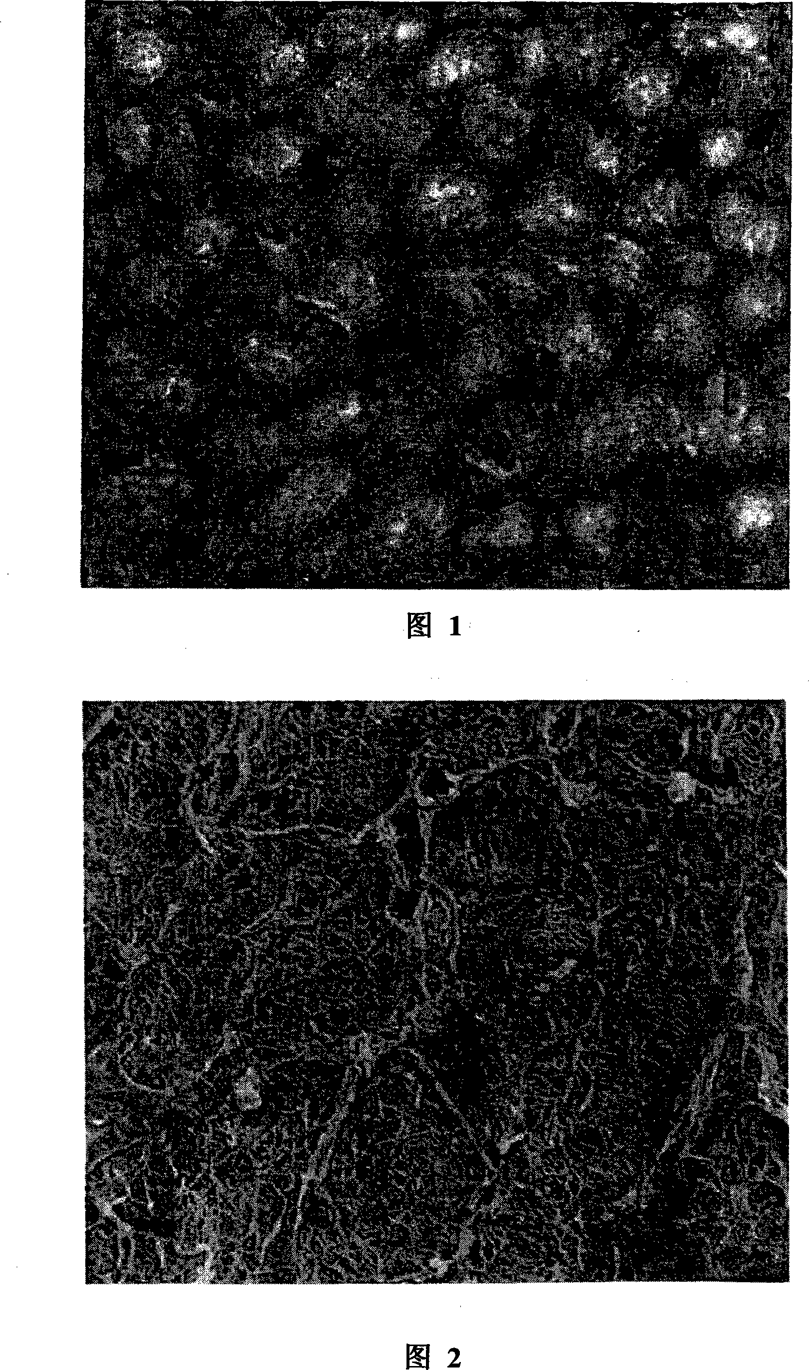
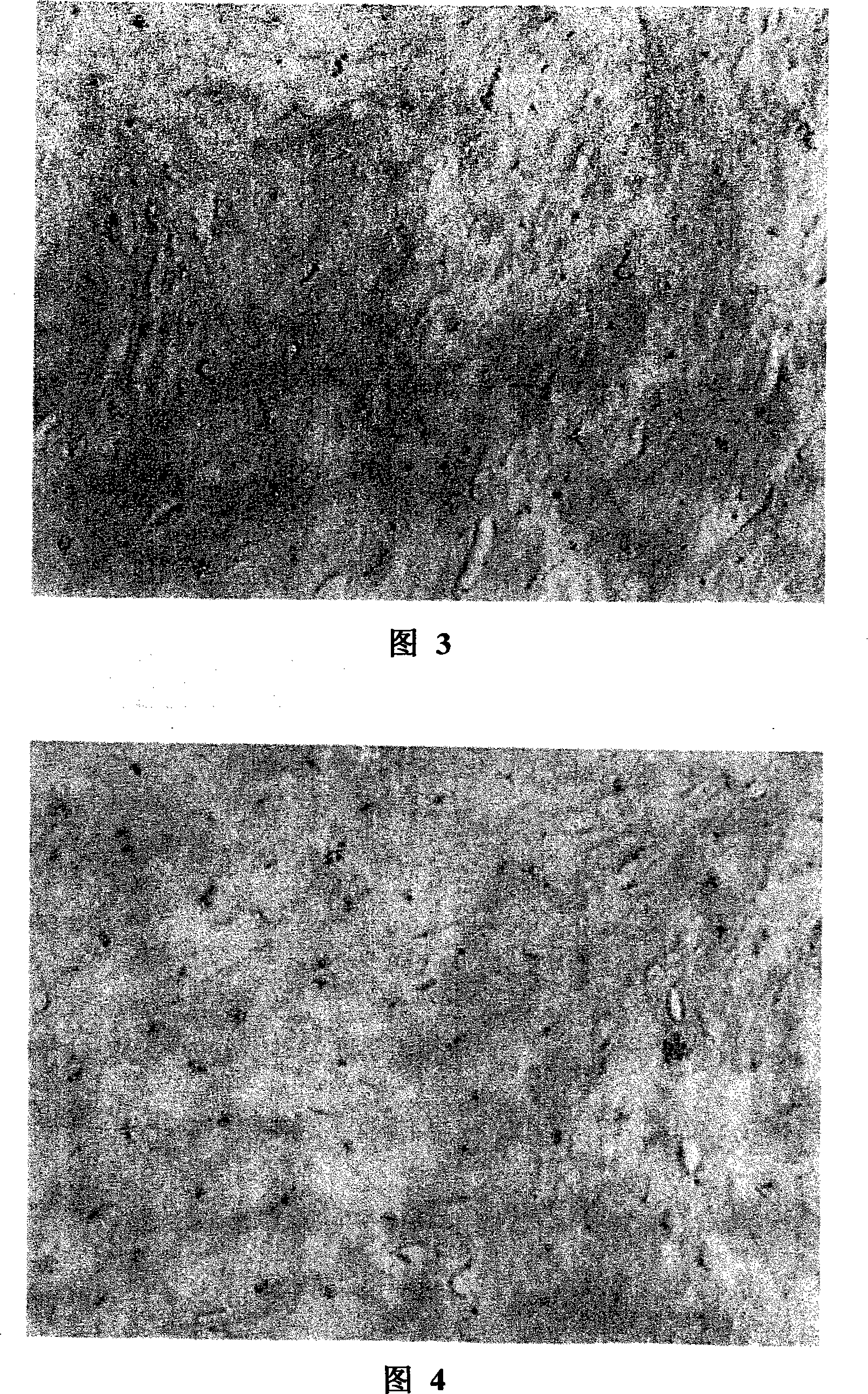
[0021] The chemical and physical combination preparation method of the porcine blood vessel decellularized scaffold of the present invention is the preferred result after various decellularized method experiments. General X-100 is example that the inventive method is described in further detail as follows:
[0022] A) Preparation: After successful anesthesia of healthy pigs, take out the ascending aorta, aortic arch and coronary arteries not shorter than 5 mm according to the requirements of aseptic surgery, keep the anterior leaflet of the mitral valve and the interventricular septal muscle with a thickness of about 3 mm, rinse with normal saline, and separate The inner diameter of the artery at 10mm and 30mm on the annulus was measured. After washing with PBS buffer, put them into a carbon dioxide dry ice incubator for disinfection. Take out the sterilized material on the ultra-clean workbench, put it into a vacuum packaging bag and seal it to be a pig blood vessel, and ind...
PUM
 Abstract
Description
Claims
Application Information