Dendritic structure mark and preparation method thereof
A dendritic structure and marker technology, which is applied in biochemical equipment and methods, microbial determination/inspection, fluorescence/phosphorescence, etc., to reduce non-specific reactions, simplify preparation methods, and enhance sensitivity
- Summary
- Abstract
- Description
- Claims
- Application Information
AI Technical Summary
Problems solved by technology
Method used
Image
Examples
preparation example Construction
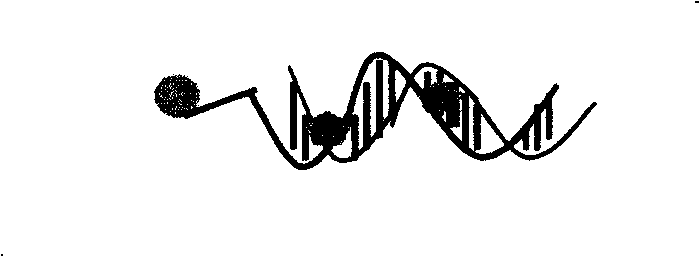
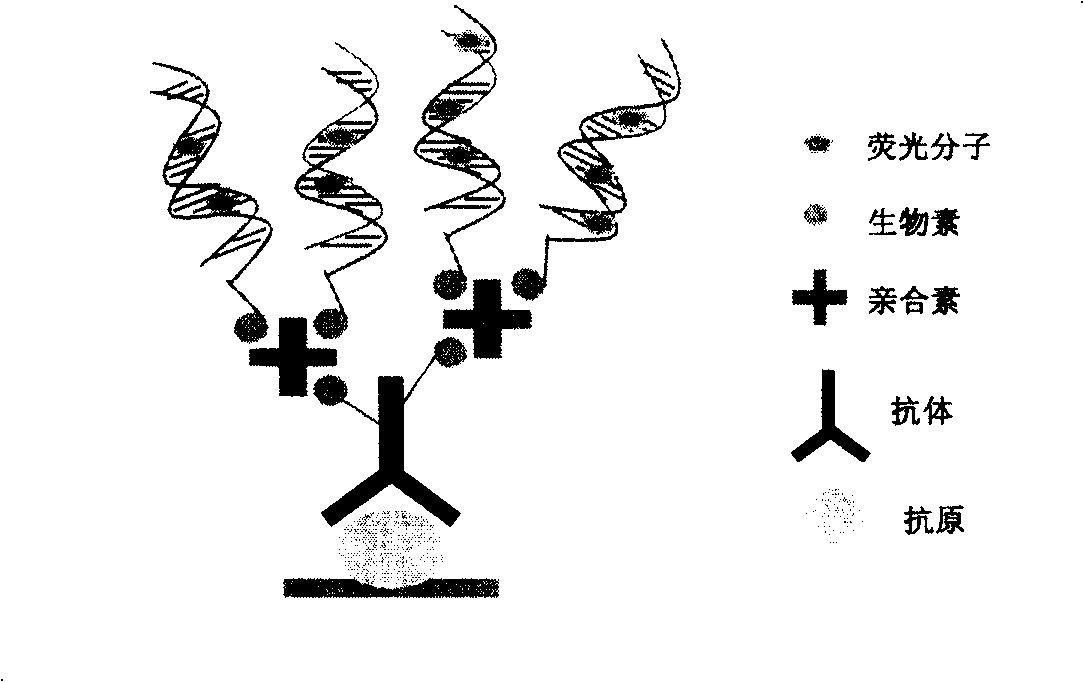
[0039] 1. Preparation of biotin-labeled double-stranded oligonucleotide
[0040] Use 2×SSC (0.3M sodium citrate, 30mM sodium chloride, pH7.0) buffer solution to mix biotin-labeled single-stranded deoxyribonucleic acid (Invitrogen, Carlsbad, CA) and its complementary nucleic acid chain (Invitrogen, Carlsbad, CA), for example, the two chains are: chain A: biotin-5'-TTT TTT TTT GCG GGT AAC GTC AAT ATT AAC TTT ACT CCC-3', chain B: 5'-GGG AGT AAA GTT AAT ATT GAC GTT ACC CGC-3'. Denature at 95°C for 5 minutes, cool to room temperature naturally, and determine the concentration of nucleic acid after hybridization with a spectrophotometer.
[0041] 2. Fluorescein isothiocyanate (FITC) labeled antibody or protein molecule
[0042] Mouse immunoglobulin G (mIgG), goat anti-mouse immunoglobulin G and streptavidin (SA) dissolved in carbonate buffer (50mM sodium carbonate, 50mM sodium bicarbonate, pH 9.15), respectively Mix with FITC (5 mg FITC / mL DMSO) dissolved in dimethyl sulfoxide (DMSO) at...
Embodiment 1
[0045] Example 1. Optimization of coating conditions for mouse immunoglobulin G
[0046] Take 100 μl of FITC-labeled mouse immunoglobulin G (FITC-mIgG) with concentrations of 0, 0.3, 1, 3, 10, 30, 100 μg / mL, respectively, and dissolve it in a buffer containing 50 mM sodium bicarbonate and a pH of 9.6 In the medium, a 96-well plate was coated according to the amount of 100μl per well at 4°C overnight. After washing 3 times with a buffer containing 50mM 3-hydroxymethyl-aminomethane, 50mM sodium chloride and 0.1% Tween 20, pH 8.0, the optimal fluorescence detection conditions of FITC (i.e. the optimal fluorescence excitation wavelength , Excitation slit width, fluorescence emission wavelength, emission slit width), detect the change of fluorescence intensity to indicate the saturation concentration of FITC-mIgG, and use this saturation concentration as the coating concentration of unlabeled mIgG.
Embodiment 2
[0047] Example 2. Optimization of experimental conditions for biotin-labeled goat anti-mouse immunoglobulin G
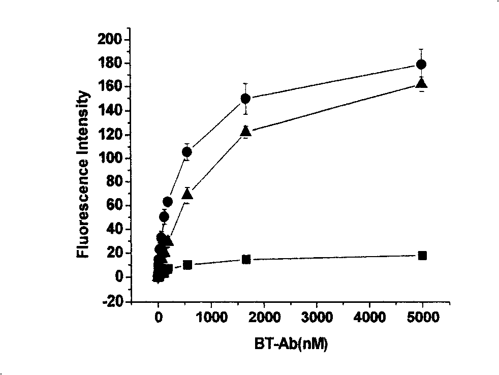
[0048] Coat mIgG with optimized mIgG coating conditions at 4°C overnight. After washing 3 times, add 300 μl of 1% bovine serum albumin-containing phosphate buffer at 4°C overnight. After washing 3 times, add 100μl of FITC-labeled goat anti-mouse IgG with concentrations of 0, 1.0, 3.2, 9.7, 29.0, 87.1, 261.3, 784.0, 2352.0μg / mL, 37℃, air bath constant temperature 200rpm shaking for 2 hours; washing After 3 times, the optimal fluorescence detection conditions of FITC were used to detect the change of fluorescence intensity to indicate the saturation concentration of FITC-mIgG, and the saturation concentration was used as the work of biotin-labeled goat anti-mouse IgG (BT-Ab) concentration.
PUM
 Abstract
Description
Claims
Application Information
 - R&D
- Intellectual Property
- Life Sciences
- Materials
- Tech Scout
- Unparalleled Data Quality
- Higher Quality Content
- 60% Fewer Hallucinations
Browse by: Latest US Patents, China's latest patents, Technical Efficacy Thesaurus, Application Domain, Technology Topic, Popular Technical Reports.
© 2025 PatSnap. All rights reserved.Legal|Privacy policy|Modern Slavery Act Transparency Statement|Sitemap|About US| Contact US: help@patsnap.com


