Elisa kit for detecting early breast cancer and method for making same
An early-stage enzyme-linked immunosorbent assay technology, applied in the fields of molecular biology and cellular immunology, can solve the problems of complex sample processing equipment and other problems, and achieve the effects of shortening detection time, improving stability and convenient detection.
- Summary
- Abstract
- Description
- Claims
- Application Information
AI Technical Summary
Problems solved by technology
Method used
Image

Examples
Embodiment 1
[0038] Example 1 Preparation of hGTA antigen
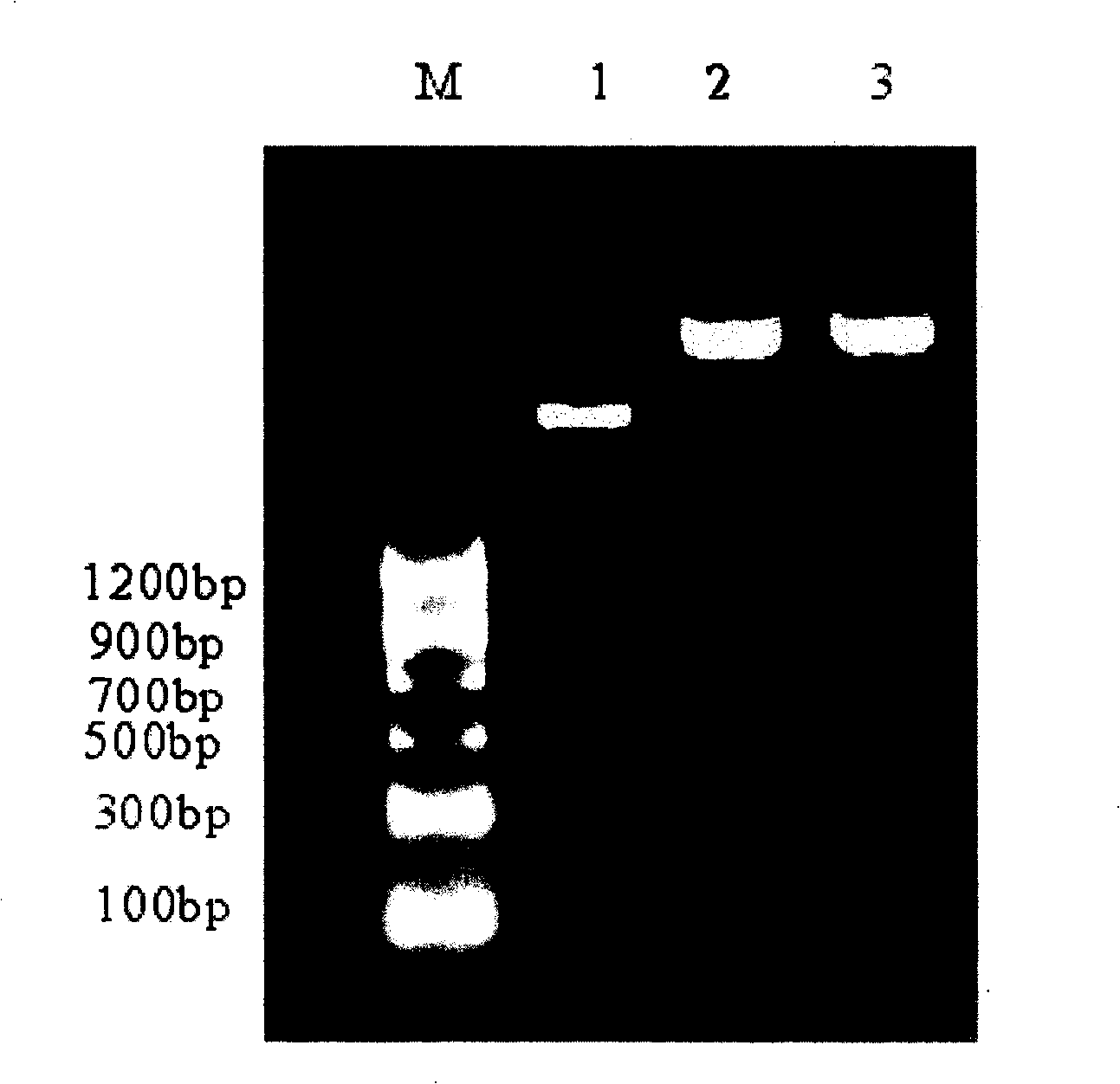
[0039] 1. Reverse transcription of human hGTA cDNA and construction of expression plasmid
[0040] ①Extraction of total RNA from breast cancer cells
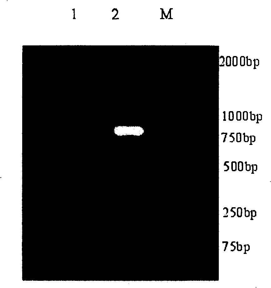
[0041] Breast cancer cells MDA-MB453 (Breast Center, The First Affiliated Hospital of Harbin Medical University) were cultured in complete RPMI 1640 medium at 37°C, 5% CO 2 , cultured to the logarithmic growth phase under saturated humidity, digest the cells, pipet the cells, prepare the cell suspension, count, 2000r / min, discard the culture medium, suck the cells (5-10×10 6) in 1 mL of TRIZOL Reagent and placed at room temperature for 5 min to completely separate the nucleic acid-protein complex, centrifuged at 12,000 r / min for 5 min, took the supernatant, transferred it to a new RNase-free centrifuge tube, added 0.3 mL of chloroform, and shaken vigorously for 20 seconds. Place at room temperature for 5min, centrifuge at 13000r / min for 5min, the sample will be divided into three ...
Embodiment 2
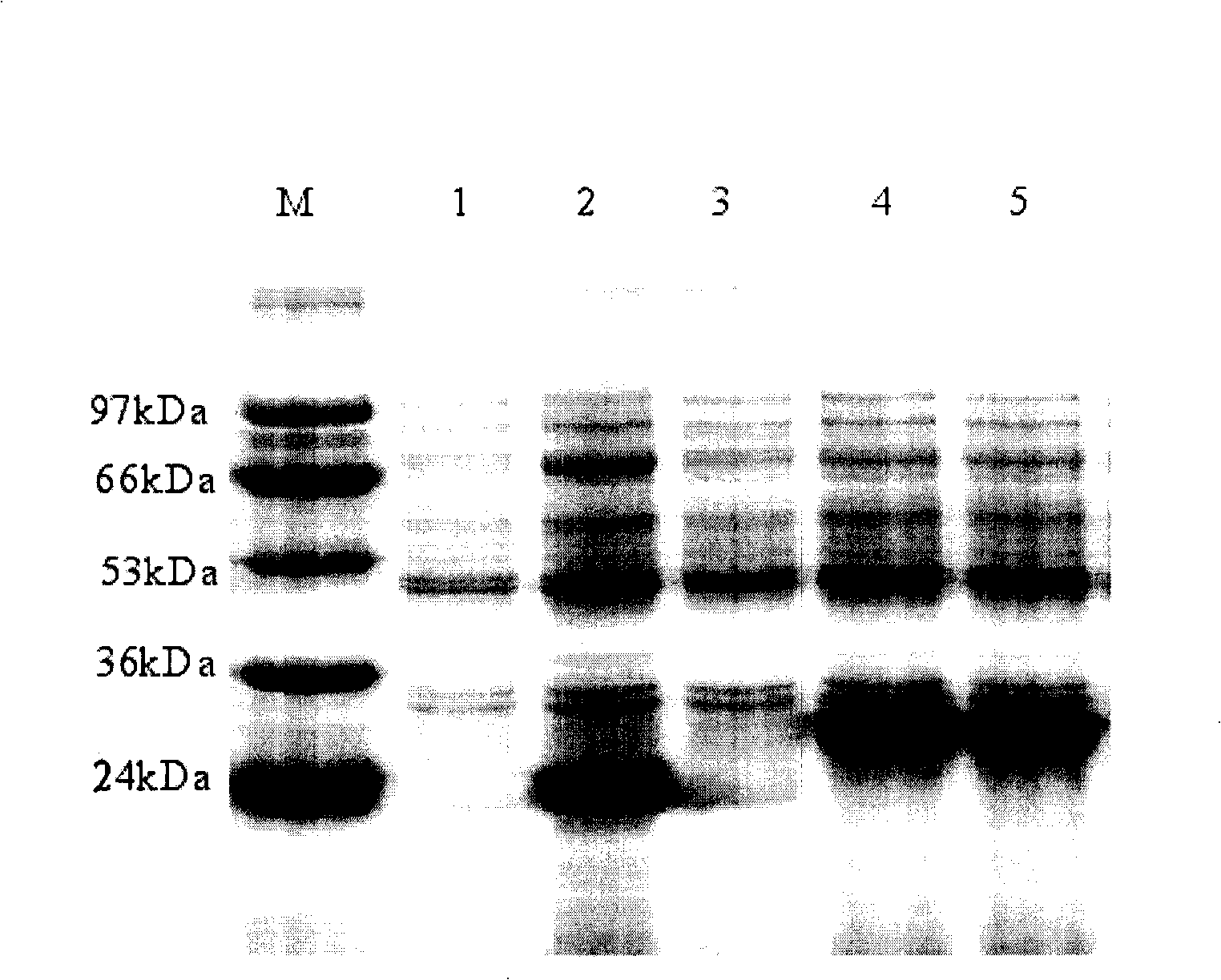
[0076] Example 2 Preparation of anti-hGTA monoclonal antibody
[0077] 1) Preparation of monoclonal antibody against ALB antigen
[0078] 1. Test material:
[0079] 1) Human hGTA antigen: prepared in Example 1.
[0080] 2), experimental animals: six-week-old BALB / c mice were purchased from the Animal Experiment Center of Harbin Medical University;
[0081] 3), DMEM sugar medium: Hclone company
[0082] 4), other reagents: Freund's complete adjuvant, prepared by yourself
[0083] 2. Preparation method:
[0084] 1) Immunized animals:
[0085] Eight-week-old female BALB / C mice were immunized by intraperitoneal injection with an equal volume of human hGTA antigen and Freund's complete adjuvant, once a week, at a dose of 100ug / mice, and each mouse was intraperitoneally injected with 0.5ml. Then, the mice were immunized twice consecutively at an interval of 1 week. The dose and immunization method were the same. The adjuvant was changed to incomplete adjuvant. The antigen was ...
Embodiment 3
[0094] Example 3 Enzymatic labeling of anti-hGTA monoclonal antibodies
[0095] Weigh 2mg HRP and dissolve it in 1ml ultrapure water, add 30ul freshly prepared 0.1M NaIO 4 Put the above solution into a dialysis bag with an interception molecular weight of 8000, and dialyze the sodium acetate buffer of 2mM pH4.4 overnight; add 30 μl of 0.2M PH9.5 carbonate buffer to adjust the pH of the hydroformylation HRP to increase When it reaches 9.0~9.5, immediately add 0.5ml of anti-hGTA antibody, shake gently on a destaining shaker for 2 hours at room temperature in the dark; add 40ul of newly prepared 4mg / ml NaBH 4 solution, mix well, and set at 4°C for 2 hours. The above-mentioned solution was put into a dialysis bag with a molecular weight of 8000, dialyzed against 0.01M PH8.2 phosphate buffer overnight, and purified by the ammonium sulfate method.
PUM
| Property | Measurement | Unit |
|---|---|---|
| molecular weight | aaaaa | aaaaa |
Abstract
Description
Claims
Application Information
 - R&D
- Intellectual Property
- Life Sciences
- Materials
- Tech Scout
- Unparalleled Data Quality
- Higher Quality Content
- 60% Fewer Hallucinations
Browse by: Latest US Patents, China's latest patents, Technical Efficacy Thesaurus, Application Domain, Technology Topic, Popular Technical Reports.
© 2025 PatSnap. All rights reserved.Legal|Privacy policy|Modern Slavery Act Transparency Statement|Sitemap|About US| Contact US: help@patsnap.com