Tissue engineering tendon and vitro construction method thereof

A tissue engineering and tendon technology, applied in the fields of medicine and biomedical engineering, can solve the problems of poor seed cell survival, inability to accurately measure the magnitude and frequency of tension, and inability to obtain a high success rate.

Inactive Publication Date: 2008-12-31
SHANGHAI TISSUE ENG LIFE SCI
View PDF1 Cites 13 Cited by
  • Summary
  • Abstract
  • Description
  • Claims
  • Application Information

AI Technical Summary

Problems solved by technology

[0005] In addition, the current research on tissue engineering tendons at home and abroad is mainly focused on the repair experiments in animals, that is, a sufficient number of seed cells are amplified in vitro, and immediately after compounding with biomaterials or only cultured in vitro for one to two weeks, they are implanted into the defect in the body. Although some preliminary successes have been achieved, there are still some major defects in the way of implanting the scaffold material seeded with cells directly back into the body to form tendons: including: (1) the implanted cell-material complex is not Tendon grafts, resulting in poor survival of the seed cells, causing graft failure and not achieving a consistently high success rate
(2) The implanted scaffold is a material that has not been degraded, and its decomposition products are mostly acidic, which can directly lead to inflammatory reactions, scar formation and tendon adhesion, thus directly affecting the repair effect of tendon
(3) Since the non-tendon graft is implanted, with the degradation of the material, the mechanical properties of the cell-material composite are greatly reduced, and the tendon defect in the high-tension area cannot be repaired
Cao Dejun et al. used the absorbable biomaterial polyglycolic acid (polyglycolic acid, PGA) and tenocytes to construct tissue-engineered tendon in vitro, and then cultured the cell-PGA complex in vitro with a U-shaped spring. Tendon-like tissue with a similar structure and certain mechanical properties, but this method cannot accurately measure parameters such as the magnitude and frequency of the applied tension, and the mechanical properties of the constructed tissue are extremely weak

Method used


Image

  • Tissue engineering tendon and vitro construction method thereof
  • Tissue engineering tendon and vitro construction method thereof
  • Tissue engineering tendon and vitro construction method thereof

Examples


Embodiment 1

[0111] Isolation and culture of skin fibroblasts

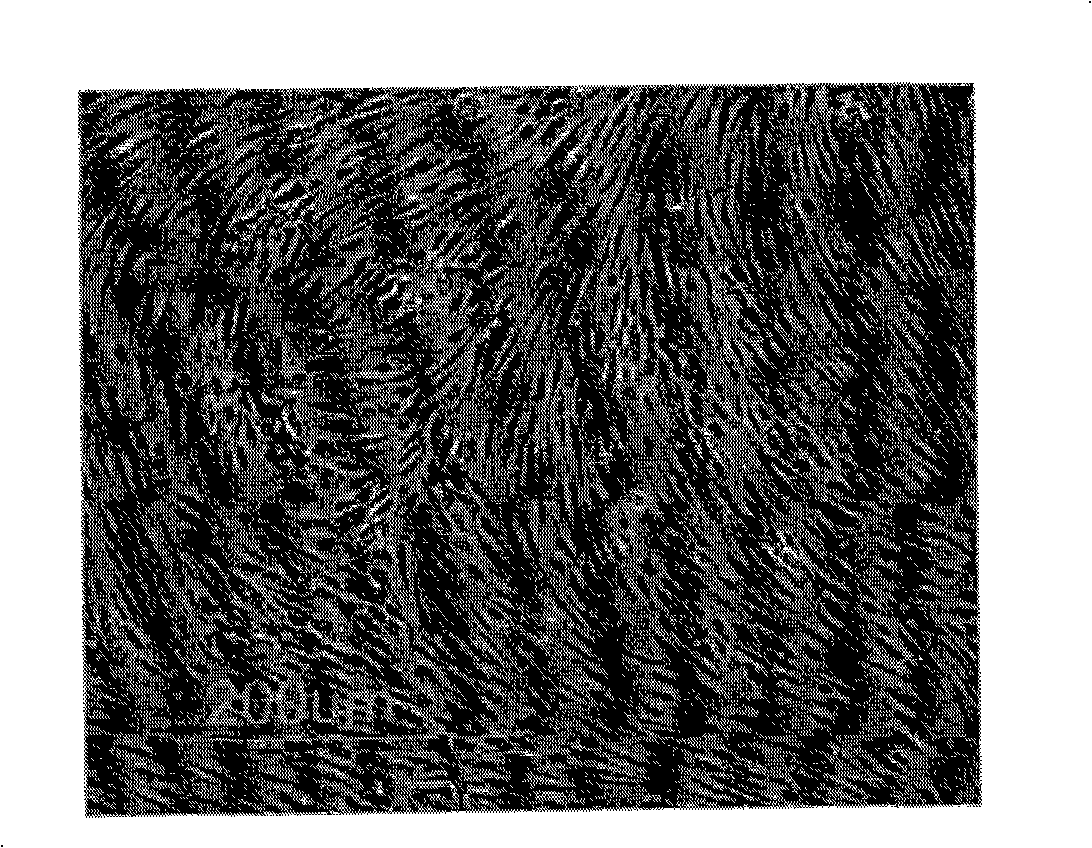
[0112] The foreskin tissue discarded during the operation was taken under aseptic conditions and cut into 2×2×2mm 3 Large and small tissue pieces were washed twice with phosphate buffer (PBS, containing 100 U / ml each of penicillin and streptomycin), and double volume of 1 mg / ml type II collagenase (purchased from Worthington, Freehold, NJ, USA) was added. Digest in a constant temperature shaker at 37°C for 4 hours, filter and centrifuge with a 150-mesh nylon mesh, wash the precipitated cells twice with PBS, count them, and check the viability of fibroblasts by staining with trypan blue at 1×10 6 / dish density (petri dish diameter 100mm) culture cell, culture fluid is DMEM (purchased from Gibco, Gland, Island, NY, USA) containing 10% fetal bovine serum, L-glutamine 300ug / ml, vitamin C50ug / ml, Each of penicillin and streptomycin was 100U / ml, and the fibroblasts were transferred to the second generation, and the cells were coll...

Embodiment 2

[0114] Isolation and culture of tenocytes

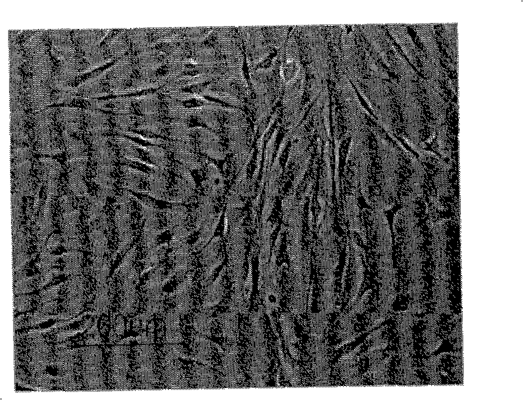
[0115] Under aseptic conditions, take the discarded human tendon tissue after fresh amputation and cut it into 2×2×2mm 3The large and small tissue pieces were washed twice with phosphate buffer solution (PBS, containing 100 U / ml each of penicillin and streptomycin), and added double volume of 0.25 mg / ml type II collagenase (purchased from Worthington, Freehold, NJ, USA) , placed in a constant temperature shaker at 37°C for digestion, after 4, 5, and 7 hours respectively, filter and centrifuge with a 150-mesh nylon mesh, wash the precipitated cells twice with PBS and count, trypan blue staining to check the viability of tenocytes, with 1× 10 6 / dish density (petri dish diameter 100mm) culture cell, culture fluid is DMEM (purchased from Gibco, Gland, Island, NY, USA) containing 10% fetal bovine serum, L-glutamine 300ug / ml, vitamin C50ug / ml, Penicillin, streptomycin each 100U / ml, tenocytes passed to the second generation, digested wi...

Embodiment 3

[0117] Isolation and culture of adipose-derived cells

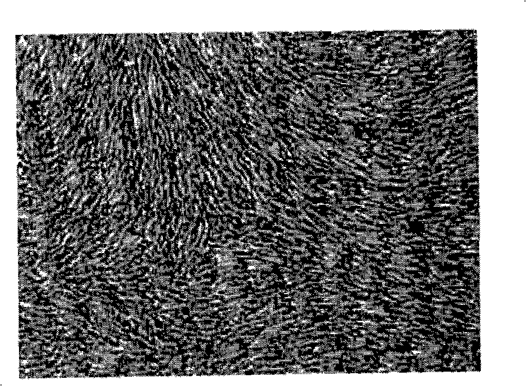
[0118] Under aseptic conditions, the discarded human tendon tissue after liposuction was transferred to a culture bottle, rinsed repeatedly with normal saline, and an equal volume of 0.075% type I collagenase (purchased from Worthington, Freehold, NJ, USA) was added. , placed in a constant temperature shaker at 37°C for digestion, and after 1 hour, centrifuge at 300g for 10 minutes to obtain a high-density cell pellet, discard the supernatant and floating adipose tissue, shake the cells, and count them as 1×10 6 Cells were cultured at a density of 1 / dish (petri dish diameter 100 mm), and the culture medium was DMEM (purchased from Gibco, Gland, Island, NY, USA) containing 10% fetal bovine serum, placed in a room at 37° C., 5% CO 2 , and 100% saturated humidity. cultured under the same conditions, the medium was changed for the first time after 24 hours, the floating cells were washed repeatedly with PBS, and the culture m...


PUM


Abstract

The invention discloses an organ engineering tendon graft which comprises (a) biodegradable materials which can be accepted in pharmacy; (b) seed cells which can be inoculated to the biodegradable materials, and the seed cells are selected from the following groups: (i) fibroblasts, (ii) adipose-derived cells, or (iii) the mixture of the dermal fibroblasts and the adipose-derived cells with the ratio of 1:10000 to 10000:1. The graft is gotten from the cultivation of a complex of the seed cells and the biodegradable materials in a bioreactor, and the complex is obtained through the mixing of the seed cells and the biodegradable materials which can be accepted in pharmacy. The graft can be used for restoring the tendon defect.

Description

technical field [0001] The invention relates to the fields of medicine and biomedical engineering, and more specifically relates to a preparation method and application of using dermal fibroblasts and (or) adipose-derived cells to construct tissue-engineered tendons in vitro. Background technique [0002] Tendon is a bundle of dense collagen fibrous connective tissue that connects the belly of skeletal muscle to bone, and is an integral part of skeletal muscle. The structure of the tendon can be regarded as the continuation and degeneration of the muscle. The tendons of fresh specimens are silvery-white, cord-like, and tough in texture. There are ripples on the surface in the relaxed state. If the tension exceeds 4% of the relaxed state, the ripples will disappear, and the ripples will reappear after the tension is removed. At the muscle-tendon junction, the collagen fibers enter the endomysium and attach to the sarcolemma of the muscle fibers. At the tendon-bone junction,...

Claims


Application Information

Patent Timeline
Patent Type & Authority Applications(China)
IPC IPC(8): A61L27/38A61L27/18A61F2/08C12N5/077
CPCA61F2/08A61L27/3804A61L27/3834A61L27/386A61L27/3886A61L27/58A61L2430/10A61P19/00A61P19/04C12N5/066
Inventor 曹谊林刘伟许锋邓丹李宏
Owner SHANGHAI TISSUE ENG LIFE SCI
Why Eureka
Try Eureka