Degradable medical hemostatic non-viscous material and preparation method thereof

An anti-adhesion and preparation process technology, applied in the field of medicine, can solve the problems of lack of hemostasis and wound repair, poor hemostasis effect, slow hemostasis, etc., to achieve hemostasis and anti-adhesion wound healing, promotion of wound healing, and less pain.

Inactive Publication Date: 2009-09-23
JINAN UNIVERSITY
View PDF1 Cites 30 Cited by
  • Summary
  • Abstract
  • Description
  • Claims
  • Application Information

AI Technical Summary

Problems solved by technology

At present, these materials still have the disadvantages of possibly causing infection of human or animal blood-borne diseases, complex use, slow hemostatic speed, and poor hemostatic effect when there is systemic coagulation dysfunction, while some anti-adhesion products on the market Does not have the effect of hemostasis and wound repair

Method used


Image

  • Degradable medical hemostatic non-viscous material and preparation method thereof
  • Degradable medical hemostatic non-viscous material and preparation method thereof
  • Degradable medical hemostatic non-viscous material and preparation method thereof

Examples


Embodiment 1

[0040] Example 1 Preparation of microsphere-type hemostatic and anti-adhesion materials

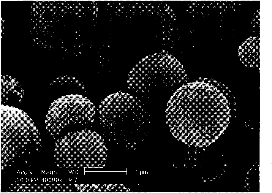
[0041] 1g chitosan was dissolved in 10mL, 10wt% hydrochloric acid aqueous solution, added 40mLNH 4 HCO 3 Aqueous solution (containing NH 4 HCO 3 9.6g), stand at 20°C for 3 days, centrifuge at 12,000 rpm, discard the supernatant, add 200mL water to the precipitate to dilute, stir at high speed for 30s, add 100ml sodium hyaluronate aqueous solution (containing 0.2g sodium hyaluronate), stir To obtain the chitosan-hyaluronic acid complex solution, the complex solution was spray-dried at 150° C. to obtain microsphere-type hemostatic and anti-adhesion materials. The results of the scanning electron microscope (Philips ESEM-30) are shown in figure 1 .

[0042] New Zealand white rabbits underwent epistaxis after partial turbinectomy, and 1 g of the microsphere-type hemostatic and anti-adhesion material prepared in this example was sprayed on the wound surface. The control group used high-exp...

Embodiment 2

[0043] Example 2 Preparation of microsphere-type hemostatic and anti-adhesion materials

[0044] Dissolve 1g of phosphorylated chitosan in 50mL of water, add 100mL of sodium hyaluronate aqueous solution (containing 0.5g of sodium hyaluronate) dropwise, stir to obtain a chitosan-hyaluronic acid complex solution, and spray the complex solution at 150°C Dry to obtain the microsphere-type hemostatic and anti-adhesion material.

[0045]A bleeding wound of about 1cm×0.5cm in size was made on the kidney surface of New Zealand rabbits, and 1g of the microsphere-type hemostatic and anti-adhesion material prepared in this example was used to stop bleeding. The blood loss was 75±12s, the blood loss was 0.8±0.1mL, and no intraoperative adhesion occurred within 30 days.

Embodiment 3

[0046] Example 3 Preparation of microsphere-type hemostatic and anti-adhesion materials

[0047] 1g of chitosan was dissolved in 50mL of acetic acid aqueous solution (containing 0.5g of acetic acid), and the chitosan solution was added dropwise to 100mL of sodium hyaluronate aqueous solution (containing 1g of sodium hyaluronate) using an electrostatic droplet generator, and centrifuged to obtain Chitosan-hyaluronic acid gel microspheres are sequentially immersed in chitosan and sodium hyaluronate aqueous solution to assemble 6 layers of polyelectrolyte membranes on the surface of the microspheres, centrifuged and freeze-dried to obtain microsphere-type hemostatic and anti-adhesion materials.

[0048] Nosebleeds occurred in New Zealand white rabbits after part of the maxillary turbinate was removed. Spray 1 g of the microsphere-type hemostatic and anti-adhesion material prepared in this example on the wound surface. The hemostatic time was 160±10s, and no nosebleed occurred with...


PUM


Abstract

The invention discloses a degradable medical hemostatic adhesion-resistant material and a preparation method thereof. The invention uses chitosan or derivatives thereof and transparent sodium hyaluronate as main raw materials, adopts methods, such as spray drying, self lamination assembly, and the like to prepare microspheric and gel-type hemostatic adhesion-resistant materials, and medicines and activated substances capable of stimulating hemostasis and antiphlogosis can be compounded and added in the preparation process. The material has high water absorption rate, can rapidly arrest bleeding to form an aquagel protective layer on a wound surface, has favorable tissue adhesiveness, adhesion resistance and bacterinertness, can suit a wound in an irregular shape, truly fully covers the wound surface to fully take the aims of arrest bleeding, resisting adhesion and stimulating wound surface healing, has remarkable postoperative hemostatic adhesion-resistant effects while being used for ear-nose-throat department operations, is convenient to use and brings little pain to patients.

Description

technical field [0001] The invention relates to the technical field of medicine, in particular to a degradable medical hemostatic and anti-adhesion material and a preparation method thereof. Background technique [0002] In the first aid treatment of sudden accidents in daily life, the hospital's wound hemostasis in the operation process of patients, and the rescue of wounded soldiers in war, it is very important to effectively and quickly hemostasis the patient locally. Tissue adhesions often occur after abdominal, gynecological, orthopedic, cardiothoracic and orthopedic surgery, and can lead to serious clinical complications, such as intestinal obstruction, abdominal and pelvic pain, infertility, etc., increasing the difficulty of reoperation. The current hemostatic materials usually accelerate the blood coagulation process through embolization, contraction of arterioles and capillaries, and enhancement of platelet function. Clinically used local hemostatic materials mainl...

Claims


Application Information

Patent Timeline
Patent Type & Authority Applications(China)
IPC IPC(8): A61L15/28A61L15/50A61L15/60A61L15/44A61L15/46C08J3/12C08L5/08
Inventor 周长忍鲁路李垒
Owner JINAN UNIVERSITY
Why Eureka
Try Eureka