Method for detecting titre of live viruses

A virus titer and virus technology, which is applied to measurement devices, material analysis, instruments and other directions by observing the impact on chemical indicators, can solve the problems of increasing virus titers, long detection time, and large differences in measured results. , to achieve the effect of shortening the culture time, fast detection speed, and low detection cost

Inactive Publication Date: 2009-09-30
BEIJING WANTAI BIOLOGICAL PHARMACY ENTERPRISE
View PDF0 Cites 10 Cited by
  • Summary
  • Abstract
  • Description
  • Claims
  • Application Information

AI Technical Summary

Problems solved by technology

There are several defects in this method: 1. The detection time is long, which generally takes 8-10 days; 2. The accuracy is not high. When the virus-infected cells are cultured for 8-10 days, adjacent plaques may unite together, reducing the If the virus titer is lowered, it is also possible that the mother plaque virus produces daughter plaques, which increases the virus titer, which leads to poor repeatability of the test; 3. The number of plaques is only the number, and there is no specificity, so it

Method used


Image

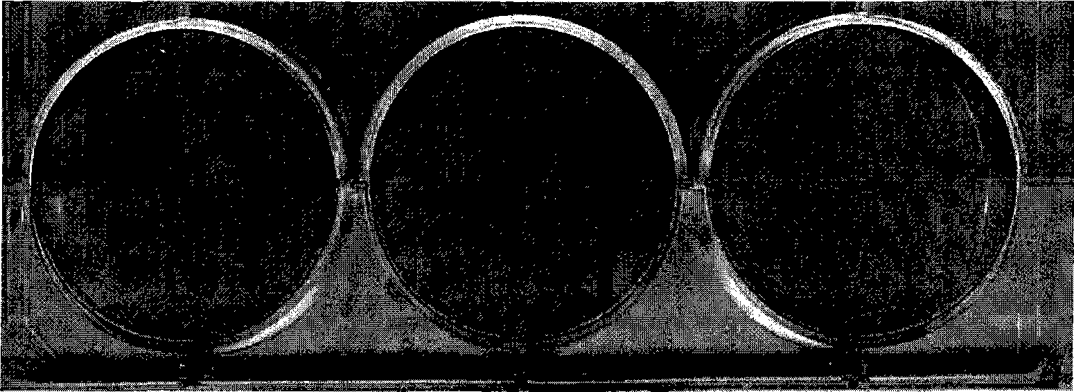
  • Method for detecting titre of live viruses
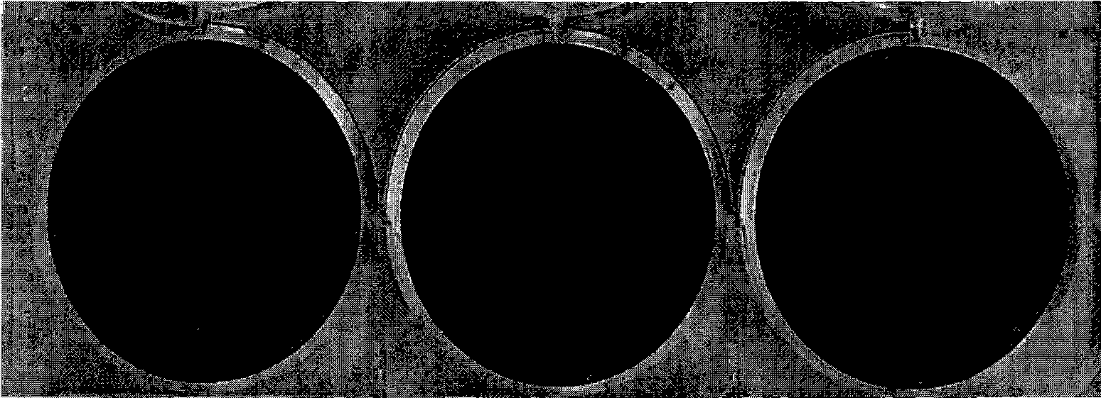
  • Method for detecting titre of live viruses

Examples


Embodiment 1

[0032] Embodiment 1 Adopt the method of the present invention to measure the titer of varicella-zoster virus

[0033] (1) Cell culture

[0034] Diploid cells (MRC-5, 2BS or WI-38 cells) were inoculated into 6-well cell culture plates at a seeding ratio of 1:2, at 37°C in 5% CO 2 The incubator cultured to a monolayer of cells.

[0035] (2) Virus infection and culture

[0036] Dilute the varicella-zoster virus to be detected (purchased from ATCC) with diluent (1xPBS, containing 5% sucrose, 1% sodium glutamate and 10% calf serum) at 1:100, take 0.1ml for infection Discard the cells in the 6-well plate of the culture medium, shake gently, and place at 37°C with 5% CO 2Incubate in an incubator for 40 minutes, shake well 1-2 times during the period, add virus maintenance solution, 3ml / well, and place the cell plate at 37°C, 5% CO 2 Incubator for 3 days.

[0037] (3) Culture fixation

[0038] The virus maintenance solution in the 6-well plate was discarded, and the culture was ...

Embodiment 2

[0050] Embodiment 2 Adopt the method of the present invention to measure the titer of rabies virus

[0051] (1) Cell culture

[0052] KMB17 cells were inoculated into 96-well cell culture plates at a seeding ratio of 1:2, at 37°C and 5% CO 2 The incubator cultured to a monolayer of cells.

[0053] (2) Virus infection and culture

[0054] After the rabies virus to be detected (quoted from Kansas State University) was serially diluted 10 times with the virus maintenance solution, the microplate cells were infected and the culture solution was discarded. Each dilution infected 6 wells, 0.1ml per well, and shook well. Afterwards, place at 37 °C 5% CO 2 Incubator for 1 day.

[0055] (3) Culture fixation

[0056] Discard the virus maintenance solution in the 96-well plate, inactivate the fixed culture with 0.1% formaldehyde and 0.1% glutaraldehyde solution, 0.1ml / well, inactivate and fix at 33°C for 26 hours.

[0057] (4) Treatment of fixed cells with hydrogen peroxide

[005...


PUM


Abstract

The invention discloses a method for detecting the titre of live viruses, which comprises the following steps: firstly, infecting diploid cells or other adherent cells with dilute viruses to be detected, culturing and fixing the infected cells; secondly, adding virus antiserum (first antibody) into the fixed cells, culturing and washing, adding a second antibody marked by horseradish peroxidase (HRP), culturing and washing, adding coloring solution for color development and producing red or brown spots, or adding virus antiserum marked by the HRP into the fixed cells, culturing the virus antiserum and washing, adding the coloring solution for color development and producing red or brown spots; thirdly, calculating the virus titre according to the quantity of the red or brown spots and the dilution multiple, or calculating the 1gTCID50 value of the viruses according to the dilution degree and the hole numbers of the spots. The detection method can be applicable to the titre detection of various live viruses, can shorten the detection time of the titre of the viruses due to fast detection speed, has good specificity as the immune spots are caused by special immune reaction, low detection cost as well as simple and convenient operation and does not need special instruments of optical or fluorescent microscopes, and the like or other reagents.

Description

technical field [0001] The invention relates to a method for detecting the titer of a live virus, in particular to a method for detecting the titer of a live virus by using an enzyme-linked immunospot method, and belongs to the field of biopharmaceuticals. Background technique [0002] So far, the detection methods for live virus titer mainly include plaque method, endpoint dilution method and enzyme-linked immunofluorescence method. The main detection process and advantages and disadvantages of the three detection methods are as follows: [0003] Plaque method: This method is the most classic method for determining virus titers. Generally speaking, one plaque is produced by one virus particle, expressed in plaque forming units (PFU). Taking varicella virus as an example, 2BS or MRC-5 cells in good growth state in the culture flask are subcultured to 6-well cell culture plates, 37°C, 5% CO 2 Culture in an incubator until the cells form a monolayer for the experiment. Disca...

Claims


Application Information

Patent Timeline
IPC IPC(8): G01N33/577G01N33/569G01N21/78
Inventor 李益民叶祥忠
Owner BEIJING WANTAI BIOLOGICAL PHARMACY ENTERPRISE
Why Eureka
Try Eureka