
Surface modification method of bio-absorbable material implanted in polyester

An implant material and surface modification technology, used in prosthesis, medical science, surgery, etc., can solve the problems of small number of holes, difficult to achieve tissue fluid, nutrient transport and exchange, unfavorable cell adhesion and proliferation, etc.

Active Publication Date: 2014-11-26
CHANGCHUN SINOBIOMATERIALS
View PDF1 Cites 0 Cited by
  • Summary
  • Abstract
  • Description
  • Claims
  • Application Information

AI Technical Summary

Problems solved by technology

[0004] At present, various medical devices are made of bioabsorbable implant materials in vivo, which have disadvantages such as poor mechanical properties and slow degradation performance, mainly because the implant materials in vivo have a very dense and smooth surface, which is not conducive to cell adhesion and proliferation
Although it is also possible to improve cell adhesion and proliferation by doping osteoinductive inorganic particles such as hydroxyapatite into it, the current general practice is to spray hydroxyapatite particles on the outer surface of medical devices to achieve surface rough purpose
However, this does not improve cell adhesion very well, and it is difficult to realize the transportation and exchange of interstitial fluid and nutrients.
[0005] In addition, there is a method of punching holes on the surface of medical devices to improve cell adhesion and proliferation. However, due to limitations such as too low porosity, a small number of holes, and no direct connection with each other, cells are not easy to uniformly flow into the inside of medical devices. growth, it is difficult to achieve synchronization between the degradation of materials and the replacement of osteoblasts

Method used


Image

  • Surface modification method of bio-absorbable material implanted in polyester
  • Surface modification method of bio-absorbable material implanted in polyester
  • Surface modification method of bio-absorbable material implanted in polyester

Examples


Embodiment 1

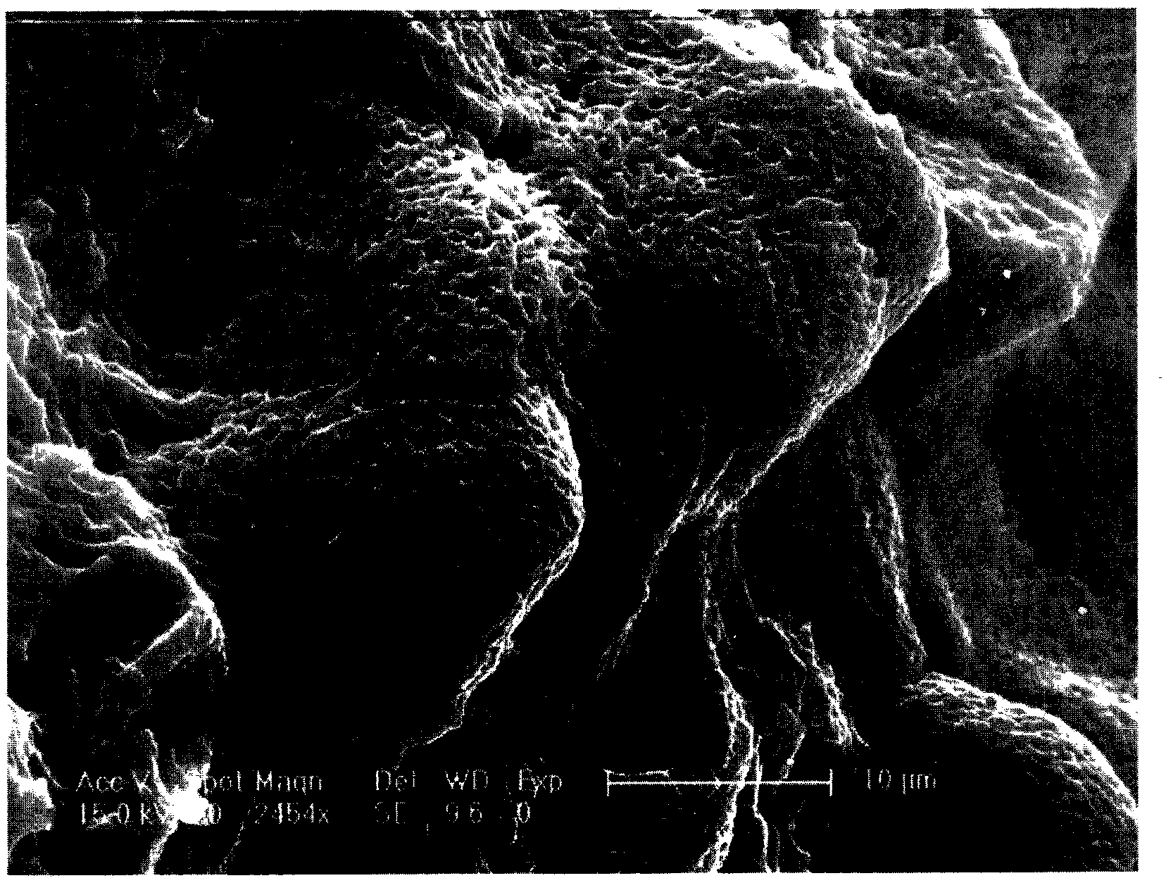
[0038] 1. Put the polylactic acid (PLA) absorbable vertebral body fusion device in the autoclave;

[0039] 2. Put the reaction kettle into the oil bath and adjust the temperature to 55°C;

[0040] 3. Open CO 2 Cylinder gas valve, open the supercritical fluid pump, and the reactor inlet valve, close the reactor vent valve; pass CO into the reactor 2 , and repeated ventilation several times;

[0041] 4. Adjust the supercritical fluid pump output CO 2 Pressure constant pressure to 20MPa;

[0042] 5. Keep the reactor at a constant temperature of 55°C and a constant pressure of 20MPa for 1 hour;

[0043] 6. After the reaction is over, depressurize and remove CO 2 , the control time is 10 minutes.

[0044] 7. Take out the surface-modified absorbable vertebral fusion cage, dry it in vacuum, and detect the surface changes by field emission scanning electron microscopy (ESEM). Such as figure 1 shown.

Embodiment 2

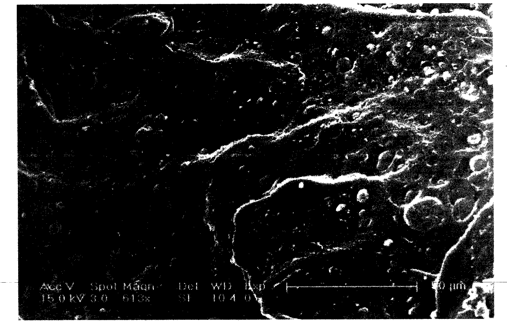
[0046] 1. Put polylactic acid (10% HA / PLA) grafted 10% hydroxyapatite absorbable thoracic fusion device in an autoclave;

[0047] 2. Put the reaction kettle into the oil bath and adjust the temperature to 55°C;

[0048] 3. Open CO 2 Cylinder gas valve, open the supercritical fluid pump, and the reactor inlet valve, close the reactor vent valve; pass CO into the reactor 2 , and repeated ventilation several times;

[0049] 4. Adjust the supercritical fluid pump output CO 2 Pressure constant pressure to 20MPa;

[0050] 5. Keep the reactor at a constant temperature of 55°C and a constant pressure of 20MPa for 1 hour;

[0051] 6. After the reaction is over, depressurize and remove CO 2 , the control time is 10 minutes.

[0052] 7. The surface-modified absorbable thoracic fusion cage was taken out, dried in vacuum, and the surface changes were detected by a field emission scanning electron microscope (ESEM). Such as figure 2 shown.

Embodiment 3

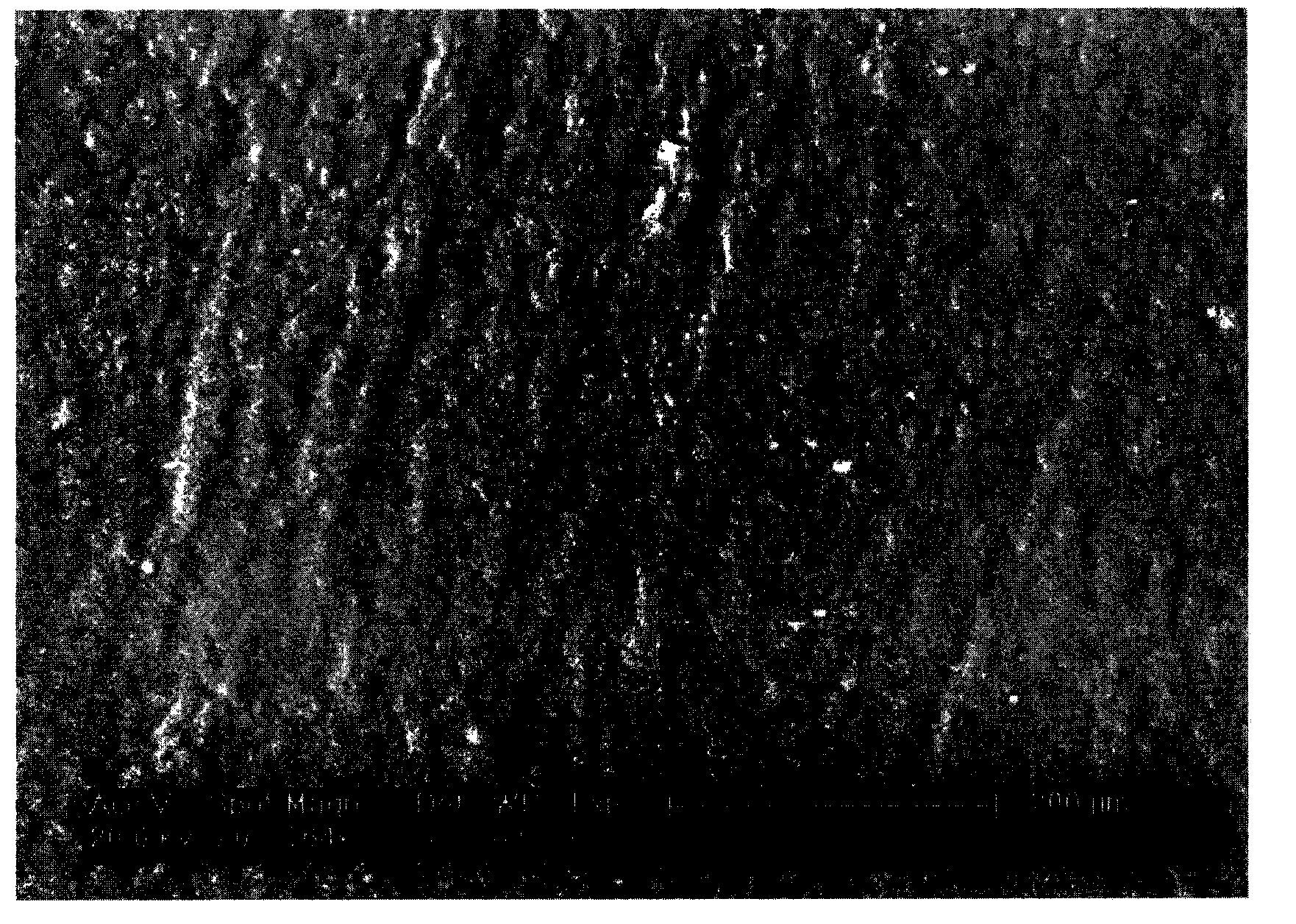
[0054] 1. Put polylactide glycolide (PLGA) in vivo absorbable vascular stent into the autoclave;

[0055] 2. Put the reaction kettle into the oil bath and adjust the temperature to 45°C;

[0056] 3. Open CO 2 Cylinder gas valve, open the supercritical fluid pump, and the reactor inlet valve, close the reactor vent valve; pass CO into the reactor 2 , and repeated ventilation several times;

[0057] 4. Adjust the supercritical fluid pump output CO 2 Pressure constant pressure to 20MPa;

[0058] 5. Keep the reactor at a constant temperature of 45°C and a constant pressure of 20MPa for 3 hours;

[0059]6. After the reaction is over, depressurize and remove CO 2 , the control time is 10 minutes.

[0060] 7. Take out the surface-modified absorbable vascular stent, dry it in vacuum, and detect the surface change by a field emission scanning electron microscope (ESEM). Such as image 3 shown.


PUM


Abstract

The invention discloses a surface modification method of a bio-absorbable material implanted in a polyester, and the bio-absorbable material implanted in the polyester is treated by supercritical fluid. The invention utilizes the supercritical carbon dioxide technology to modify the surface of various existing internally absorbable implanted materials, so as to form holes with nanometer size or micron size on the surface of the material, the invention solves the problem that the interface of existing internally absorbable implanted material is not suitable for cell adhesion and proliferation, thereby facilitating protein adsorption and deposition of extracellular matrix, and integration with the surrounding tissues; moreover, the invention can form tiny blood vessels timely, and can achieve good synchronization of material degradation and replacement of bone cells.

Description

technical field [0001] The invention relates to a surface modification method, in particular to a surface modification method for bioabsorbable polyester internal implant materials. Background technique [0002] Bioabsorbable materials for implantation in the body are materials that can automatically break molecular chains under physiological conditions, change from large molecules to small molecules, from insoluble substances to soluble substances, and finally be gradually metabolized or absorbed by the body. At present, the more commonly used in vivo absorbable materials include polyester in vivo absorbable polymer materials, such as polylactic acid, polyglycolide, polyglycolide-lactide copolymer, etc., and natural polymers, such as collagen, shell Polysaccharides, alginates, etc. [0003] Bioabsorbable implant materials in the body, which can be used to make various medical devices, such as in vivo degradable artificial teeth, skin, joints, kidneys, pancreas, heart, hear...

Claims


Application Information

Patent Timeline
Patent Type & Authority Patents(China)
IPC IPC(8): A61L27/18A61L27/56A61L27/58A61L31/06A61L31/14
Inventor 章培标崔立国陈学思崔毅高战团王宇王宗良
Owner CHANGCHUN SINOBIOMATERIALS
Learn More