
Expression vector for stably indicating cell autophagy activities, establishing method and application method thereof

A technology for expressing vectors and cells, which is applied in the field of biomedicine, can solve problems such as the distortion of fluorescent spot observation, difficulty in screening stable strains and amplification, increase the difference in biological characteristics between transfected cells and mother cells, and achieve quantitative analysis of autophagy The method is simple and easy to use, the indication of autophagy is sensitive and reliable, and the effects on a wide range of cell types

Inactive Publication Date: 2010-11-24
ZHONGSHAN HOSPITAL FUDAN UNIV
View PDF0 Cites 13 Cited by
  • Summary
  • Abstract
  • Description
  • Claims
  • Application Information

AI Technical Summary

Problems solved by technology

Since these observation methods are to observe and analyze autophagosomes or related key protein molecules in cells at a certain time point, real-time dynamic observation cannot be realized. In addition, these methods have other shortcomings: ① Transmission electron microscopy: the earliest application The classic method, the observation result is reliable, but it needs to rely on the transmission electron microscope and technicians who are proficient in autophagy to complete the image observation analysis and result judgment, and the transmission electron microscope and technicians are scarce in China, and the laboratory usually does not have it, and the observation often needs to make an appointment to wait , coupled with the cumbersome process of making observation samples, it is very inconvenient to observe
However, although the lentiviral vector system overcomes many deficiencies of traditional vectors and transfection methods, the lentiviral expression vectors used in current research reports still have shortcomings relative to the special field of autophagy research.
Its shortcomings are mainly manifested in the lack of expression plasmid function of the expression vector and the lack of fast and effective quantitative analysis and other application methods: ①The expression plasmid of the expression vector adopts CMV promoter: practical application shows that the activity of CMV promoter is too high for the expression of GFP-LC3, which is easy to Overexpression, overexpression of GFP-LC3 On the one hand, for autophagy research, due to the formation of an excessively strong fluorescent background, the observation of fluorescent spots is distorted, affecting autophagy indication and quantitative analysis; on the other hand, for cells, the GFP-LC3 gene is continuously overexpressed High intracellular pressure (stress) is formed, leading to a higher degree of cytotoxicity, affecting cell growth, and cells are prone to death after subculture, and it is difficult to screen stable strains and expand; in addition, the CMV promoter may increase the number of transfected cells and mother cells. differences in the biological characteristics of
These deficiencies of CMV require a relatively mild promoter; ② GFP is often used as a marker protein: GFP fluorescence is relatively weak. In order to obtain clear images and good observation, it is usually necessary to increase the ratio of virus / cell, and the increased amount of virus will lead to cell death The cell viability is greatly affected due to the excessive virus attack, and the background autophagy level is increased, which affects the judgment of autophagy; ③The lack of corresponding stable indicators indicates the method of establishing cell lines: different cell types are The response of lentiviral expression vectors varies greatly, and different lentiviral expression vectors have their specificity in application. Especially for the establishment of cell lines used in autophagy research, it is necessary to instruct the cell autophagy when establishing cell lines. Minimize the impact, these need to provide a method for the establishment of cell lines corresponding to the expression vector; ④ lack of rapid and effective autophagy quantitative analysis methods: the traditional autophagy quantitative method is to manually count the GFP-LC3 autophagy fluorescent bright spots with the naked eye, and then manually count Cells, calculated to obtain the average number of fluorescent bright spots / cell
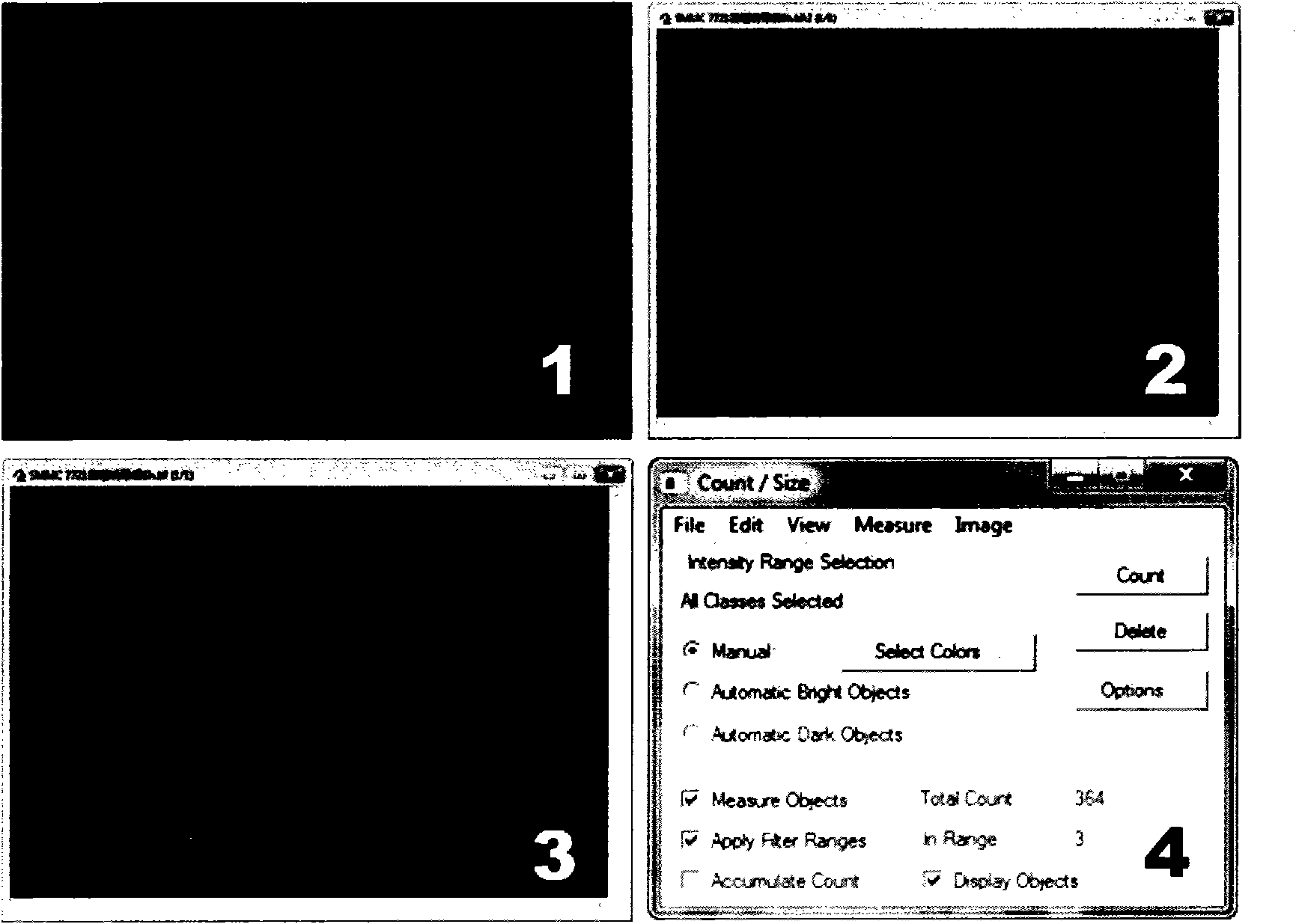
The manual counting workload is very heavy (the total number of counted cells in each treatment group is usually more than 200, and the total number of fluorescent bright spots is usually more than 1000), especially in multi-factor and large-sample experiments, the work is very heavy, and the counter is easy to see. Fatigue counting is wrong and the reliability is poor. At the same time, manual counting is subjective and arbitrary, and the quantitative analysis results have limited credibility due to lack of sufficient objectivity.
[0005] In short, the current expression vectors and related application methods cannot meet the needs of autophagy research, and there is an urgent need for new expression vectors and their application methods that can overcome the shortcomings of current expression vectors and methods. The expression vector should be able to stably and efficiently indicate cell autophagy activity. It can be used to establish a stable and reliable autophagy indicator cell line and quickly and conveniently quantitatively analyze the changes of autophagy, so that real-time dynamic observation and analysis of cell autophagy activity can be carried out, so that the research work can be carried out quickly and smoothly

Method used


Image

  • Expression vector for stably indicating cell autophagy activities, establishing method and application method thereof
  • Expression vector for stably indicating cell autophagy activities, establishing method and application method thereof
  • Expression vector for stably indicating cell autophagy activities, establishing method and application method thereof

Examples


Embodiment 1

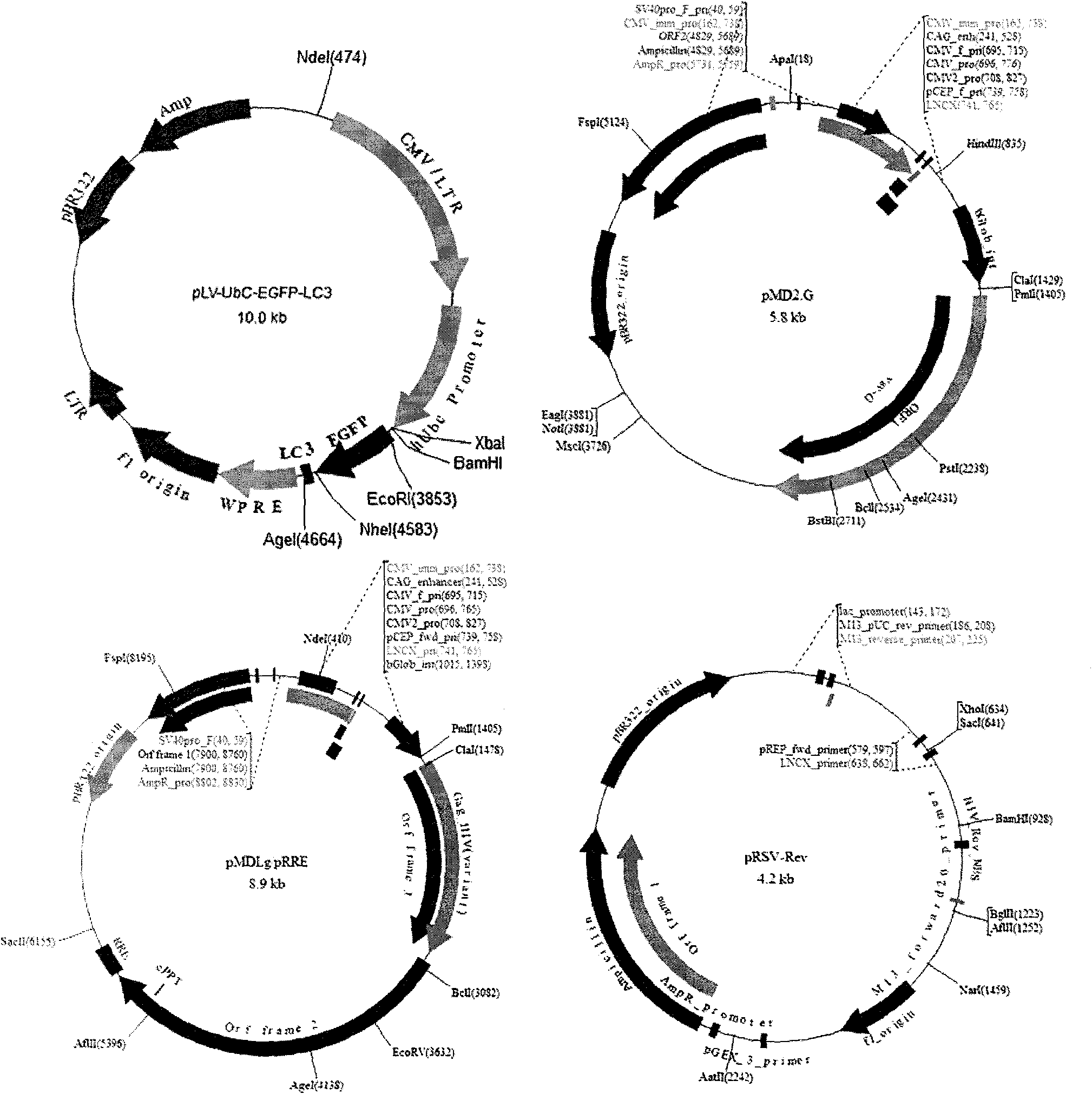
[0021] Example 1: This example is used to illustrate the construction process of an expression vector stably indicating autophagy activity of cells, and the application of expression vectors to establish autophagy indicator cells and quantitative analysis applications;

[0022] (1) Expression vector construction: ① Expression plasmid construction: NCBI obtained LC3 gene sequence information and artificially synthesized LC3 gene. EcoR V digested the pUC57 plasmid and synthesized the LC3 gene, and inserted the LC3 gene fragment into the pUC57 vector to construct pUC57-LC3. Transform competent cells, screen clones, extract plasmids by electrophoresis and KpnI, HindIII double enzyme digestion identification, and sequence to confirm the insertion product. The clones with correct sequencing were re-inoculated and amplified for plasmid extraction. pUC57-LC3 was double digested with Nhe I and Age I, and the 384bp LC3 gene fragment was recovered by gel after electrophoresis. The pLV-...

Embodiment 2

[0025] Embodiment 2 to 5 are in order to further illustrate the concrete application of the present invention

[0026] Example 2

[0027] Apply the expression vector of the present invention to infect difficult-to-transfect cells to establish corresponding autophagy indicator cell lines

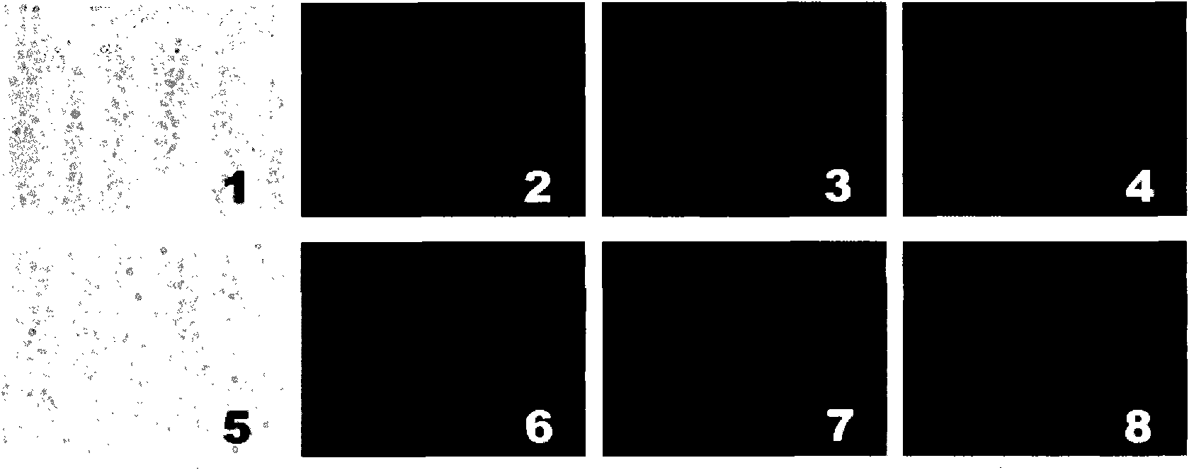
[0028] Some cell lines are difficult to transfect by common transfection methods (such as liposome method), and the transfection rate is usually less than 10%. If the cell line is sensitive to the toxicity of the transfection reagent, the transfection is successful. It is difficult to carry out follow-up experiments. At the same time, the background autophagy of the traditional transfection method is high ( Figure 4- left). In this example, HCCLM3 liver cancer cells, Caco-2 colon cancer cells and NIH3T3 fibroblasts are represented by three difficult-to-transfect cells, and the expression vectors of the present invention are used to establish corresponding autophagy indicator cells (while ...

Embodiment 3

[0030] Application of the present invention to carry out research on the correlation between drug resistance of liver cancer cells and autophagy

[0031] Tumor resistance to chemotherapeutic drugs is an important obstacle in tumor treatment, and its related research has always been a hot spot. Studies in recent years have shown that autophagy is an important mechanism of tumor cell chemotherapy resistance, and inhibition of autophagy increases chemotherapy sensitivity. The in vitro study of tumor drug resistance needs to detect the changes of autophagy of different cell lines under the action of different chemotherapeutic drugs and different drug doses. The workload of detection by conventional Western blot, transmission electron microscopy and other methods is very large, and the application of the present invention The workload can be greatly reduced and rapid screening can be completed. In this case, the effects of 8 different chemotherapeutic drugs at standard blood level...


PUM


Abstract

The invention belongs to the biomedicine field, and aims at providing an expression vector for stably indicating cell autophagy activities, an establishing method and an application method thereof. By constructing a slow-virus expression plasmid containing an EGFP-LC3 autophagy reporter gene and adopting a four-plasmid system, the invention establishes a slow-virus expression vector capable of enabling cells to stably express an EGFP-LC3 gene. By using the expression vector to infect a host cell, the EGFP-LC3 gene can be stably integrated in a genome of the host cell, and the host cell can stably and efficiently express the EGFP-LC3 and indicate cell autophagy changes. A stable autophagy indicating cell can be established by using the expression vector, and the cell autophagy changes are quickly and quantitatively analyzed by an image analysis method based on Top-hat operators and morphological filtration. The invention has the characteristics of stable and reliable autophagy indication, real-time sensitivity, quick and accurate autophagy quantification, simply and convenient operation and the like, greatly facilitates autophagy observation and analysis, and can be widely used for autophagy-correlated researches.

Description

technical field [0001] The invention belongs to the technical field of biomedicine, and relates to an expression vector stably indicating cell autophagy activity and its establishment and application method. Background technique [0002] Autophagy is a process of cell self-degradation and digestion, and is an important life activity of cells. Under normal physiological conditions, cells rely on autophagy to remove aging or damaged organelles and renew some important proteins; when cells are in a state of stress, autophagy activation is enhanced to maintain energy and metabolic homeostasis by recycling intracellular components , remove damaged proteins and organelles, limit oxidative damage, etc., are important mechanisms for maintaining cell homeostasis and survival. Because it is closely related to many human diseases, especially in the occurrence and development of major diseases such as tumors, it has become a research hotspot in recent years, and related research is inc...

Claims


Application Information

Patent Timeline
IPC IPC(8): C12N15/867C12N15/66C12N5/10C12Q1/70C12Q1/02G01N21/64G01N15/10
Inventor 樊嘉史颖弘彭远飞丁振斌
Owner ZHONGSHAN HOSPITAL FUDAN UNIV
Learn More