Flow cytometer apparatus for three dimensional diffraction imaging and related methods

A flow cytometer and cytometer technology, which is applied in the field of flow cytometer systems that can measure diffraction image signals and three-dimensional structural parameters, and can solve problems such as low signal-to-noise ratio, inability to apply image flow cytometers, and high particle flow rates. question

Inactive Publication Date: 2011-04-27
EAST CAROLINA UNIVERISTY
View PDF5 Cites 29 Cited by
  • Summary
  • Abstract
  • Description
  • Claims
  • Application Information

AI Technical Summary

Problems solved by technology

However, this technique generally requires many images, so it cannot be applied to image flow cytometry with fast particle flow
Additionally, existing flow cytometers have high particle flow rates and low signal-to-noise ratios, which limit the amount of information that can be extracted from scattered light and / or fluorescent signals

Method used


Image

  • Flow cytometer apparatus for three dimensional diffraction imaging and related methods
  • Flow cytometer apparatus for three dimensional diffraction imaging and related methods
  • Flow cytometer apparatus for three dimensional diffraction imaging and related methods

Examples


example 1

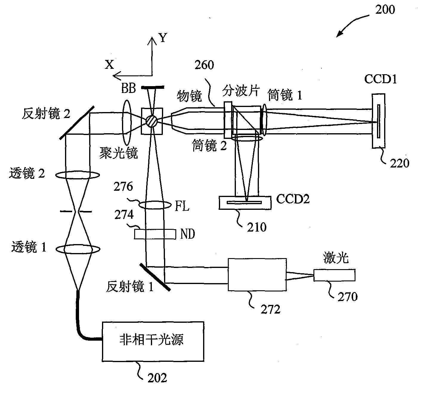
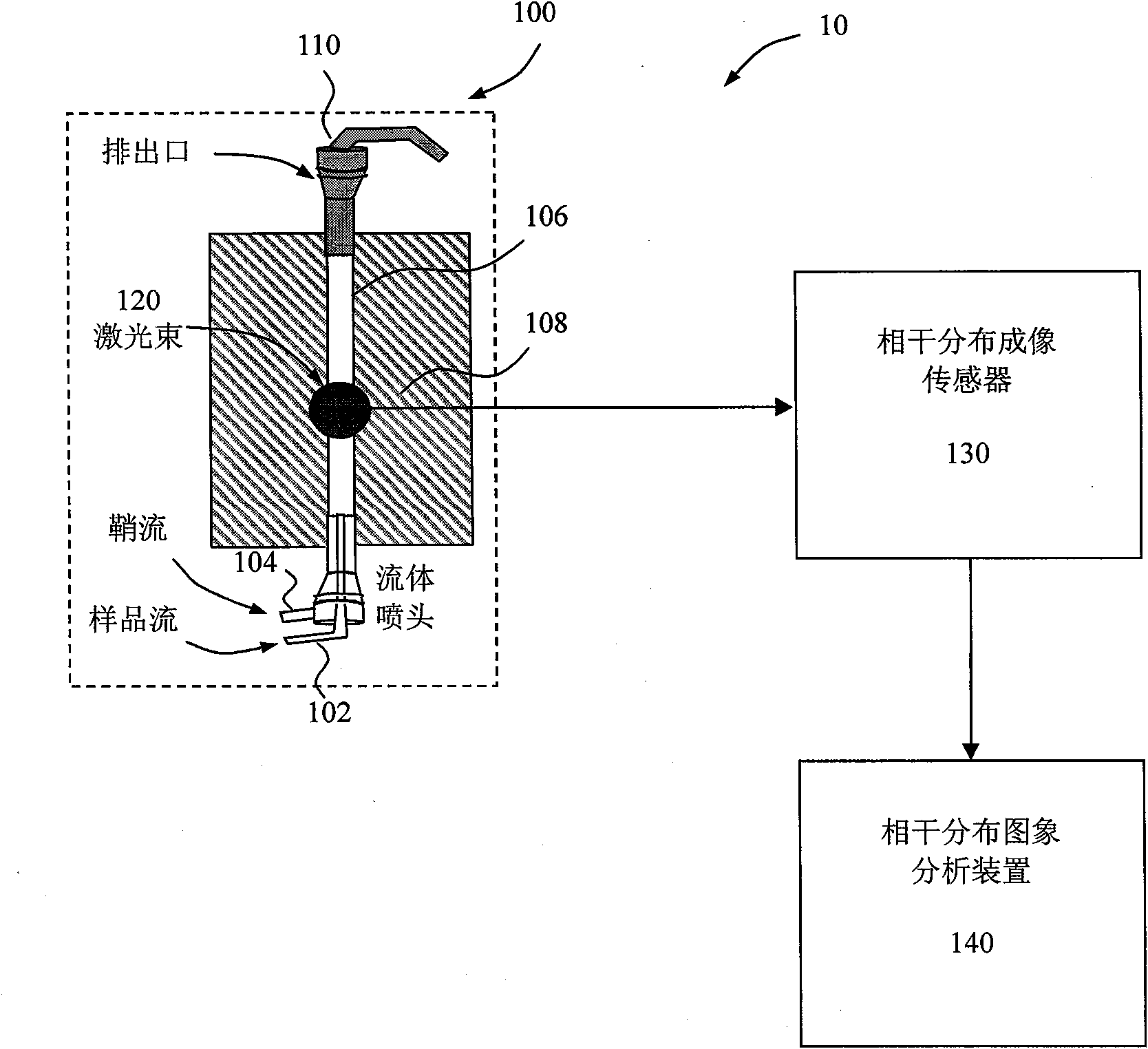
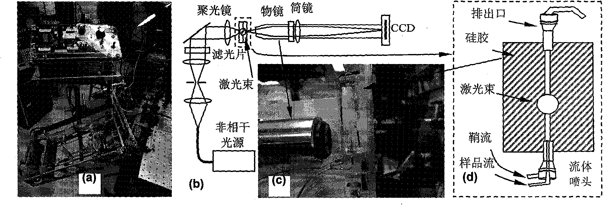
[0078] In order to extract more structural information from the elastically scattered light signal generated by particles excited by coherent scattered light, we fabricated a prototype microfluidic device-based flow cytometer to verify the use of a standard charge-coupled device A camera (Alta 2000, Apogee) measures the concept of diffraction images. The prototype is as Figure 6 a to Figure 6 As shown in d, its performance can reach the indicators listed in Table 1.

[0079] Table 1: Expected indicators of a dual-image microfluidic device-based flow cytometer

[0080] Flow rate adjustment range

0.01 to 0.50 m / s

Sample flow diameter adjustment range

20 to 150 microns

Half-angle width value of diffraction image measurement field of view*

32 degrees

Diffraction image measurement center angle value of field of view

5 degrees, 45 degrees to 135 degrees, 180 degrees

Lateral Resolution of Incoherent Image Measurement Field of V...

example 2

[0086] Figure 9 It is a schematic diagram of scattered light generated when a monochromatic coherent light beam is incident on a particle with a non-uniform internal optical refractive index n(r, λ), where r is the position coordinate vector, and λ is the incident and scattered light wavelengths (both are the same). The spatial coherence between scattered light fields results in a characteristic scattered light distribution, which can be measured as a diffraction (spatial domain) or speckle (spatial or temporal domain) image. Utilizing accurate wave models based on solutions of Maxwell's equations or wave equations can train pattern recognition software and potentially extract the light-refractive index distribution and associated 3D structural information inside scattered particles or cells from distributions representing elastically scattered light or from diffraction image data. The refractive index distribution function n(r, λ) inside the cell is related to its structure ...

example 3

[0090] We randomly selected 40 NALM-6 cultured cells at different growth cycle stages to establish a database that can be used as a training cell classification. Fluorescent microscopic image data of these cells were obtained after staining using a confocal microscope (model LSM 510, Zeiss). The reconstructed three-dimensional structure of cells can be used in two ways. One is to input into the differential time-domain parallel computing software to simulate and calculate the angular distribution of scattered light of cells excited by coherent light beams. The second is to analyze and define a variety of structural feature categories of cells, which are used to develop image pattern recognition software with cell classification functions. The light scattering process of a single biological cell can be considered as a problem of a plane electromagnetic wave incident on a dielectric particle placed in the host medium. To account for the change in polarization of scattered ligh...


PUM

PropertyMeasurementUnit
diameteraaaaaaaaaa
diameteraaaaaaaaaa
refractive indexaaaaaaaaaa

Abstract

The invention relates to a flow cytometer system for detection of three dimensional diffraction image and a method program product. The system includes a fluid controller configured to form a hydrodynamically focused flow stream including an outer sheath fluid and an inner core fluid; a coherent light source configured to illuminate a particle in the inner core fluid; a detector configured to detect a spatially coherent distribution of elastically scattered light from the particle excited by the coherent light source; an analyzing module configured to extract a three-dimensional morphology parameter of the particle from a spatially coherent distribution of the elastically scattered light. The method includes forming the hydrodynamically focused flow stream including the outer sheath fluid and the inner core fluid; illuminating one particle in the inner core fluid with the coherent light source; elastically detecting scattered light from the particle that is excited by the coherent light source; and extracting a three-dimensional morphology parameter of the particle from a spatially coherent distribution of the elastically scattered light. According to the invention, it is possible to perform single-particle analysis to a polulation containing a plurality of particles in a multi-dimensional feature space.

Description

[0001] related application [0002] This patent application claims priority from US Provisional Patent Application Serial No. 61 / 060,993, filed June 12, 2008, the entire disclosure of which is incorporated into this patent application. technical field [0003] The invention relates to a flow cytometer system, in particular to a flow cytometer system capable of measuring diffraction image signals and three-dimensional structural parameters. Background technique [0004] Flow cytometry systems are used in life science research to quantify communities containing large numbers of biological cells and particles. Consider illuminating a fluid that is hydrodynamically focused with a beam of light (usually a single wavelength). The fluid generally includes a sheath flow as a carrier and a sample flow containing many particles. Such fluids typically allow individual particles to flow through the illumination beam. Multiple sensors can be used to collect signals from the position o...

Claims


Application Information

Patent Timeline
Patent Type & Authority Applications(China)
IPC IPC(8): G01N15/14G01N33/487
CPCG01N2015/0222G01N15/0211G01N2015/1497G01N15/1475G01N15/147G01N2015/025
Inventor 胡新华肯尼思.M.雅可布卢军O.
Owner EAST CAROLINA UNIVERISTY
Why Eureka
Try Eureka