Adeno-associated virus capsid protein gene, corresponding protein and application of protein

A technology of capsid protein gene and virus, applied in the fields of biomedicine and genetic engineering

Inactive Publication Date: 2011-05-25
SUN YAT SEN UNIV
View PDF2 Cites 10 Cited by
  • Summary
  • Abstract
  • Description
  • Claims
  • Application Information

AI Technical Summary

Problems solved by technology

At the same time, AAV has not been found to be related to any human disease, which makes AAV vector one of the safest gene therapy and gene vaccine vectors so far
In all clinical trials using AAV vectors as gene transfer tools that have been conducted so far, no significant side effects related to AAV vectors have been found

Method used


Image

  • Adeno-associated virus capsid protein gene, corresponding protein and application of protein
  • Adeno-associated virus capsid protein gene, corresponding protein and application of protein
  • Adeno-associated virus capsid protein gene, corresponding protein and application of protein

Examples


Embodiment 1

[0026] Cloning of the new AAV capsid protein gene of embodiment 1

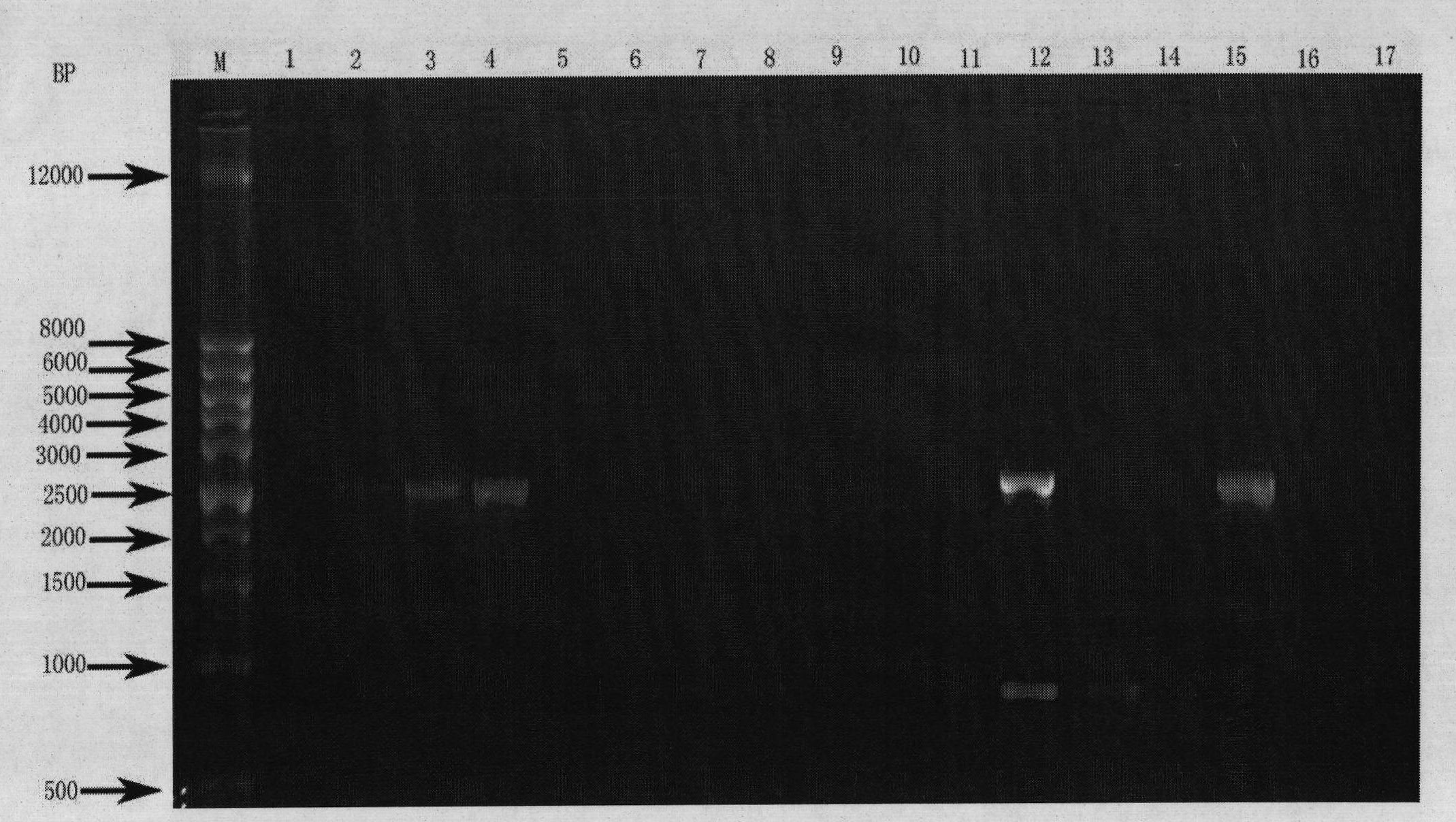
[0027] Human tissue samples were taken, and genomic DNA was extracted with a genomic DNA extraction kit (Qiagen, USA, catalog number: 69504), followed by PCR reaction. The PCR primers used are: the forward primer is shown in SEQ ID NO:3, and the reverse primer is shown in SEQ ID NO:4. PCR primers were synthesized by Shanghai Sangon Bioengineering Technology Service Co., Ltd. The PCR reaction system is: PCR enzyme 0.25uL (Bao Biological Engineering (Dalian) Co., Ltd., China, product number: DR001A), 10×PCR buffer 5uL, dNTP mixture 4uL (each final concentration is 0.2mM), forward primer (final concentration 0.2uM), reverse primer (final concentration 0.2uM), genomic DNA (100ng), add water to 50uL. The PCR program is: 94 degrees for 30 seconds, 65 degrees for 30 seconds, 72 degrees for 3 minutes, 30 cycles. Take 5uL PCR product for electrophoresis, and the expected band size is about 3.0kb. The result is as ...

Embodiment 2

[0028] Example 2 Preparation of AAV vector using the new AAV capsid protein packaging of the present invention

[0029] Preparation of AAV vectors was performed using a triple plasmid co-transfection method. Specifically: take 1000ug of the auxiliary plasmid ΔF6 carrying the adenovirus fragment, 500ug of the cis plasmid pAAV.CMV.EGFP, and 500ug of the plasmid containing the new capsid protein gene, and add CaCl 2 solution to a final concentration of 0.25M, with an equal volume of HBS buffer (5g HEPES, 8g NaCl, 0.37g KCl, 0.14g Na 2 HPO 4 .2H 2 O dissolved in 1 liter of double distilled water) mixed, transfected human embryonic kidney cell line-293 cells. The cells were harvested 3 days after transfection, and after repeated freezing and thawing three times, they were purified according to the literature method (Fisher, Gao, et al., 1996): using cesium chloride isopycnic gradient centrifugation to purify, and adding sterile glycerol after purification (final Concentration: ...

Embodiment 3

[0030] Example 3 In vitro infection experiment of the AAV vector packaged by the new AAV capsid protein of the present invention

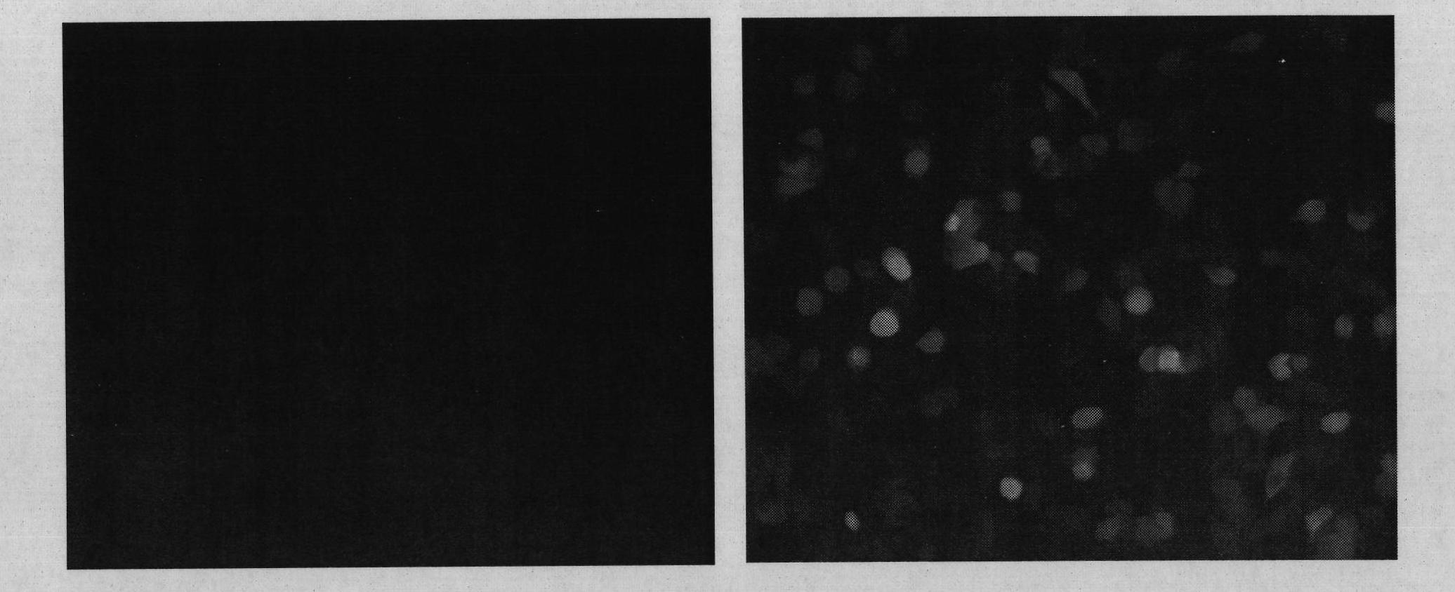
[0031]One day before infection, 293 cells were taken, digested with trypsin / EDTA solution, and plated on a 24-well cell plate at a ratio of 1:2.5. On the day of infection, the culture medium in the cell plate was replaced, and the AAV vector particles prepared according to the above method (Example 2) were added to each well of the cells (1×10 gc of the vector particles per well), and PBS was used as a negative control. Place in cell culture incubator. After culturing for 24 hours, use a fluorescence microscope to observe the expression of green fluorescent protein (EGFP), and take pictures ( figure 2 ). The results show that the AAV vector packaged with the novel AAV capsid protein disclosed by the invention can effectively introduce genes into cells cultured in vitro.


PUM


Abstract

The invention discloses an adeno-associated virus capsid protein gene, a corresponding protein and the applications of the protein. The nucleotide sequence of the adeno-associated virus capsid protein gene is shown in SEQ ID NO:1 and the amino acid sequence of the protein coded by the gene is shown in SEQ ID NO:2. The adeno-associated virus vector packaged by the adeno-associated virus capsid protein can be used to effectively introduce foreign genes in cells outside the body or in the body and prepare the medicines related to gene therapy or gene vaccines.

Description

technical field [0001] The invention relates to the fields of genetic engineering and biomedicine, in particular to an adeno-associated virus capsid protein gene, a corresponding protein and applications thereof. Background technique [0002] Adeno-associated virus (Adeno-associated virus, AAV, also translated as adeno-associated virus or adeno-associated virus) belongs to the Parvoviridae dependent virus genus. AAV virus particles consist of two parts: first, an icosahedron-like protein capsid, in which single-stranded viral genomic DNA is wrapped. Both ends of the genomic DNA are inverted repeats (Invert Terminal Repeat, ITR). There are two genes between the ITRs: Rep gene and Cap gene, the former is related to virus replication and encodes four Rep proteins, and the latter encodes three capsid proteins VP1, VP2 and VP3. VP1, VP2, and VP3 are located in the same reading frame and are translated by ribosomes from different start codons. In terms of amino acid sequence, V...

Claims


Application Information

Patent Timeline
Patent Type & Authority Applications(China)
IPC IPC(8): C12N15/35C12N15/864C12N1/15C12N1/19C12N1/21C07K14/015A61K48/00A61K39/00
Inventor 卢建溪王强李刚
Owner SUN YAT SEN UNIV
Why Eureka
Try Eureka